Review
. 2015 Feb;78(3):036601.
doi: 10.1088/0034-4885/78/3/036601. Epub 2015 Feb 26.

Material properties of biofilms-a review of methods for understanding permeability and mechanics

Affiliations
Review

Material properties of biofilms-a review of methods for understanding permeability and mechanics

Nicole Billings et al. Rep Prog Phys. 2015 Feb.

Abstract

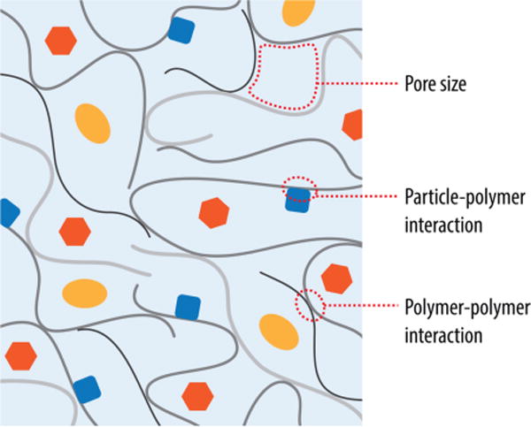
Microorganisms can form biofilms, which are multicellular communities surrounded by a hydrated extracellular matrix of polymers. Central properties of the biofilm are governed by this extracellular matrix, which provides mechanical stability to the 3D biofilm structure, regulates the ability of the biofilm to adhere to surfaces, and determines the ability of the biofilm to adsorb gases, solutes, and foreign cells. Despite their critical relevance for understanding and eliminating of biofilms, the materials properties of the extracellular matrix are understudied. Here, we offer the reader a guide to current technologies that can be utilized to specifically assess the permeability and mechanical properties of the biofilm matrix and its interacting components. In particular, we highlight technological advances in instrumentation and interactions between multiple disciplines that have broadened the spectrum of methods available to conduct these studies. We review pioneering work that furthers our understanding of the material properties of biofilms.


Figures

Figure 1
Figure 1
The biofilm matrix is comprised of entangled polymers (polysaccharides, DNA, proteins) that affect the permeability and mechanical properties of the entire biofilm. To understand the biophysical properties of the biofilm several questions need to be addressed. For example, what is the pore size of the matrix? Does a specific substrate interact with the matrix components? Which structural components of the matrix regulate the permeability properties? Is the matrix a static arrangement or do the individual components engage in dynamic rearrangements?
Figure 2
Figure 2
Conceptual scheme of a biofilm with an illustration of tools currently used for studies of biofilm permeability and mechanics. Fluorescence microscopy techniques such as FRAP, FCS, and fluorescence intensity measurements offer methods to visualize the transport of solutes and particles. Microelectrodes can be directly inserted into regions of interest to quantify concentration gradients of gasses and solutes within biofilms. Macroscale rheological measurements provide insight into the mechanical properties of a biofilm in total, where as a microrheological assessment of specific regions within a biofilm from single particle tracking provides information on the local properties of materials.
Figure 3
Figure 3
Fluorescence microscopy and nuclear magnetic resonance (NMR) can be used to observe channel development and fluid transport in biofilms. Bacillus subtilis biofilms grown on an agar surface where channels are visualized with fluorescent dye (a,b) Reprinted with permission from (Wilking et al., 2013). Copyright (2012) National Academy of Sciences, U.S.A. An NMR setup for imaging biofilms in situ (c) Horizontal and vertical 2D MRI sections of a Shewanella oneidensis biofilm (d). 3D MRI rendering (left) and transmitted light image (right) of a S. oneidensis biofilm (e). Reprinted by permission from Macmillan Publishers Ltd: The ISME Journal (McLean et al., 2008b), copyright (2008).
Figure 4
Figure 4
Examples of techniques that can be used to determine biofilm material properties. A rheometer setup in which a natural biofilm sample attached to a membrane can be tested (Korstgens et al., 2001a) (a). Copyright (2001) Institute of Physics Publishing. Staphylococcus aureus biofilm at 8 hours, with the tracks of bacterial motion (b). Scale bar is 5 mm. Reprinted with permission from (Rogers et al., 2008). Copyright (2008) American Chemical Society. A magnetic tweezers setup for monitoring biofilms grown in flow cells (c) Reprinted with permission from (Galy et al., 2012). Copyright (2012) Elsevier. SEM images of biofilm coated beads used for AFM measurements (d). The bead on the left is surrounded by younger biofilm than the bead on the right. Scale bars are 30 μm. Reprinted with permission from (Lau et al., 2009) Copyright (2009) Elsevier.

References

    1. AGGARWAL S, HOZALSKI RM. Determination of biofilm mechanical properties from tensile tests performed using a micro-cantilever method. Biofouling. 2010;26:479–486. - PubMed
    1. AGGARWAL S, POPPELE EH, HOZALSKI RM. Development and testing of a novel microcantilever technique for measuring the cohesive strength of intact biofilms. Biotechnology and Bioengineering. 2010;105:924–934. - PubMed
    1. AHIMOU F, SEMMENS MJ, NOVAK PJ, HAUGSTAD G. Biofilm cohesiveness measurement using a novel atomic force microscopy methodology. Appl Environ Microbiol. 2007;73:2897–904. - PMC - PubMed
    1. AMSDEN B. Solute diffusion within hydrogels. Mechanisms and models. Macromolecules. 1998;31:8382–8395.
    1. AXELROD D, KOPPEL DE, SCHLESSINGER J, ELSON E, WEBB WW. Mobility measurement by analysis of fluorescence photobleaching recovery kinetics. Biophysical Journal. 1976;16:1055–69. - PMC - PubMed

Publication types

LinkOut - more resources