Review
. 2015 May;396(5):415-44.
doi: 10.1515/hsz-2015-0102.

Thiol-based redox switches in prokaryotes

Review

Thiol-based redox switches in prokaryotes

Melanie Hillion et al. Biol Chem. 2015 May.

Abstract

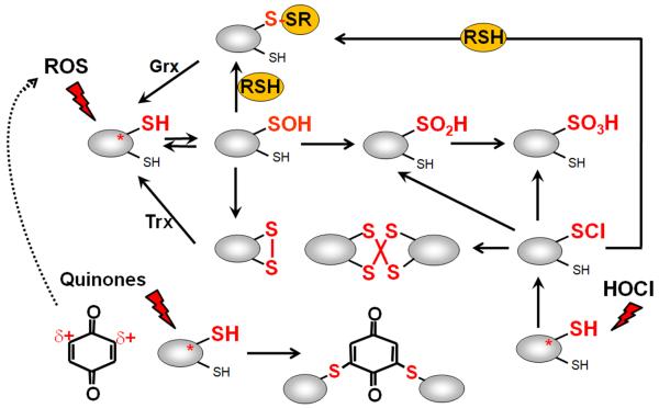
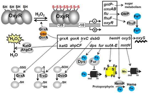
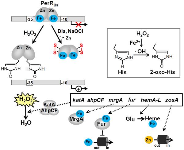
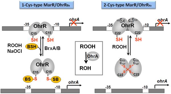
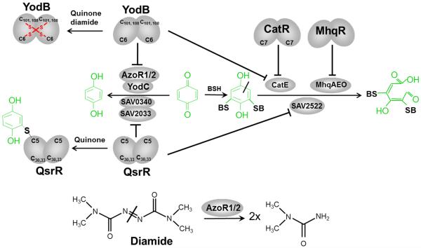
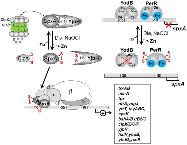
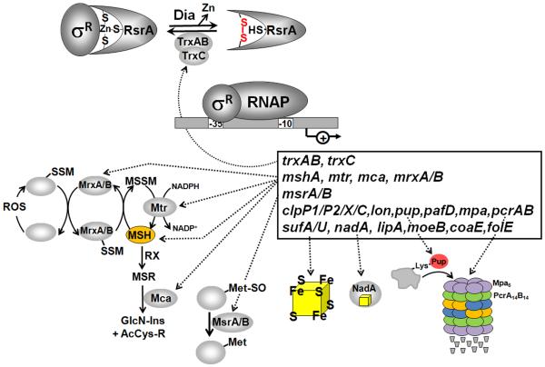
Bacteria encounter reactive oxygen species (ROS) as a consequence of the aerobic life or as an oxidative burst of activated neutrophils during infections. In addition, bacteria are exposed to other redox-active compounds, including hypochloric acid (HOCl) and reactive electrophilic species (RES) such as quinones and aldehydes. These reactive species often target the thiol groups of cysteines in proteins and lead to thiol-disulfide switches in redox-sensing regulators to activate specific detoxification pathways and to restore the redox balance. Here, we review bacterial thiol-based redox sensors that specifically sense ROS, RES and HOCl via thiol-based mechanisms and regulate gene transcription in Gram-positive model bacteria and in human pathogens, such as Staphylococcus aureus and Mycobacterium tuberculosis. We also pay particular attention to emerging widely conserved HOCl-specific redox regulators that have been recently characterized in Escherichia coli. Different mechanisms are used to sense and respond to ROS, RES and HOCl by 1-Cys-type and 2-Cys-type thiol-based redox sensors that include versatile thiol-disulfide switches (OxyR, OhrR, HypR, YodB, NemR, RclR, Spx, RsrA/RshA) or alternative Cys phosphorylations (SarZ, MgrA, SarA), thiol-S-alkylation (QsrR), His-oxidation (PerR) and methionine oxidation (HypT). In pathogenic bacteria, these redox-sensing regulators are often important virulence regulators and required for adapation to the host immune defense.


Figures

Figure 1
Figure 1. Thiol-chemistry of ROS, RES and HOCl with redox-sensing regulators
Reversible thiol-oxidation by ROS leads first to a Cys sulfenic acid intermediate (R-SOH) that is unstable and reacts further to form intramolecular and intermolecular disulfides or mixed disulfides with LMW thiols, such as glutathione, bacillithiol, cysteine or CoASH, termed as S-thiolations. The Cys sulfenic acid can be also overoxidized to Cys sulfinic and sulfonic acids. Reactive electrophiles (RES) such as quinones have been shown to act via the S-alkylation and oxidation mode with quinone-sensing redox regulators. HOCl causes first chlorination of Cys thiol goups to the unstable sulfenylchloride which react further to form protein disulfides and S-thiolations in the presence of proximal thiols. In the absence of another thiol the sulfenylchloride rapidly forms irreversible Cys sulfinic or sulfonic acids (Hawkins et al, 2003).
Figure 2
Figure 2. The thiol-disulfide-switch model of E. coli OxyR and functions of the OxyR regulon
OxyR responds to hydrogen peroxide (H2O2) in E. coli and other bacteria. The conserved C199 and C208 residues of OxyR are essential for redox-sensing of OxyR. C199 is initially oxidized to the sulfenic acid intermediate that rapidly reacts further to form an intramolecular disulfide with C208. Oxidized OxyR binds as a tetramer to promoter regions of target genes and activates transcription of peroxide detoxification genes by contact with αCTD of RNA polymerase. OxyR positively controls genes for peroxide detoxification, such as catalase and peroxiredoxin (katG, ahpCF), Fe-storage miniferritin (dps), glutaredoxin, thioredoxin and glutathione reductase (grxA, trxC, gor), sulfenic acid oxidoreductase (dsbG), ferric uptake regulator (fur), Fe-S-cluster assembly machinery (sufABCDE), ferrochelatase (hemH), manganese import (mntH) and the small RNA (oxyS). OxyR negatively regulates its own expression and that of the genes for the ferric ion reductase (fhuF), the outer membrane protein (flu), the mannonate hydrolase (uxuAB) and gluconate permease (gntP). OxyR is regenerated by the glutaredoxin/GSH/Gor system upon return to non-stress conditions. Examples for OxyR regulon genes and their functions are also listed in Table 1.
Figure 3
Figure 3. Redox-regulation of B. subtilis PerRBs by peroxides (metal-catalyzed histidine oxidation) and by diamide stress (intramolecular disulfides) and functions of the PerR regulon members
PerR has a regulatory Fe2+or Mn2+-binding site with Asp and His residues as ligands and a structural Zn2+-binding site coordinated by four cysteine residues. Reaction of PerR-Fe with H2O2 leads to a Fenton reaction generating HO with subsequent oxidation of His37 and His91 to the 2-oxo-His derivatives that inactivate the PerR repressor under H2O2 stress leading to up-regulation of the PerR regulon genes. Under disulfide stress conditions provoked by diamide and NaOCl, PerR is inactivated by intramolecular disulfide formation in the Zn-binding site that also lead to derepression of the PerR regulon genes. The PerR regulon includes genes with antioxidant functions, such as the catalase and peroxiredoxin (katA, ahpCF), Fe-storage miniferritin (mrgA), ferric uptake regulator (fur), heme biosynthesis enzymes (hemAXCDBL) and zinc uptake systems (zosA). Examples for PerR regulon genes of B. subtilis and their functions are also listed in Table 2.
Figure 4
Figure 4. Proposed redox-regulation of PerRCa in the strict anaerobe Clostridium acetobutylicum by oxygen, peroxides, superoxide and functions of the PerR regulon members
The PerR repressor is proposed to sense O2, O2 and H2O2 by oxidation of two His residues in the conserved regulatory Fe-binding site leading to 2-oxo-His generation. This causes PerR inactivation and derepression of the PerR regulon genes. The PerR regulon controls genes for an anaerobic oxygen and ROS detoxification pathway, including the oxygen-reducing flavodiiron proteins (fprA2), reverse rubrerythrins and peroxidases (rbr3A, rbr3B), peroxiredoxin (bcp), thiol peroxidase (tpx), glutathione peroxidase (bsa2), glutaredoxin (grx), superoxide-reducing desulfoferrodoxin (dfx), rubredoxin (rd) and the NADH-dependent rubredoxin oxidoreductase (nror). Examples for PerR regulon genes of C. acetobutylicum and their functions are also listed in Table 2.
Figure 5
Figure 5. Thiol-based redox sensing of organic hydroperoxides by 1-Cys and 2-Cys-type MarR/OhrR regulators
OhrR controls the OhrA thiol-dependent peroxiredoxin and contains one conserved Cys15 residue in B. subtilis and three Cys residues (C22, C127 and C131) in X. campestris. The 1-Cys OhrR protein of B. subtilis is initially oxidized by CHP and NaOCl to the Cys sulfenic acid, which reacts further with bacillithiol (BSH) to form S-bacillithiolated OhrR. The 2-Cys OhrR protein of X. campestris is regulated by intersubunit disulfide formation between the redox-sensing C22 and C127′ of opposing subunits. These different thiol-disulfide switches inactivate OhrR proteins leading to derepression of the peroxidase OhrA that functions in detoxification of organic hydroperoxides. Regeneration of S-bacillithiolated OhrR involves the bacilliredoxins BrxA and BrxB in B. subtilis (Gaballa et al, 2014).
Figure 6
Figure 6. Redox-sensing by the MarR/OhrR-family regulator SarZ of S. aureus
In S. aureus, the MarR/OhrR-family regulator SarZ functions as global regulator for ROS detoxification, antibiotic resistance and virulence functions and contains a single Cys13 required for redox-sensing. The DNA-binding activity of SarZ was shown to be reversibly redox-regulated by S-thiolation with a synthetic benzene thiol (Poor et al, 2009) and by cysteine phosphorylation via the eukaryotic-like serine/threonine kinase (Stk1) and phosphatase (Stp1)(Sun et al, 2012). SarZ controls genes for the ohr peroxiredoxin, cell surface proteins, antibiotic resistance efflux pumps (norB, tet38), amino acid, sugar, fatty acid and anaerobic metabolism (pflAB). Examples for SarZ regulon genes are listed in Table 3.
Figure 7
Figure 7. Redox-sensing of RES by the MarR/DUF24-regulators YodB in B. subtilis and QsrR in S. aureus
Exposure of B. subtilis to quinones induces quinone detoxification regulons controlled by the MarR-type repressors MhqR, YodB and CatR. In S. aureus, the homologous quinone-sensing YodB (QsrR) repressor responds to quinones and controls paralogous quinone reductases, dioxygenases and nitroreductases. The redox-sensing YodB and CatR repressors are inactivated by the oxidative mode of quinone leading to disulfide formation that involves the conserved Cys6 or Cys7 residues (Antelmann et al, 2008; Antelmann & Helmann, 2011; Chi et al, 2010a; Chi et al, 2010b; Towe et al, 2007). In S. aureus, YodB (QsrR) with mutated C-terminal Cys residues senses quinones by thiol-S-alkylation of Cys5 leading to up-regulation of the dioxygenase SAV2522, the quinone reductase SAV0340 and the nitroreductase SAV2033 (Ji et al, 2013). The thiol-dependent dioxygenases MhqA, MhqE, MhqO, CatE of B. subtilis and SAV2522 of S. aureus are involved in specific ring-cleavage of quinones-S-adducts. The quinone reductases AzoR1, AzoR2 of B. subtilis and SAV0340 of S. aureus and the nitroreductases YodC, MhqN of B. subtilis and SAV2033 of S. aureus catalyze the reduction of quinones to redox stable hydroquinones.
Figure 8
Figure 8. Post-translational and transcriptional control of SpxA by disulfide stress in B. subtilis
Under non-stressed conditions, SpxA is unstable and targeted by the YjbH adaptor protein to the ClpXP machinery for proteolytic degradation. The stability of SpxA is increased by diamide due to oxidation of SpxA, YjbH and ClpX that prevents SpxA degradation. SpxA is oxidized in its redox-active CXXC motif and binds to the αCTD of RNAP resulting in transcriptional activation of the SpxA disulfide stress regulon. Transcription of spxA is regulated by the repressors YodB and PerR that are oxidized and inactivated under diamide stress leading to increased spxA transcription. SpxA controls a large regulon of genes encoding thioredoxin/thioredoxin reductase (trxAB), thiol peroxidase (tpx), FMN-dependent oxidoreductases (nfrA, yugJ), methionine sulfoxide reductase (msrA), cysteine biosynthesis enzymes (yrrT-operon, cysK, tcyABC), bacillithiol biosynthesis enzymes (bshA, bshB1, bshB2 and bshC), the protein quality control Clp machinery (clpX, clpE, clpC, clpP, yjbH) and several thiol-based redox regulators (hxlR, yodB, yhdQ, yceK). Examples for SpxA regulon genes and their functions are listed in Table 5.
Figure 9
Figure 9. Redox regulation of the ZAS factor RsrA and its cognate sigma factor SigR in S. coelicolor and role of the SigR regulon
RsrA is a redox-sensitive zinc-binding anti sigma (ZAS) factor in S. coelicolor that sequesters its cognate sigma factor SigR under reducing conditions. Diamide stress leads to intramolecular disulfide formation in RsrA, resulting in Zn release and relief of SigR. Free SigR activates transcription of the SigR regulon that functions to restore the thiol-redox balance. The SigR regulon includes genes for thioredoxins and thioredoxin reductase (trxAB, trxC), enzymes for mycothiol biosynthesis and recycling (mshA, mca, mtr), mycoredoxins (mrxA, mrxB), methionine sulfoxide reductase (msrA, msrB), protein quality control machinery (pepN, ssrA, clpP1P2, clpX, clpC, lon), ubiquitin-like protein-conjugation pathway and proteasomal components (pup, mpa, pafD, prcAB), Fe-S assembly components (sufA, sufU) and Fe-S containing enzymes (nadA, lipA), biosynthesis enzymes for the cofactors Fe-S, folate, CoASH and lipoic acid (moeB, coaE, folE, lipA). Examples for SigR regulon genes and their functions are also listed in Table 6.

Similar articles

Cited by

References

    1. Antelmann H, Engelmann S, Schmid R, Sorokin A, Lapidus A, Hecker M. Expression of a stress- and starvation-induced dps/pexB-homologous gene is controlled by the alternative sigma factor sigmaB in Bacillus subtilis. J Bacteriol. 1997;179:7251–7256. - PMC - PubMed
    1. Antelmann H, Hecker M, Zuber P. Proteomic signatures uncover thiol-specific electrophile resistance mechanisms in Bacillus subtilis. Expert Rev Proteomics. 2008;5:77–90. - PubMed
    1. Antelmann H, Helmann JD. Thiol-based redox switches and gene regulation. Antioxid Redox Signal. 2011;14:1049–1063. - PMC - PubMed
    1. Atichartpongkul S, Loprasert S, Vattanaviboon P, Whangsuk W, Helmann JD, Mongkolsuk S. Bacterial Ohr and OsmC paralogues define two protein families with distinct functions and patterns of expression. Microbiology. 2001;147:1775–1782. - PubMed
    1. Bae JB, Park JH, Hahn MY, Kim MS, Roe JH. Redox-dependent changes in RsrA, an anti-sigma factor in Streptomyces coelicolor: zinc release and disulfide bond formation. J Mol Biol. 2004;335:425–435. - PubMed

Publication types

Substances

LinkOut - more resources