

doi: 10.1039/c5cc00082c.
Light sensitization of DNA nanostructures via incorporation of photo-cleavable spacers
Affiliations
- PMID: 25720373
- PMCID: PMC4394657
- DOI: 10.1039/c5cc00082c
Item in Clipboard
Light sensitization of DNA nanostructures via incorporation of photo-cleavable spacers
Chem Commun (Camb).
.
Abstract
Using light irradiation as a trigger, large-scale structural reconfiguration of DNA nanostructures is demonstrated. We incorporated photo-cleavable spacers at strategic locations within the short oligonucleotide strands connecting adjacent helices within a DNA origami sphere, and then used light to transform the sphere into two tethered hemispheres.
Figures
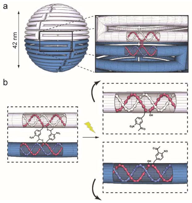
(a) Schematic diagram of the light reconfigurable DNA sphere. The cylinder represents the scaffold DNA strand, and the top and bottom hemispheres are shown as white and blue respectively. The excess scaffold is omitted. Insert: The red helices represent the crossover strands that connect the top (white) and bottom (blue) scaffold helices. (b) Depiction of the separation of adjacent helices that would result when photo-crossovers were cleaved with light.
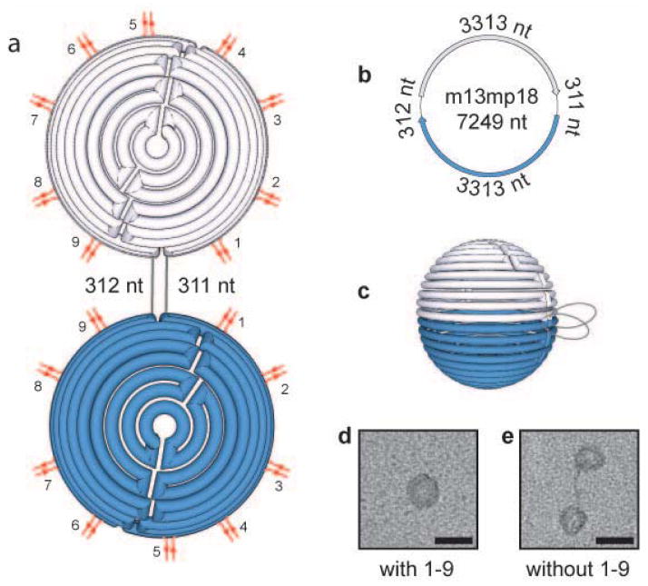
DNA sphere reconfiguration upon modification of staple crossovers at strategic locations connecting the two equator scaffold strands. (a) Flattened, (b) unfolded, and (c) full depiction of the DNA sphere. Red double lines indicate the location of the crossovers at the hemisphere equator. Grey lines indicate the location of the unfolded scaffold DNA. TEM images of the nanostructure created in the presence (d) and absence (e) of the staple crossover pairs 1–9. Scale bars equal 50 nm. nt = nucleotides
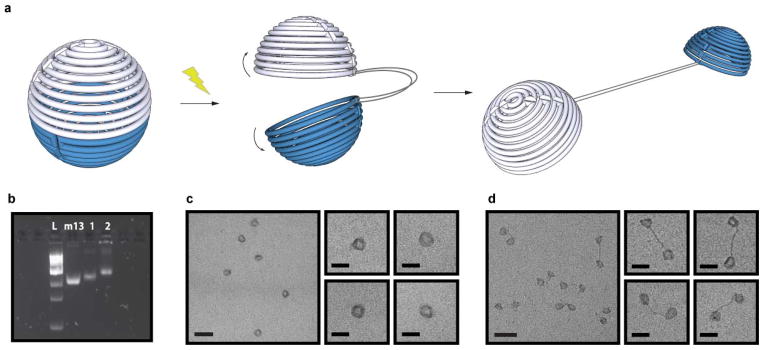
Light-triggered transformation of the DNA sphere into to two hemispheres. (a) Schematic depiction of the structural reconfiguration of the sphere upon exposure to light. (b) Fluorescent image of SYBR Safe stained 1.8% agarose gel showing successful photo-reconfiguration of the DNA sphere. L = 1kb ladder, m13 = m13 DNA scaffold, 1 = Closed sphere containing o-nb photo-crossovers, 2 = Sphere containing o-nb photo-crossovers after 10 min light irradiation. TEM images of the nanostructures before (c) and after light irradiation (d). Scale bars are 100 nm (zoom out) and 50 nm (zoom in).
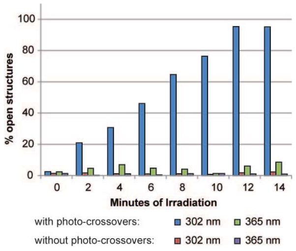
Kinetics of light induced structural reconfiguration. Fraction of total structures that were identified as in open configurations were quantified upon irradiation with varying period of light. Spheres containing either unmodified or photo-crossovers were irradiated with either 302 nm or 365 nm light. Only nanostructures containing photo-crossovers irradiated at 302 nm showed increased opening over time.
References
Publication types
MeSH terms
Substances
Grants and funding
LinkOut - more resources
Full Text Sources
Other Literature Sources