Randomized Controlled Trial
. 2015 Jul;64(7):2624-35.
doi: 10.2337/db14-0976. Epub 2015 Feb 26.

Exenatide Protects Against Glucose- and Lipid-Induced Endothelial Dysfunction: Evidence for Direct Vasodilation Effect of GLP-1 Receptor Agonists in Humans

Affiliations
Randomized Controlled Trial

Exenatide Protects Against Glucose- and Lipid-Induced Endothelial Dysfunction: Evidence for Direct Vasodilation Effect of GLP-1 Receptor Agonists in Humans

Juraj Koska et al. Diabetes. 2015 Jul.

Abstract

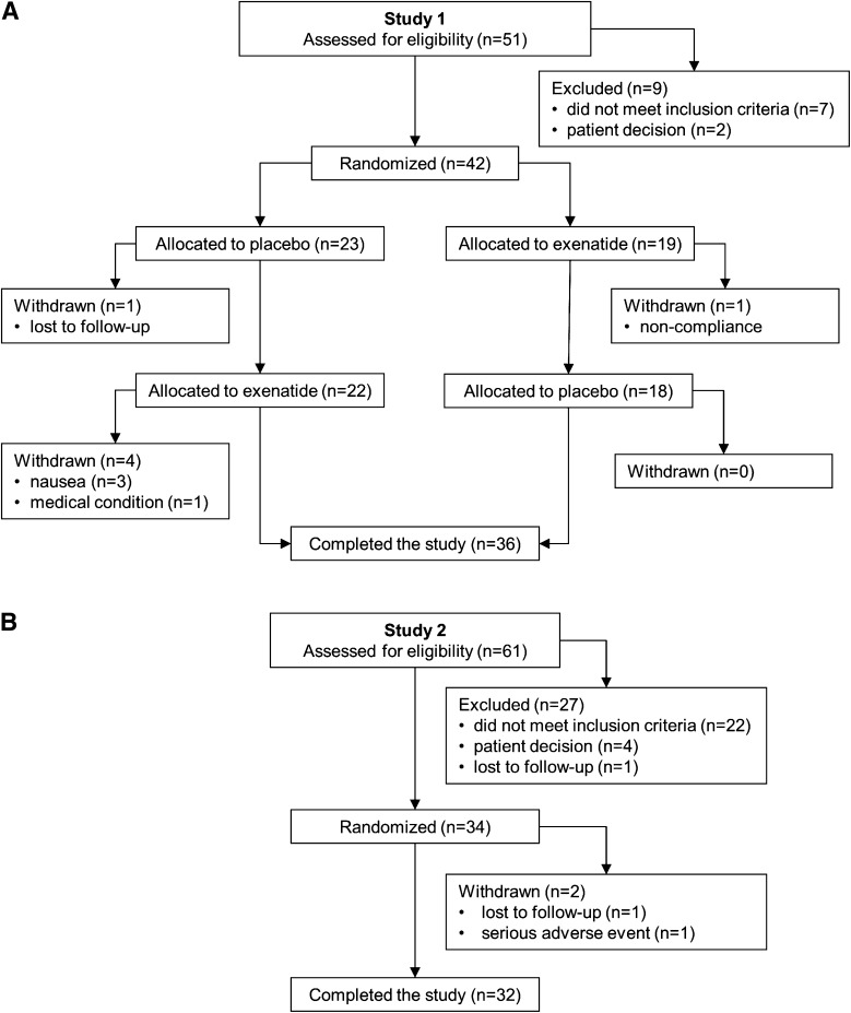
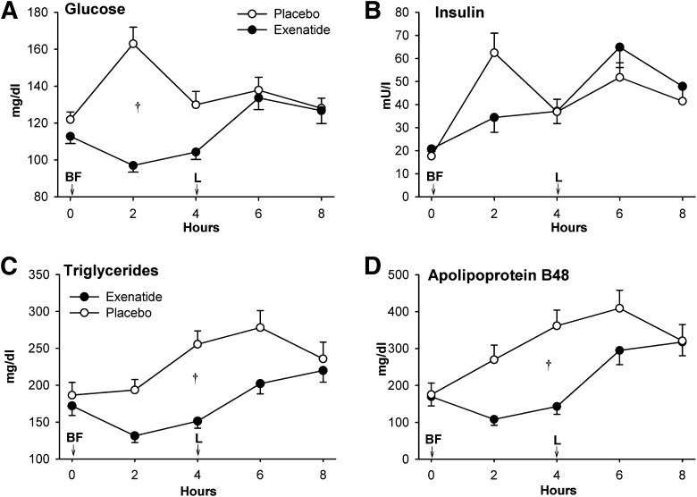
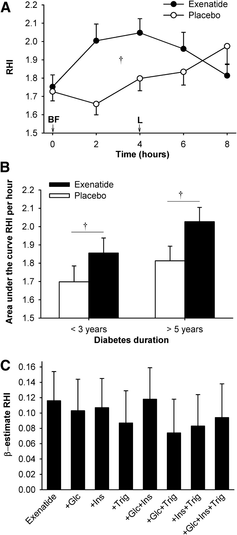
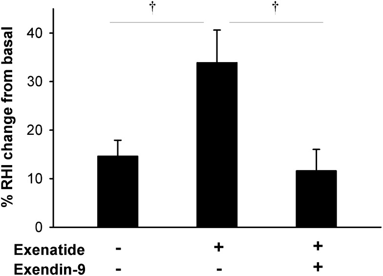
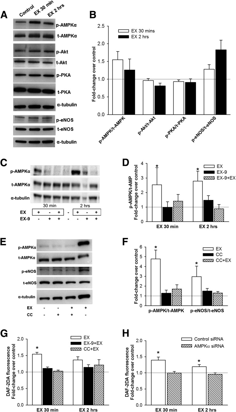
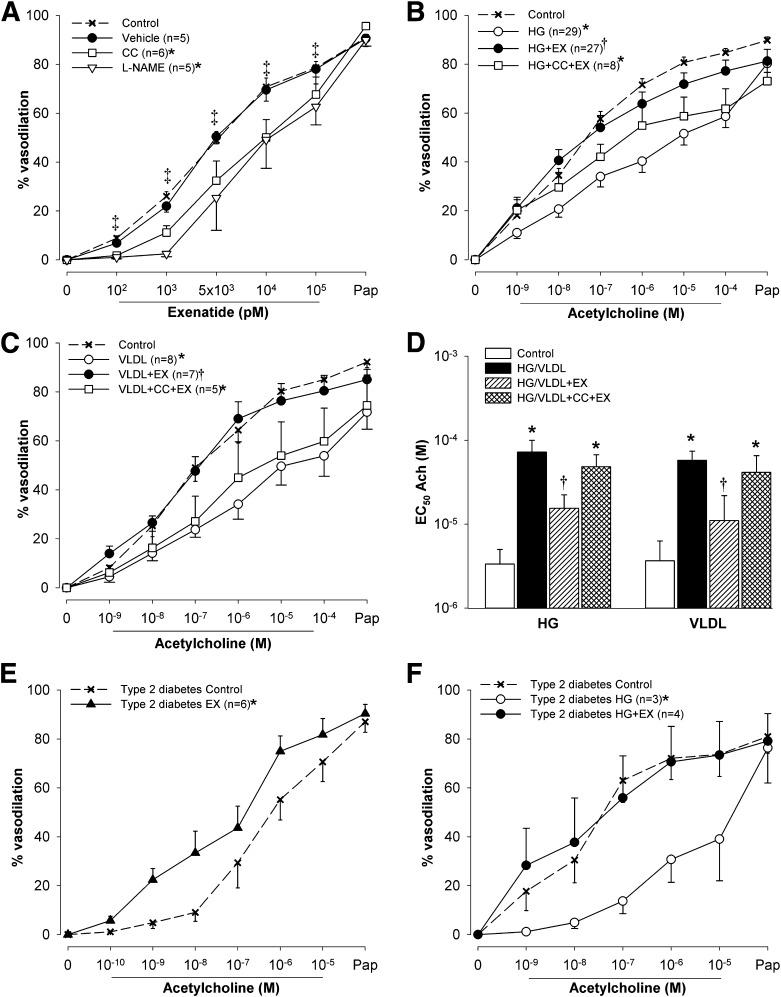
GLP-1 receptor (GLP-1R) agonists may improve endothelial function (EF) via metabolic improvement and direct vascular action. The current study determined the effect of GLP-1R agonist exenatide on postprandial EF in type 2 diabetes and the mechanisms underlying GLP-1R agonist-mediated vasodilation. Two crossover studies were conducted: 36 participants with type 2 diabetes received subcutaneous exenatide or placebo for 11 days and EF, and glucose and lipid responses to breakfast and lunch were determined; and 32 participants with impaired glucose tolerance (IGT) or diet-controlled type 2 diabetes had EF measured before and after intravenous exenatide, with or without the GLP-1R antagonist exendin-9. Mechanisms of GLP-1R agonist action were studied ex vivo on human subcutaneous adipose tissue arterioles and endothelial cells. Subcutaneous exenatide increased postprandial EF independent of reductions in plasma glucose and triglycerides. Intravenous exenatide increased fasting EF, and exendin-9 abolished this effect. Exenatide elicited eNOS activation and NO production in endothelial cells, and induced dose-dependent vasorelaxation and reduced high-glucose or lipid-induced endothelial dysfunction in arterioles ex vivo. These effects were reduced with AMPK inhibition. In conclusion, exenatide augmented postprandial EF in subjects with diabetes and prevented high-glucose and lipid-induced endothelial dysfunction in human arterioles. These effects were largely direct, via GLP-1R and AMPK activation.


Figures

Figure 1
Figure 1
Participant flow for studies 1 (A) and 2 (B).
Figure 2
Figure 2
The effect of exenatide on plasma glucose (A), insulin (B), triglycerides (C), and apoB48 (D) concentrations in study 1. On day 11 of therapy, immediately after initial blood sampling (time 0), study drug was injected and participants ingested a breakfast meal (BF) and 4 h later a lunch meal (L). Data are means ± SE. †P < 0.05, exenatide vs. placebo.
Figure 3
Figure 3
The effect of exenatide on EF (RHI) in study 1. RHI was calculated as the ratio of the average amplitude of the PAT signal over a 30 s time interval starting 90 s after blood pressure cuff deflation divided by the average amplitude of the PAT signal of a 3.5-min time period before cuff inflation. On day 11 of therapy, immediately after initial blood sampling (time 0), study drug was injected and participants ingested a breakfast meal (BF) and 4 h later a lunch meal (L). A: RHI over the 8-h test period. B: RHI area under the curve (normalized to 1 h, by trapezoid method) according to the duration of diabetes. Data are means ± SE. †P < 0.05, exenatide vs. placebo. C: Multivariate models of exenatide’s effect on RHI. The modeled effect is shown as β estimates and SE of exenatide’s effect on RHI before and after adjustment for plasma glucose (Glc), insulin (Ins), and triglyceride (Trig) concentrations. Adjustments for individual or combinations of these variables did not significantly reduce the effect of exenatide.
Figure 4
Figure 4
Percent change in RHI after intravenous infusion of exenatide, placebo, or exenatide + GLP-1R inhibitor exendin-9 in study 2. RHI was calculated as the ratio of the average amplitude of the PAT signal over a 30-s time interval starting 90 s after blood pressure cuff deflation divided by the average amplitude of the PAT signal of a 3.5-min time period before cuff inflation. Data are means ± SE. †P < 0.05 between treatments.
Figure 5
Figure 5
The effects of exenatide in vitro in human endothelial cells. A and B: Phosphorylation of AMPKα (Thr172), PKA (Thr197), Akt kinase (Ser473), and eNOS (Ser1177) in HAECs after treatment with 10 nmol/L exendin-4 (EX) for 30 min and 2 h (A: representative Western blot; B: densitometry analysis [means ± SE]; n = 6). C and D: AMPKα phosphorylation in HAECs after EX (30 min and 2 h) with or without pretreatment for 30 min with 1 μmol/L GLP-1R inhibitor EX-9 (C: representative Western blot; D: densitometry analysis; n = 7–8). E and F: AMPKα and eNOS phosphorylation in HAECs after EX (2 h) with or without 1-h pretreatment with 5 mol/L of AMPKα inhibitor CC (E: representative Western blot; F: densitometry analysis; n = 8–9). The effect of EX on NO production (by DAF-2DA fluorescence) in HAECs with or without pretreatment with EX-9 or CC (n = 4–7) (G) and in HUVECs with knocked-down AMPKα gene expression (siRNA, n = 6) (H). Phosphorylated bands were normalized to total bands and α-tubulin. Control, untreated cells. Data are means ± SE. *P < 0.05 vs. control.
Figure 6
Figure 6
The effects of exenatide (EX) ex vivo in isolated human adipose tissue arterioles. A: Vasodilation responses to increasing doses of EX followed by papaverine before (control) and after treatment with vehicle, eNOS inhibitor l-NAME (5 µmol/L), or AMPK inhibitor CC (1 µmol/L). B: Vasodilation responses to acetylcholine before (control) and after exposure to high glucose for 2 h (HG, 33 mmol/L), HG with addition of 10 nmol/L EX after 1 h (HG+EX), and HG+EX pretreated with 1 µmol/L CC (HG+EX+CC). C: Vasodilation responses to acetylcholine before (control) and after exposure to VLDL lipolysis products mixture for 2 h (VLDL, 150 μmol/L fatty acids), VLDL with addition of 10 nmol/L EX after 1 h (VLDL+EX), and VLDL+EX pretreated with 1 µmol/L CC. D: EC50 of acetylcholine from experiments shown in panels B and C. EC50 was calculated by nonlinear regression and variable slope (four parameters) and least squares fit (GraphPad Prism 5.0, San Diego CA). If the vessels dilated to <50% with maximum dose of acetylcholine, EC50 was set at 10−4 mol/L. Acetylcholine induced vasodilation in adipose tissue arterioles from subjects with type 2 diabetes before (control) and after 1-h exposure to 10 nmol/L EX (E) and HG and HG+EX (F). Data are means ± SE. *P < 0.05 vs. control; †P < 0.05 vs. HG or VLDL; ‡P < 0.05 vs. EX 0 pmol/L (tested in control only).

Comment in

References

    1. Halcox JP, Schenke WH, Zalos G, et al. . Prognostic value of coronary vascular endothelial dysfunction. Circulation 2002;106:653–658 - PubMed
    1. Gokce N, Keaney JF Jr, Hunter LM, Watkins MT, Menzoian JO, Vita JA. Risk stratification for postoperative cardiovascular events via noninvasive assessment of endothelial function: a prospective study. Circulation 2002;105:1567–1572 - PubMed
    1. Yeboah J, Crouse JR, Hsu FC, Burke GL, Herrington DM. Brachial flow-mediated dilation predicts incident cardiovascular events in older adults: the Cardiovascular Health Study. Circulation 2007;115:2390–2397 - PubMed
    1. McVeigh GE, Brennan GM, Johnston GD, et al. . Impaired endothelium-dependent and independent vasodilation in patients with type 2 (non-insulin-dependent) diabetes mellitus. Diabetologia 1992;35:771–776 - PubMed
    1. Williams SB, Cusco JA, Roddy MA, Johnstone MT, Creager MA. Impaired nitric oxide-mediated vasodilation in patients with non-insulin-dependent diabetes mellitus. J Am Coll Cardiol 1996;27:567–574 - PubMed

Publication types

MeSH terms