

Hypoxia induces an undifferentiated phenotype of oral keratinocytes in vitro
- PMID: 25720390
- PMCID: PMC4410785
- DOI: 10.1159/000371342
Hypoxia induces an undifferentiated phenotype of oral keratinocytes in vitro
Abstract
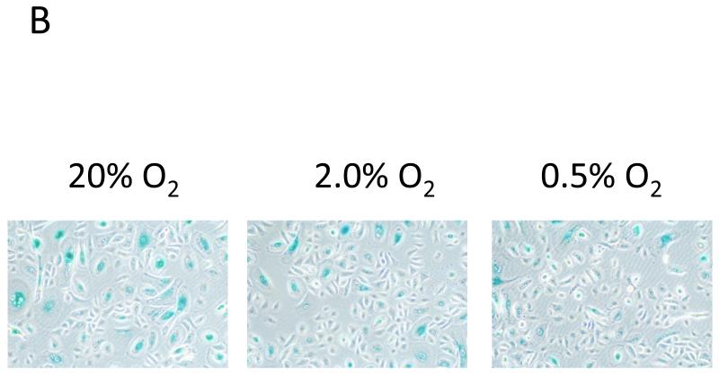
The aim of this study was to determine the effects of hypoxia on the proliferating potential and phenotype of primary human oral keratinocytes cultured at ambient oxygen tension (20%) or at different levels of hypoxia (2 and 0.5% O2). The effects of oxygen tensions on cellular metabolic activity, cell proliferation, clonogenicity and proliferation heterogeneity were measured. Cell cycle profiles were analyzed by a fluorescent-activated cell sorter, and p21(WAF1/CIP1) expression in the G0/G1 phase was also concomitantly quantitated. The expression levels of cell cycle regulatory proteins were examined by immunoblotting, and the cellular senescence was assessed by senescence-associated β-galactosidase staining. Basal and suprabasal keratinocyte phenotypes were determined by the expression levels of 14-3-3σ, p75(NTR) and α6 integrin. Despite having a lower metabolism, the proliferation rate and clonogenic potential were remarkably enhanced in hypoxic cells. The significantly higher percentage of cells in the G0/G1 phase under hypoxia and the expression patterns of cell cycle regulatory proteins in hypoxic cells were indicative of a state of cell cycle arrest in hypoxia. Furthermore, a decrease in the expression of p21(WAF1/CIP1) and p16(INK4A) and fewer β-galactosidase-positive cells suggested a quiescent phenotype rather than a senescent one in hypoxic cells. Compared with normoxic cells, the differential expression patterns of keratinocyte phenotypic markers suggest that hypoxic cells that generate minimal reactive oxygen species, suppress the mammalian target of rapamycin activity and express hypoxia-inducible factor-1α favor a basal cell phenotype. Thus, regardless of the predisposition to the state of cell cycle arrest, hypoxic conditions can maintain oral keratinocytes in vitro in an undifferentiated and quiescent state.
© 2015 S. Karger AG, Basel.
Figures


References
-
- Bath C, Yang S, Muttuvelu D, Fink T, Emmersen J, Vorum H, Hjortdal J, Zachar V. Hypoxia is a key regulator of limbal epithelial stem cell growth and differentiation. Stem Cell Res. 2013;10:349–360. - PubMed
-
- Baris OR, Klose A, Kloepper JE, Weiland D, Neuhaus JF, Schauen M, et al. The mitochondrial electron transport chain is dispensable for proliferation and differentiation of epidermal progenitor cells. Stem Cells. 2011;29:1459–1468. - PubMed
-
- Buravkova LB, Rylova YV, Andreeva ER, Kulikov AV, Pogodina MV, Zhivotovsky B, et al. Low ATP level is sufficient to maintain the uncommitted state of multipotent mesenchymal stem cells. Biochim Biophys Acta. 2013;1830:4418–4425. - PubMed
Publication types
MeSH terms
Substances
Grants and funding
LinkOut - more resources
Full Text Sources
Other Literature Sources
Research Materials
