. 2015 Apr 10;290(15):9727-37.
doi: 10.1074/jbc.M115.645689. Epub 2015 Feb 26.

Structural basis of the stereospecificity of bacterial B12-dependent 2-hydroxyisobutyryl-CoA mutase

Affiliations

Structural basis of the stereospecificity of bacterial B12-dependent 2-hydroxyisobutyryl-CoA mutase

Nadya Kurteva-Yaneva et al. J Biol Chem. .

Abstract

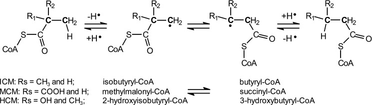
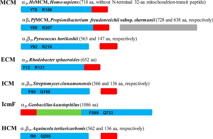
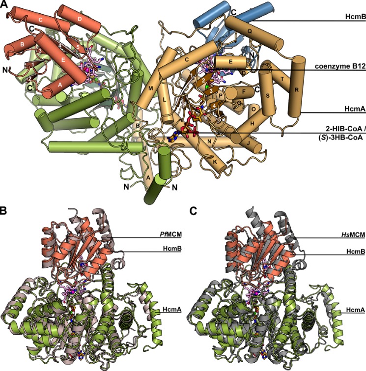
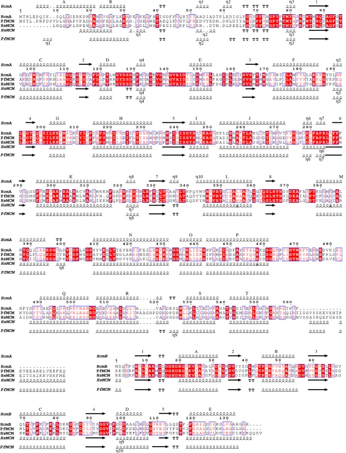
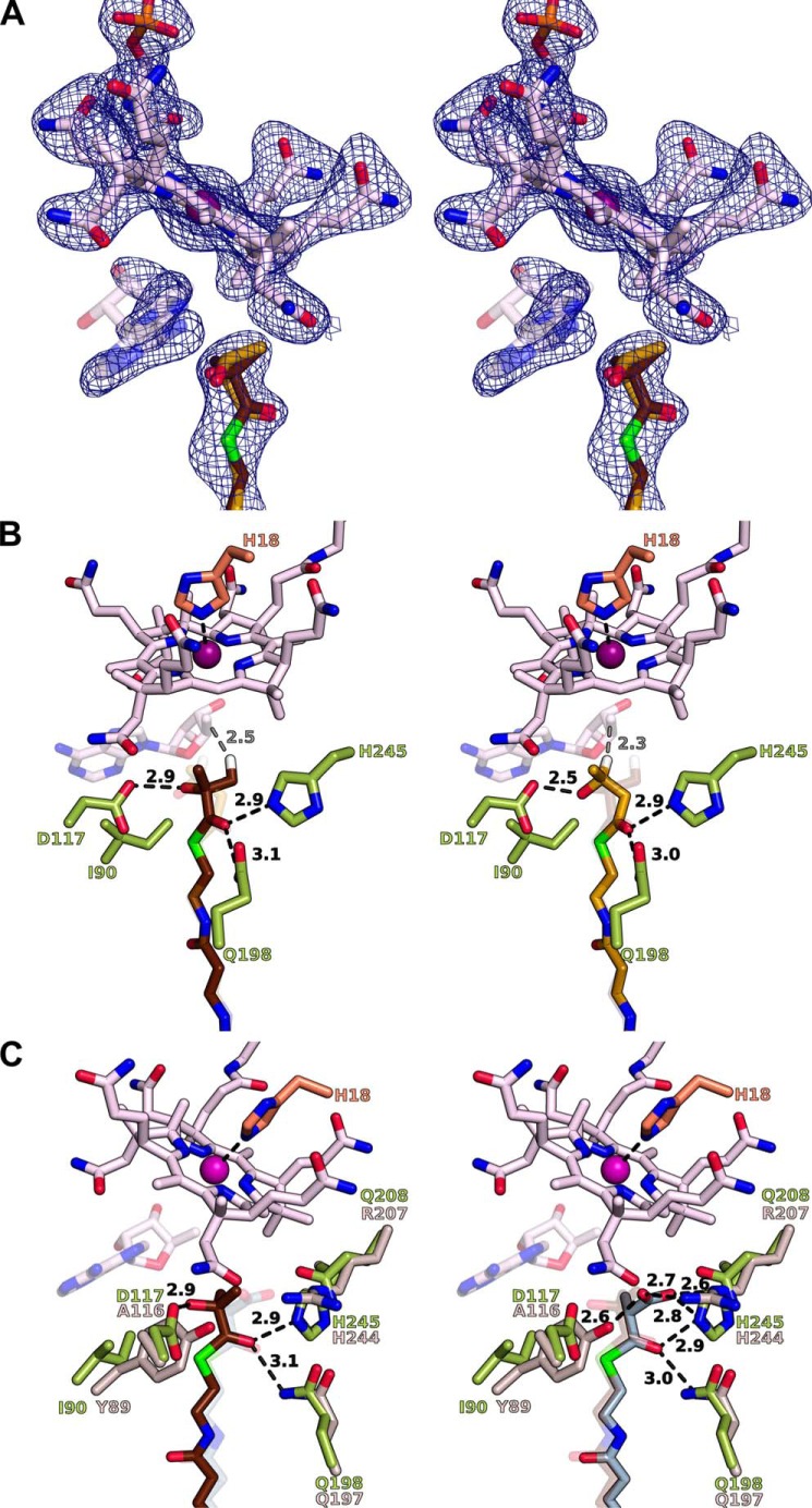
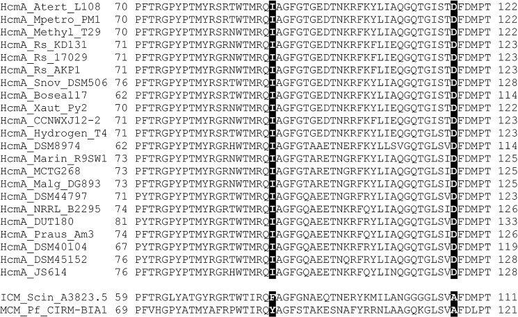
Bacterial coenzyme B12-dependent 2-hydroxyisobutyryl-CoA mutase (HCM) is a radical enzyme catalyzing the stereospecific interconversion of (S)-3-hydroxybutyryl- and 2-hydroxyisobutyryl-CoA. It consists of two subunits, HcmA and HcmB. To characterize the determinants of substrate specificity, we have analyzed the crystal structure of HCM from Aquincola tertiaricarbonis in complex with coenzyme B12 and the substrates (S)-3-hydroxybutyryl- and 2-hydroxyisobutyryl-CoA in alternative binding. When compared with the well studied structure of bacterial and mitochondrial B12-dependent methylmalonyl-CoA mutase (MCM), HCM has a highly conserved domain architecture. However, inspection of the substrate binding site identified amino acid residues not present in MCM, namely HcmA Ile(A90) and Asp(A117). Asp(A117) determines the orientation of the hydroxyl group of the acyl-CoA esters by H-bond formation, thus determining stereospecificity of catalysis. Accordingly, HcmA D117A and D117V mutations resulted in significantly increased activity toward (R)-3-hydroxybutyryl-CoA. Besides interconversion of hydroxylated acyl-CoA esters, wild-type HCM as well as HcmA I90V and I90A mutant enzymes could also isomerize pivalyl- and isovaleryl-CoA, albeit at >10 times lower rates than the favorite substrate (S)-3-hydroxybutyryl-CoA. The nonconservative mutation HcmA D117V, however, resulted in an enzyme showing high activity toward pivalyl-CoA. Structural requirements for binding and isomerization of highly branched acyl-CoA substrates such as 2-hydroxyisobutyryl- and pivalyl-CoA, possessing tertiary and quaternary carbon atoms, respectively, are discussed.

Keywords: (S)-3-Hydroxybutyryl-CoA; 2-Hydroxyisobutyryl-CoA; Adenosylcobalamin (AdoCbl); Crystal Structure; Pivalyl-CoA; Stereoselectivity; Substrate Specificity; X-ray Crystallography; acyl-CoA Mutase.


Figures

FIGURE 1.
FIGURE 1.
Mechanistic scheme for coenzyme B12-dependent acyl-CoA mutase-catalyzed 1,2-rearrangements. Vicinal carbon atoms exchanging a hydrogen atom with a thioester group are highlighted in bold. Residues R1 and R2 are not involved in the isomerization reaction but may interact specifically with active site amino acids.
FIGURE 2.
FIGURE 2.
Domain/subunit organization of coenzyme B12-dependent acyl-CoA mutases according to Ref. . Color code is as follows: acyl-CoA-binding domain (blue); adenosylcobalamin-binding domain (red); G-protein chaperone (green); and catalytically inactive mutase subunit of PfMCM (gray). Amino acid (aa) residues specifically interacting with the acyl moiety of CoA ester substrates in PfMCM (15, 16) and homologous residues are indicated in the acyl-CoA-binding domains. Tyr78 and Arg196 of HsMCM correspond to residues Tyr110 and Arg228, respectively, in the unprocessed 750-amino acid mutase protein (UniProt P22033), including the N-terminal 32-amino acid mitochondrion-transit peptide. ECM, ethylmalonyl-CoA mutase.
FIGURE 3.
FIGURE 3.
Crystal structure of the bacterial HCM from A. tertiaricarbonis. A, complex structure of the α2β2-heterotetramer of HCM (α-subunits in green and orange, β-subunits in red and blue, cobalamin in magenta, 2-hydroxyisobutyryl-CoA in brown, (S)-3-hydroxybutyryl-CoA in orange). B, superposition of one αβ-HCM-heterodimer (HcmA in green and HcmB in red) with the catalytically active subunit of PfMCM (gray, PDB entry 4REQ). C, superposition of one αβ-HCM-heterodimer (HcmA in green and HcmB in red) with HsMCM (gray, PDB entry 2XIQ).
FIGURE 4.
FIGURE 4.
Clustal Omega (30) and ESPript (31) sequence alignment of HcmA and HcmB with MCM including secondary structure elements. For the alignment with HCM, the catalytically active MCM subunit of P. freudenreichii (PfMCM, PDB 4REQ, chain A) and human MCM (HsMCM, PDB 2XIQ, alignment without N-terminal 32-amino acid mitochondrion-transit peptide) were used. α-Helices and β-strands of HCM are labeled as indicated in Fig. 3A.
FIGURE 5.
FIGURE 5.
Active site architecture of the bacterial HCM from A. tertiaricarbonis. A, stereo figure of the omit electron density map (blue, 3.0 σr.m.s. where r.m.s. indicates root mean square) for the coenzyme B12 (light pink) and the substrates 2-hydroxyisobutyryl- (dark brown) and (S)-3-hydroxybutyryl-CoA (orange). The adenosyl radical (light pink), which is part of the coenzyme B12 complex, is depicted in dim colors because it is only partially occupied (occupancy factor 0.5). B, stereoscopic view of the interaction of coenzyme B12 and the substrates 2-hydroxyisobutyryl- (dark brown) and (S)-3-hydroxybutyryl-CoA (orange) with HCM. The substrates are shown in different color intensities in each figure part (left or right stereo part). When viewed in stereo, alternating eye-switching results in an optimal impression of the binding modes of the two substrates. The adenosyl radical is depicted in dim colors as in panel A. Hydrogen atoms best positioned for abstraction by the adenosyl radical are colored white. Residues of HCM that interact with the acyl groups of the substrates or coenzyme B12 are colored in green or red, respectively. C, stereoscopic view of the superposition of HCM (green carbon atoms) in complex with 2-hydroxyisobutyryl-CoA (brown) with PfMCM with bound (R)-methylmalonyl-CoA (light blue). For a better view, the substrates are shown in different color intensities as in panel B. Interacting PfMCM residues are colored in gray.
FIGURE 6.
FIGURE 6.
Clustal Omega (30) multiple sequence alignment of the acyl-CoA binding domain of bacterial HCMs. Comparison of HcmA subunit from A. tertiaricarbonis ((ATert) AFK77668) with orthologous sequences from Methylibium petroleiphilum PM1 ((MPetro) Mpe_B0541), Methylibium sp. T29 ((Methyl) EWS54629), Rhodobacter sphaeroides KD131 ((Rs) RSKD131_3116), R. sphaeroides ATCC 17029 (Rsph17029_3657), R. sphaeroides AKP1 (D516_2161), Starkeya novella DSM 506 (Snov_2770), Bosea sp. 117 (WP_029354926), Xanthobacter autotrophicus Py2 (Xaut_5021), Mesorhizobium alhagi CCNWXJ12-2 (ZP_09295256), Hydrogenophaga sp. T4 ((Hydrogen) EWS66439), Arhodomonas aquaeolei DSM 8974 (WP_018719064), Marinobacter sp. R9SW1 ((Marin) AHI32051), Marinobacter algicola DG893 ((Malg) MDG893_09606), Marinobacter sp. MCTG268 (WP_036206499.1), Pseudonocardia spinosispora DSM 44797 (WP_028936951), Prauserella rugosa NRRL_B2295 (WP_030531194), Streptomyces sulphureus DSM 40104 (WP_026328509), Streptomyces xiaopingdaonensis DUT180 (WP_016910155), Prauserella sp. Am3 ((Praus) WP_039501511.1), Sciscionella marina DSM 45152 (WP_020495773), and Nocardioides sp. JS614 (Noca_2131). In addition, the paralogous domains of ICM from S. cinnamonensis A3823.5 (AAC08713) and MCM from P. freudenreichii CIRM-BIA1 (YP_003687736) were aligned. Highlighted amino acids indicate the positions of IleA90 and AspA117 identified to determine stereospecificity in HCM.

Similar articles

Cited by

References

    1. Banerjee R. (2003) Radical carbon skeleton rearrangements: catalysis by coenzyme B12-dependent mutases. Chem. Rev. 103, 2083–2094 - PubMed
    1. Birch A., Leiser A., Robinson J. A. (1993) Cloning, sequencing, and expression of the gene encoding methylmalonyl-coenzyme A mutase from Streptomyces cinnamonensis. J. Bacteriol. 175, 3511–3519 - PMC - PubMed
    1. Erb T. J., Rétey J., Fuchs G., Alber B. E. (2008) Ethylmalonyl-CoA mutase from Rhodobacter sphaeroides defines a new subclade of coenzyme B12-dependent acyl-CoA mutases. J. Biol. Chem. 283, 32283–32293 - PubMed
    1. Ratnatilleke A., Vrijbloed J. W., Robinson J. A. (1999) Cloning and sequencing of the coenzyme B12-binding domain of isobutyryl-CoA mutase from Streptomyces cinnamonensis, reconstitution of mutase activity, and characterization of the recombinant enzyme produced in Escherichia coli. J. Biol. Chem. 274, 31679–31685 - PubMed
    1. Cracan V., Padovani D., Banerjee R. (2010) IcmF is a fusion between the radical B12 enzyme isobutyryl-CoA mutase and its G-protein chaperone. J. Biol. Chem. 285, 655–666 - PMC - PubMed

MeSH terms

Associated data

LinkOut - more resources