

Measurement of perihematomal edema in intracerebral hemorrhage
- PMID: 25721012
- PMCID: PMC5340416
- DOI: 10.1161/STROKEAHA.114.007565
Measurement of perihematomal edema in intracerebral hemorrhage
Abstract
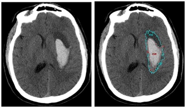
Background and purpose: Perihematomal edema (PHE) is a marker of secondary injury in intracerebral hemorrhage (ICH). PHE measurement on computed tomography (CT) is challenging, and the principles used to detect PHE have not been described fully. We developed a systematic approach for CT-based measurement of PHE.
Methods: Two independent raters measured PHE volumes on baseline and 24-hour post-ICH CT scans of 20 primary supratentorial ICH subjects. Boundaries were outlined with an edge-detection tool and adjusted after inspection of the 3 orthogonal planes. PHE was delineated with the additional principle that it should be (a) more hypodense than the corresponding area in the contralateral hemisphere and (b) most hypodense immediately surrounding the hemorrhage. We examined intra- and interrater reliability using intraclass correlation coefficients and Bland-Altman plots for interrater consistency. CT-based PHE was also compared using magnetic resonance imaging-based PHE detection for 18 subjects.
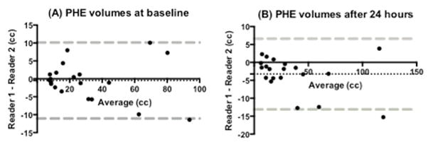
Results: Median PHE volumes were 22.7 cc at baseline and 20.4 cc at 24 hours post-ICH. There were no statistically significant differences in PHE measurements between raters. Interrater and intrarater reliability for PHE were excellent. At baseline and 24 hours, interrater intraclass correlation coefficients were 0.98 (0.96-1.00) and 0.98 (0.97-1.00); intrarater intraclass correlation coefficients were 0.99 (0.99-1.00) and 0.99 (0.98-1.00). Bland-Altman analysis showed the bias for PHE measurements at baseline and 24 hours, -0.5 cc (SD, 5.4) and -3.2 cc (SD, 5.0), was acceptably small. PHE volumes determined by CT and magnetic resonance imaging were similar (23.9±16.9 cc versus 23.9±16.0 cc, R(2) = 0.98, P<0.0001).
Conclusions: Our method measures PHE with excellent reliability at baseline and 24 hours post-ICH.
Keywords: computed tomography; intracerebral hemorrhage.
© 2015 American Heart Association, Inc.
Figures
References
-
- Broderick JP, Brott TG, Duldner JE, Tomsick T, Huster G. Volume of intracerebral hemorrhage. A powerful and easy-to-use predictor of 30-day mortality. Stroke. 1993;24:987–993. - PubMed
-
- Li N, Liu YF, Ma L, Worthmann H, Wang YL, Wang YJ, et al. Association of molecular markers with perihematomal edema and clinical outcome in intracerebral hemorrhage. Stroke. 2013;44:658–663. - PubMed
-
- Appelboom G, Bruce SS, Hickman ZL, Zacharia BE, Carpenter AM, Vaughan KA, et al. Volume-dependent effect of perihaematomal oedema on outcome for spontaneous intracerebral haemorrhages. J Neurol Neurosurg Psychiatry. 2013;84:488–493. - PubMed
Publication types
MeSH terms
Grants and funding
LinkOut - more resources
Full Text Sources
