

TCF4 Triplet Repeat Expansion and Nuclear RNA Foci in Fuchs' Endothelial Corneal Dystrophy
- PMID: 25722209
- PMCID: PMC4373545
- DOI: 10.1167/iovs.14-16222
TCF4 Triplet Repeat Expansion and Nuclear RNA Foci in Fuchs' Endothelial Corneal Dystrophy
Abstract
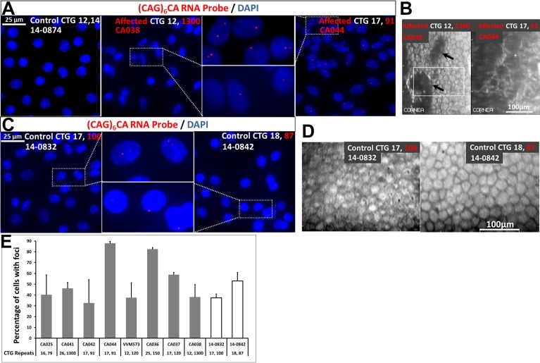
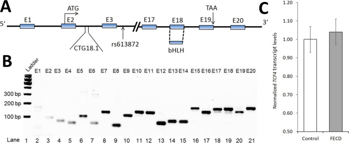
Purpose: Expansion of the intronic CTG18.1 triplet repeat locus within TCF4 contributes significant risk to the development of Fuchs' endothelial corneal dystrophy (FECD) in Eurasian populations, but the mechanisms by which the expanded repeats result in degeneration of the endothelium have been hitherto unknown. The purpose of this study was to examine FECD endothelial samples for the presence of RNA nuclear foci, the hallmark of toxic RNA, as well as evidence of haploinsufficiency of TCF4.
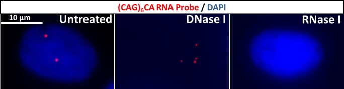
Methods: Using fluorescence in situ hybridization, we examined for the presence of nuclear RNA foci containing expanded CUG transcripts in corneal endothelial samples from FECD subjects with CTG18.1 expansion. We also examined for any changes in expression levels of TCF4 by quantitative real-time PCR.
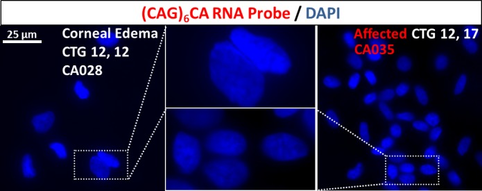
Results: Numerous discrete nuclear RNA foci were identified in endothelial samples of FECD subjects (n = 8) harboring the CTG18.1 expansion, but not in controls lacking the expansion (n = 5) (P = 7.8 × 10(-4)). Percentage of cells with foci in expansion-positive endothelial samples ranged from 33% to 88%. RNA foci were absent in endothelial samples from an FECD subject without CTG18.1 expansion and a subject with endothelial dysfunction without FECD. Expression of the constitutive TCF4 exon encoding the basic helix-loop-helix domain was unaltered with CTG18.1 expansion.
Conclusions: Our findings suggest that the RNA nuclear foci are pathognomonic for CTG18.1 expansion-mediated endothelial disease. The RNA nuclear foci have been previously found only in rare neurodegenerative disorders caused by repeat expansions. Our detection of abundant ribonuclear foci in FECD implicates a role for toxic RNA in this common disease.
Keywords: CTG18.1; Fuchs' endothelial corneal dystrophy; RNA nuclear foci; TCF4; triplet repeat expansion.
Copyright 2015 The Association for Research in Vision and Ophthalmology, Inc.
Figures
References
-
- Lorenzetti DW, Uotila MH, Parikh N, Kaufman HE. Central cornea guttata. Incidence in the general population. Am J Ophthalmol. 1967; 64: 1155–1158. - PubMed
-
- Eye Bank Association of America. 2013 Eye Banking Statistical Report. Washington: Eye Bank Association of America; 2013. Available at: http://www.restoresight.org/wpcontent/uploads/2014/04/2013_Statistical_R.... Accessed December 8, 2014.
-
- Zoega GM, Fujisawa A, Sasaki H, et al. Prevalence and risk factors for cornea guttata in the Reykjavik Eye Study. Ophthalmology. 2006; 113: 565–569. - PubMed
-
- Kitagawa K, Kojima M, Sasaki H, et al. Prevalence of primary cornea guttata and morphology of corneal endothelium in aging Japanese and Singaporean subjects. Ophthalmic Res. 2002; 34: 135–138. - PubMed
-
- Chi HH, Teng CC, Katzin HM. Histopathology of primary endothelial-epithelial dystrophy of the cornea. Am J Ophthalmol. 1958; 45: 518–535. - PubMed
Publication types
MeSH terms
Substances
Grants and funding
LinkOut - more resources
Full Text Sources
Other Literature Sources
