. 2015 Jun;138(Pt 6):1613-28.
doi: 10.1093/brain/awv045. Epub 2015 Feb 25.

PI3K/AKT pathway mutations cause a spectrum of brain malformations from megalencephaly to focal cortical dysplasia

Affiliations

PI3K/AKT pathway mutations cause a spectrum of brain malformations from megalencephaly to focal cortical dysplasia

Laura A Jansen et al. Brain. 2015 Jun.

Abstract

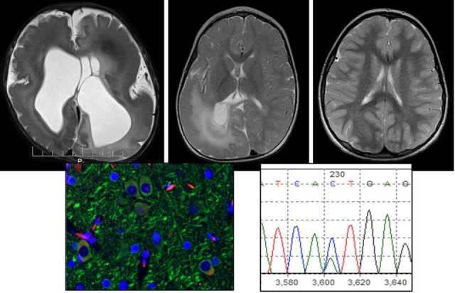
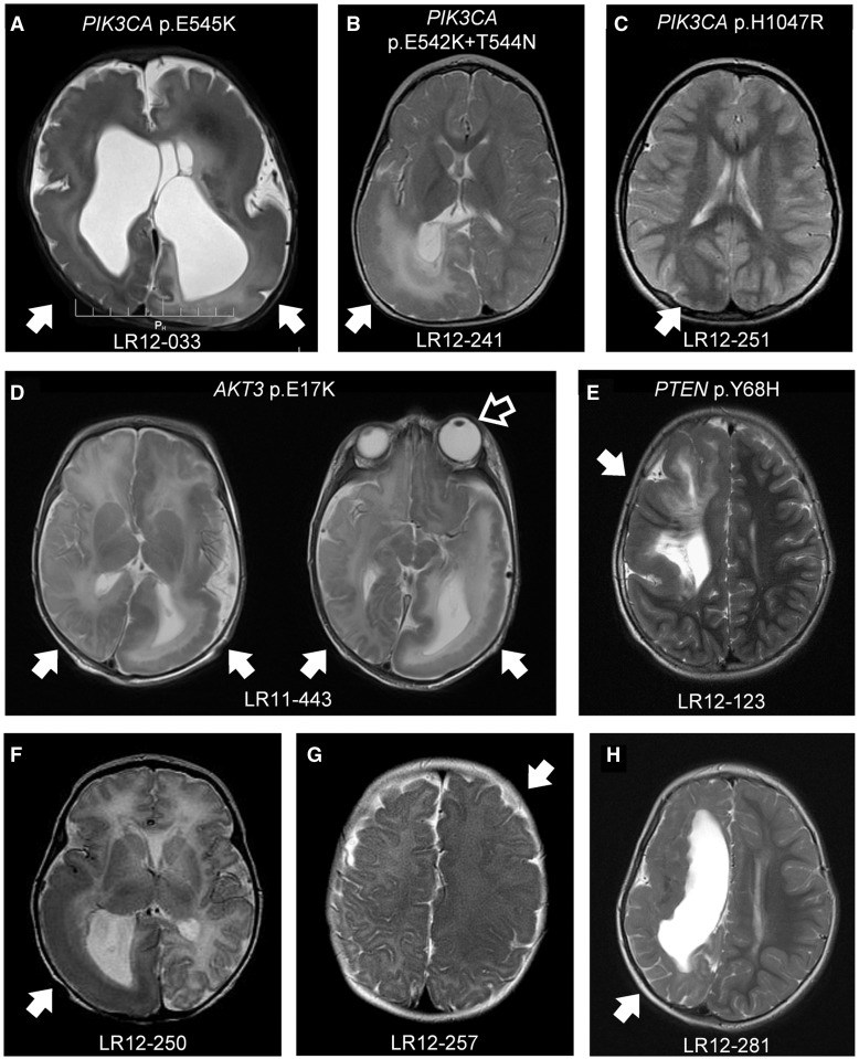
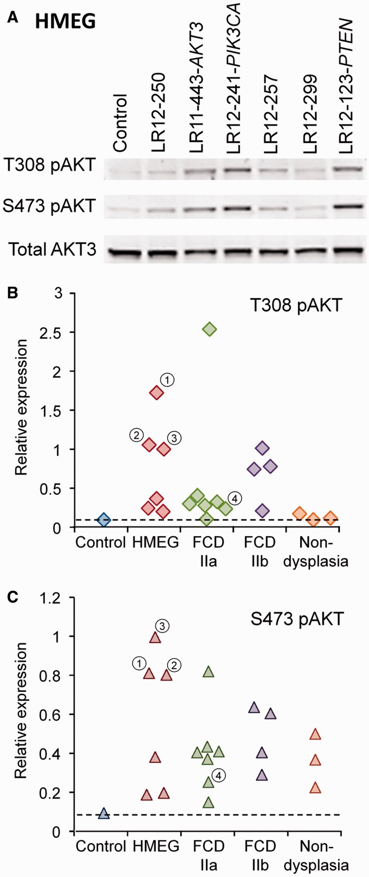
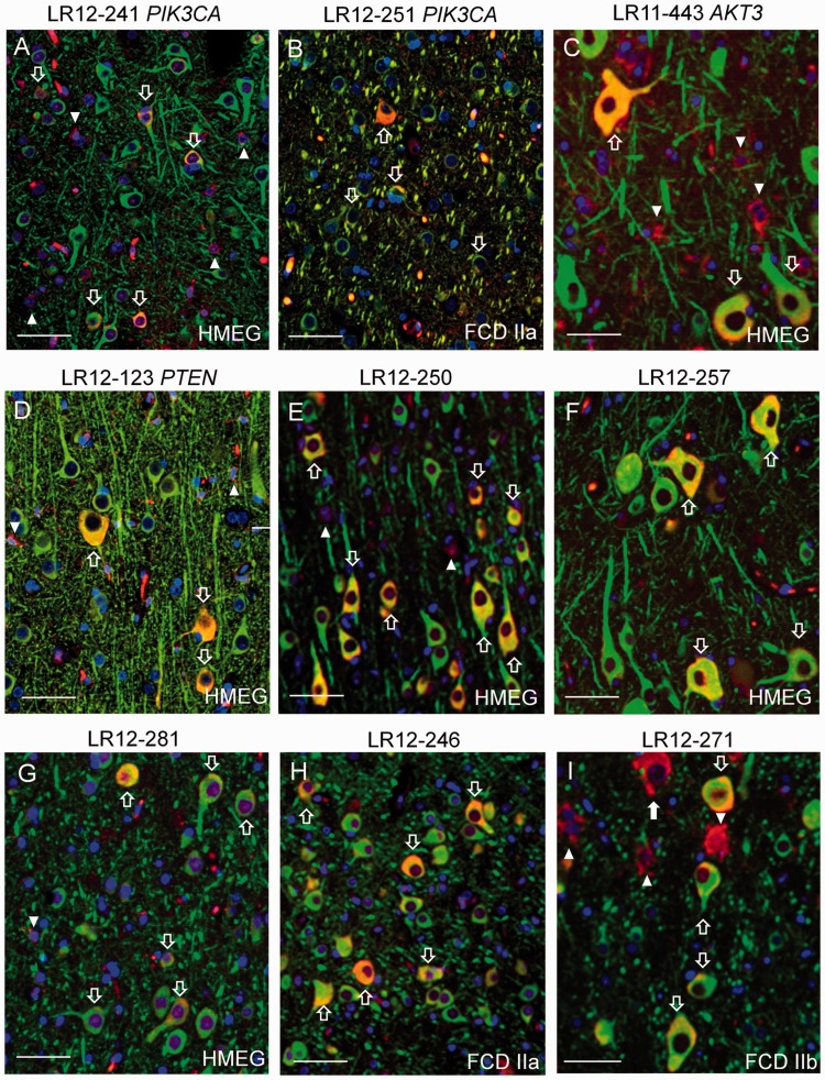
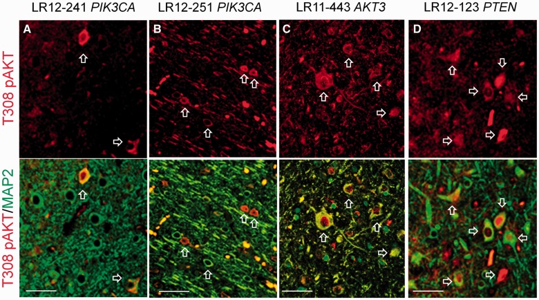
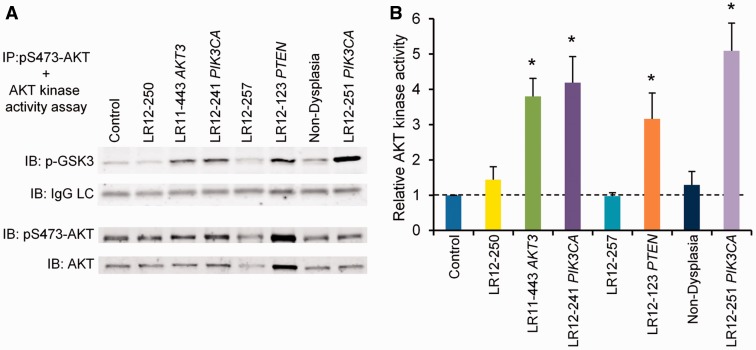
Malformations of cortical development containing dysplastic neuronal and glial elements, including hemimegalencephaly and focal cortical dysplasia, are common causes of intractable paediatric epilepsy. In this study we performed multiplex targeted sequencing of 10 genes in the PI3K/AKT pathway on brain tissue from 33 children who underwent surgical resection of dysplastic cortex for the treatment of intractable epilepsy. Sequencing results were correlated with clinical, imaging, pathological and immunohistological phenotypes. We identified mosaic activating mutations in PIK3CA and AKT3 in this cohort, including cancer-associated hotspot PIK3CA mutations in dysplastic megalencephaly, hemimegalencephaly, and focal cortical dysplasia type IIa. In addition, a germline PTEN mutation was identified in a male with hemimegalencephaly but no peripheral manifestations of the PTEN hamartoma tumour syndrome. A spectrum of clinical, imaging and pathological abnormalities was found in this cohort. While patients with more severe brain imaging abnormalities and systemic manifestations were more likely to have detected mutations, routine histopathological studies did not predict mutation status. In addition, elevated levels of phosphorylated S6 ribosomal protein were identified in both neurons and astrocytes of all hemimegalencephaly and focal cortical dysplasia type II specimens, regardless of the presence or absence of detected PI3K/AKT pathway mutations. In contrast, expression patterns of the T308 and S473 phosphorylated forms of AKT and in vitro AKT kinase activities discriminated between mutation-positive dysplasia cortex, mutation-negative dysplasia cortex, and non-dysplasia epilepsy cortex. Our findings identify PI3K/AKT pathway mutations as an important cause of epileptogenic brain malformations and establish megalencephaly, hemimegalencephaly, and focal cortical dysplasia as part of a single pathogenic spectrum.

Keywords: brain development; childhood epilepsy; malformations of cortical development; molecular genetics.


Figures

None
Dysplastic cortical malformations are common causes of paediatric epilepsy. Using techniques including histopathology, immunohistochemistry, and deep sequencing in resected tissue from affected children, Jansen et al. show that megalencephaly, hemimegalencephaly and focal cortical dysplasia are overlapping phenotypes associated with upregulation of the PI3K/AKT/mTOR pathway.
Figure 1
Figure 1
MRI findings in dysplastic brain malformations with and without detected mutations. T2-weighted images are shown. Filled arrows indicate regions of abnormality, including ventricular enlargement, variable degrees of cortical dysmorphism (i.e. pachygyria, polymicrogyria, or undersulcation), periventricular nodular heterotopia, abnormal white matter signal, and/or widening of the extra-axial space. (A) DMEG; (B, E–H) HMEG; (C) FCD type IIa; (D) bihemispheric asymmetric dysplasia. The open arrow indicates unilateral ocular enlargement in Patient LR11-443, who exhibits severe abnormality of the left hemisphere (HMEG) along with milder abnormalities of the posterior portion of the right hemisphere.
Figure 2
Figure 2
Sanger sequencing confirmation of mutations detected in HMEG brain by molecular inversion probe capture technology. Sequence changes are highlighted by the blue bars. (A) Mosaic PIK3CA p.E542K and p.T544N mutations from HMEG brain LR12-241. (B) Mosaic AKT3 p.E17K mutation from HMEG brain LR11-443 operculum and hippocampus, which was absent in overlying dura. (C) Germline PTEN p.Y68H heterozygous mutation detected in LR12-123 brain.
Figure 3
Figure 3
Western blot analysis of T308 pAKT, S473 pAKT, and total AKT3 levels in dysplastic brain specimens. (A) Representative western blot showing increased expression of T308 pAKT and S473 pAKT in six HMEG brain specimens as compared to control, non-epileptic brain (all surgical specimens). The highest levels of pAKT were seen in the HMEG specimens with detected PIK3CA, AKT3 and PTEN mutations. (B and C) Summary of relative T308 pAKT (B) and S473 pAKT (C) levels in 17 dysplastic brain specimens (HMEG, FCD IIa, and FCD IIb) as compared with control brain and three non-dysplasia epilepsy brain specimens. The ratio of T308 pAKT or S473 pAKT band intensity to total AKT3 band intensity was determined for each specimen and is expressed relative to that of specimen LR12-241, which was run concurrently on each blot. 1LR11-443 AKT3 mutation; 2LR12-123 PTEN mutation; 3LR12-241 PIK3CA mutation; 4LR12-251 PIK3CA mutation.
Figure 4
Figure 4
Phosphorylated ribosomal S6 protein (phospho-S6) immunofluorescence in neurons from dysplastic brain specimens with and without detected mutations. Red = phospho-S6; Green = neuronal marker MAP2; Blue = nuclear marker DAPI. Neurons co-localizing phospho-S6 and MAP2 appear orange-yellow in colour, and are indicated by the open arrows. Arrowheads indicate phospho-S6 positive cells with astrocytic morphology. The closed arrow in I indicates a phospho-S6 positive balloon cell in FCD IIb. Scale bars = 50 µm.
Figure 5
Figure 5
Neuronal T308 pAKT immunofluorescence in dysplastic brain specimens with PI3K/AKT pathway mutations. Red = T308 pAKT; Green = MAP2. Open arrows indicate neurons colocalizing T308 pAKT and MAP2. In many neurons T308 pAKT is visible in both the cytoplasm and nucleus. Scale bars = 50 µm.
Figure 6
Figure 6
In vitro AKT kinase assay of frozen brain extracts showing highest levels of kinase activity in specimens with detected PI3K/AKT pathway mutations. (A) Representative western blot showing phosphorylated GSK 3 fusion protein (p-GSK3) levels after incubation with immunoprecipitated pAKT and ATP. IgG LC = IgG light chain from immunoprecipitating antibody, used for normalization of p-GSK3. (B) Average relative kinase activity ± SEM as determined from three to five separate blots per sample. Note that other than specimen LR12-123, AKT kinase activity is not correlated with pS473-AKT levels, consistent with a requirement for phosphorylation at both the S473 and T308 sites for full AKT kinase activity. *P < 0.05 versus control, one-way ANOVA followed by post-tests.
Figure 7
Figure 7
The PI3K/AKT kinase pathway and associated brain malformations. Wide green arrows indicate disorders caused by activating mutations, whereas wide red arrows indicate disorders caused by loss of function mutations. See text for references.

Comment in

References

    1. Alessi DR, Andjelkovic M, Caudwell B, Cron P, Morrice N, Cohen P, et al. Mechanism of activation of protein kinase B by insulin and IGF-1. EMBO J. 1996;15:6541–51. - PMC - PubMed
    1. Arch EM, Goodman BK, Van Wesep RA, Liaw D, Clarke K, Parsons R, et al. Deletion of PTEN in a patient with Bannayan-Riley-Ruvalcaba syndrome suggests allelism with Cowden disease. Am J Med Genet. 1997;71:489–93. - PubMed
    1. Aronica E, Boer K, Baybis M, Yu J, Crino P. Co-expression of cyclin D1 and phosphorylated ribosomal S6 proteins in hemimegalencephaly. Acta Neuropathol (Berl) 2007;114:287–93. - PubMed
    1. Backman SA, Stambolic V, Suzuki A, Haight J, Elia A, Pretorius J, et al. Deletion of Pten in mouse brain causes seizures, ataxia and defects in soma size resembling Lhermitte-Duclos disease. Nat Genet. 2001;29:396–403. - PubMed
    1. Barkovich AJ, Chuang SH. Unilateral megalencephaly: correlation of MR imaging and pathologic characteristics. Am J Neuroradiol. 1990;11:523–31. - PMC - PubMed

Publication types

MeSH terms

Substances