. 2015 Mar 11;43(5):2625-37.
doi: 10.1093/nar/gkv133. Epub 2015 Feb 26.

Unraveling the mechanistic features of RNA polymerase II termination by the 5'-3' exoribonuclease Rat1

Affiliations

Unraveling the mechanistic features of RNA polymerase II termination by the 5'-3' exoribonuclease Rat1

Jieun Park et al. Nucleic Acids Res. .

Abstract

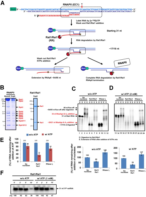
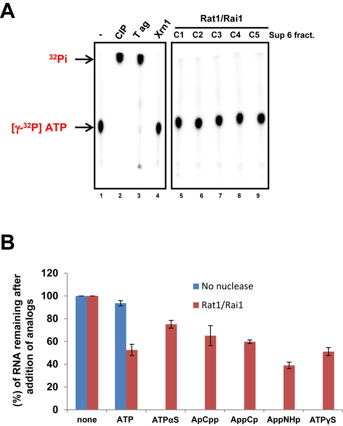
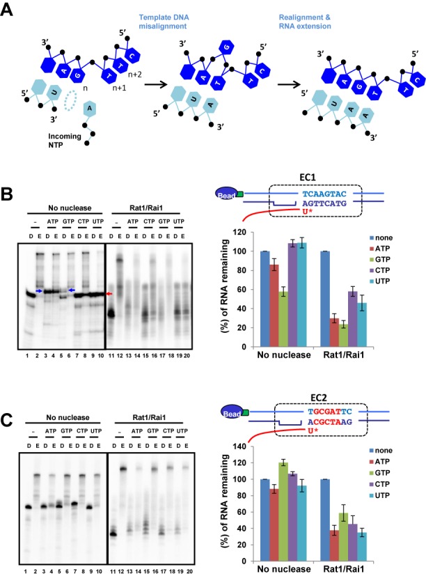
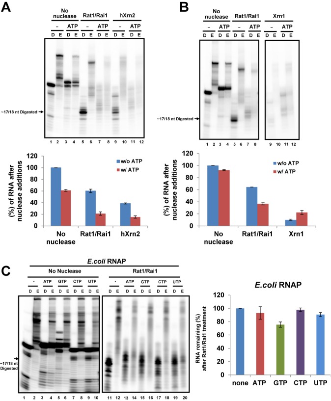
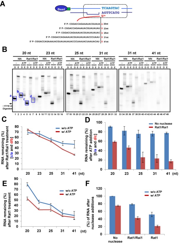
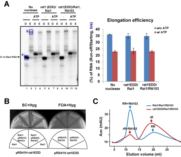
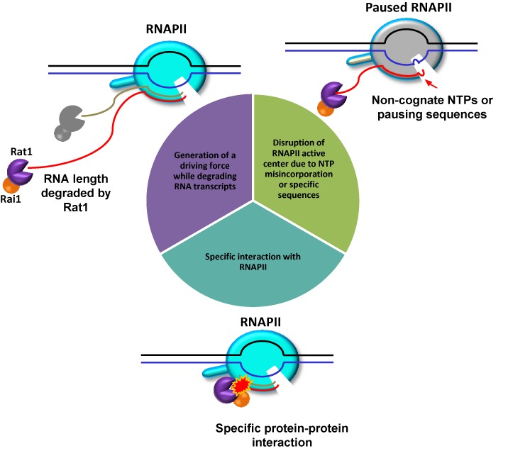
Within a complex with Rai1, the 5'-3' exoribonuclease Rat1 promotes termination of RNA polymerase II (RNAPII) on protein-coding genes, but its underlying molecular mechanism is still poorly understood. Using in vitro transcription termination assays, we have found that RNAPII is prone to more effective termination by Rat1/Rai1 when its catalytic site is disrupted due to NTP misincorporation, implying that paused RNAPII, which is often found in vivo near termination sites, could adopt a similar configuration to Rat1/Rai1 and trigger termination. Intriguingly, yeast Rat1/Rai1 does not terminate Escherichia coli RNAP, implying that a specific interaction between Rat1/Rai1 and RNAPII may be required for termination. Furthermore, the efficiency of termination increases as the RNA transcript undergoing degradation by Rat1 gets longer, which suggests that Rat1 may generate a driving force for dissociating RNAPII from the template while degrading the nascent transcripts to catch up to the polymerase. These results indicate that multiple mechanistic features contribute to Rat1-mediated termination of RNAPII.


Figures

Figure 1.
Figure 1.
Rat1/Rai1 terminates RNAPII in vitro in the presence of ATP. (A) In vitro transcription termination assay scheme. The EC was assembled with double-stranded DNA (EC1), 5′ phosphorylated RNA and purified RNAPII and was subsequently coupled to magnetic beads. The 3′-end of RNA was radioactively labeled by RNAPII. Rat1/Rai1 digested RNA from 5′ to 3′ up to the surface of RNAPII. If Rat1/Rai1 failed to terminate RNAPII, the polymerase would elongate further using NTPs. However, if Rat1/Rai1 terminated RNAPII, the remaining RNA, which was protected by RNAPII, would be completely degraded by Rat1/Rai1. The red arrow specifies the position (n) of the first incoming NTP. (B) Purified RNAPII complex and Rat1/Rai1 on gels stained with Coomassie. (C) Representative gel image of the in vitro transcription termination assay with Rat1/Rai1 treatment in the absence of ATP. Rat1/Rai1 did not terminate RNAPII by itself. Black arrows indicate the RNAs predicted in (A). Red arrows show ATP-misincorporated RNAs. (D) digestion by Rat1/Rai1. (E) Extension of RNA after addition of NTPs mixture. Quantification of the remaining RNA compared with the control lacking nuclease (set to 100%) is shown below. (D) Rat1/Rai1 terminates RNAPII efficiently in the presence of 2 mM ATP. (E) Quantification of the remaining RNAs after ATP treatment compared with the no ATP control (set to 100%). (F) Time course showing the degradation of 31-nt single strand RNA substrate by Rat1/Rai1 in the absence or presence of 1 mM ATP. The amount of remaining RNA is presented as a percentage of that from the 0 min reaction after Rat1/Rai1 treatment. Asterisk represents radioactive labeling at 5′end of 31-nt RNA.
Figure 2.
Figure 2.
Rat1/Rai1 does not have ATPase activity. (A) ATPase activity assay of Rat1/Rai1. Calf intestinal phosphatase (CIP), SV40 T antigen (T ag) and Xrn1 were used as controls. Highly purified Rat1/Rai1 fractions from size-exclusion chromatography (Superose 6) show no ATPase activity. (B) Quantification of the remaining RNAs from the in vitro transcription termination assay with various non-hydrolyzable ATP analogs.
Figure 3.
Figure 3.
NTP misincorporation induces RNAPII pausing and enhances termination by Rat1/Rai1. (A) Schematic detail for NTP misincorporation via template misalignment. The RNA strand is shown in sky blue and the DNA strand is in dark blue. (B) EC1 scaffold tested. DNA sequences are shown on top of the quantification graph. An asterisk specifies the incorporation site of an incoming NTP (n position). In a control group lacking nuclease, the addition of non-cognate ATP or GTP generated misincorporated RNA bands via template misalignment (blue arrows, ∼34/35 nt), whereas cognate CTP does not. Notably, Rat1/Rai1 more efficiently terminates RNAPII when non-cognate NTP (ATP or GTP) rather than cognate CTP was added. Addition of UTP to EC1 induces strong pausing of RNAPII that does not support further elongation (red arrow), which results in slight inhibition of termination by Rat1/Rai1. (B) EC2 scaffold tested. Different sequences are shown in red. Rat1/Rai1 more effectively terminates RNAPII when a non-cognate NTP (ATP, CTP or UTP), rather than cognate GTP, was added.
Figure 4.
Figure 4.
Other 5′-3′ exoribonucleases and Escherichia coli RNAP. In vitro assays were performed using EC1 scaffold. (A) ATP enhances RNAPII termination by recombinant hXrn2 in vitro. Quantification of the remaining RNAs is shown below. (B) Yeast Xrn1, Rat1's cytoplasmic counterpart, terminates RNAPII regardless of the presence of ATP in vitro. Quantification of the remaining RNAs is shown below. (C) Rat1/Rai1 cannot terminate E. coli RNAP in vitro and addition of each NTP had no effect either. Representative gel image is presented on the left and quantification of the remaining RNA is presented in the right panel.
Figure 5.
Figure 5.
The length of RNA degraded by Rat1 affects RNAPII termination. (A) Five ECs harboring different lengths of RNA were tested in the assay. (B) Representative gel images of the Rat1/Rai1-treated in vitro transcription termination assay. (C) Quantification of the remaining RNAs after Rat1/Rai1 treatment without or with ATP. The remaining RNA amounts without or with ATP addition in the no nuclease (NN) groups (a or b, respectively; blue box in gel images) were set to 100% for each EC and the percentage of remaining RNA amounts in the Rat1/Rai1-treated groups without or with ATP treatment (c/a or d/b, respectively) were calculated for each EC. In the presence of ATP, the remaining RNA level after Rat1/Rai1 treatment rapidly decreased as the RNA lengthened. However, this level moderately decreased in the absence of ATP. (D) Quantification of the remaining RNAs after ATP addition in no nuclease (NN) or Rat1/Rai1-treated groups. The remaining RNA amounts without ATP addition (a or c, respectively) were set to 100% and the percentage of remaining RNA amounts after ATP addition (b/a or d/c, respectively) were calculated for each EC. The remaining RNA amounts specifically and gradually decreased in the presence of Rat1/Rai1 as the length of the RNA increased. (E) Quantification of the remaining RNA amounts without or with ATP addition after Rat1 treatment. There is a dramatic decrease in the remaining RNA level as the RNA length increases but little difference in the remaining RNA levels, regardless of ATP addition, at the ECs harboring 31 and 41 nt RNAs. (F) Rat1 terminates RNAPII in a similar fashion to Rat1/Rai1. Addition of ATP also enhances termination by Rat1 alone.
Figure 6.
Figure 6.
Exoribonucleolytic-deficient rat1 (rat1EDD) cannot terminate RNAPII and Rtt103 does not rescue the termination defect of rat1EDD. (A) (Left) rat1EDD/Rai1, regardless of Rtt103 addition, does not terminate RNAPII. (Right) Quantification of the extended run-off RNA amounts relative to the initial starting RNAs without or with ATP addition. rat1EDD does not reduce the RNA elongation efficiency by RNAPII compared with the no nuclease control. (B) Multiple copies of the Rtt103 gene cannot rescue the lethality of the rat1EDD mutation. (C) Elution profiles of size-exclusion chromatography (Superose 6) of Rat1/Rai1/Rtt103 (RRR) and rat1EDD/Rai1/Rtt103 (rRR) show that most of the Rat1/Rai1 is co-eluted with Rtt103, whereas most of rat1EDD/Rai1 is not. These profiles indicate that rat1EDD shows reduced binding to Rtt103 compared with wild-type Rat1.
Figure 7.
Figure 7.
Multiple mechanistic features contribute to Rat1-mediated RNAPII termination. Disruption of the RNAPII active center due to NTP misincorporation or specific sequences facilitated termination by Rat1/Rai1 in vitro. A specific interaction between Rat1/Rai1 and RNAPII is critical because Rat1/Rai1 cannot terminate Escherichia coli RNAP. Furthermore, Rat1 must degrade RNA transcripts to build up a driving force for termination. Thus, 5′-3′ exonuclease activity is essential for Rat1 not only to gain access to RNAPII but also to accumulate a sufficient driving force to execute termination. Active RNAPII, cyan; paused RNAPII, gray.

References

    1. Kim M., Vasiljeva L., Rando O.J., Zhelkovsky A., Moore C., Buratowski S. Distinct pathways for snoRNA and mRNA termination. Mol. Cell. 2006;24:723–734. - PubMed
    1. Steinmetz E.J., Conrad N.K., Brow D.A., Corden J.L. RNA-binding protein Nrd1 directs poly(A)-independent 3′-end formation of RNA polymerase II transcripts. Nature. 2001;413:327–331. - PubMed
    1. Steinmetz E.J., Warren C.L., Kuehner J.N., Panbehi B., Ansari A.Z., Brow D.A. Genome-wide distribution of yeast RNA polymerase II and its control by Sen1 helicase. Mol. Cell. 2006;24:735–746. - PubMed
    1. Kim M., Krogan N.J., Vasiljeva L., Rando O.J., Nedea E., Greenblatt J.F., Buratowski S. The yeast Rat1 exonuclease promotes transcription termination by RNA polymerase II. Nature. 2004;432:517–522. - PubMed
    1. West S., Gromak N., Proudfoot N.J. Human 5′ –> 3′ exonuclease Xrn2 promotes transcription termination at co-transcriptional cleavage sites. Nature. 2004;432:522–525. - PubMed

Publication types

MeSH terms