Review
. 2015 Mar;9(1):1-10.
doi: 10.1007/s11701-015-0496-1. Epub 2015 Feb 7.

Role of intra-operative contrast-enhanced ultrasound (CEUS) in robotic-assisted nephron-sparing surgery

Affiliations
Review

Role of intra-operative contrast-enhanced ultrasound (CEUS) in robotic-assisted nephron-sparing surgery

Ahmad N Alenezi et al. J Robot Surg. 2015 Mar.

Abstract

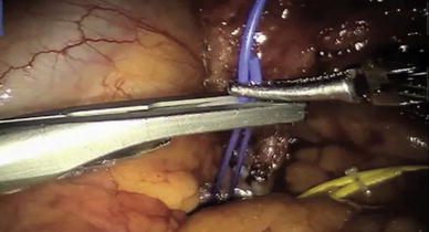
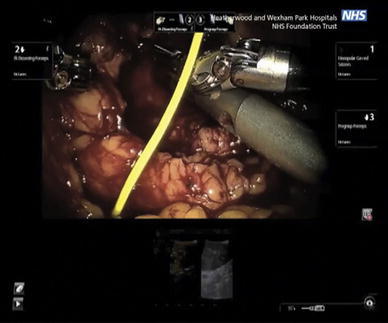
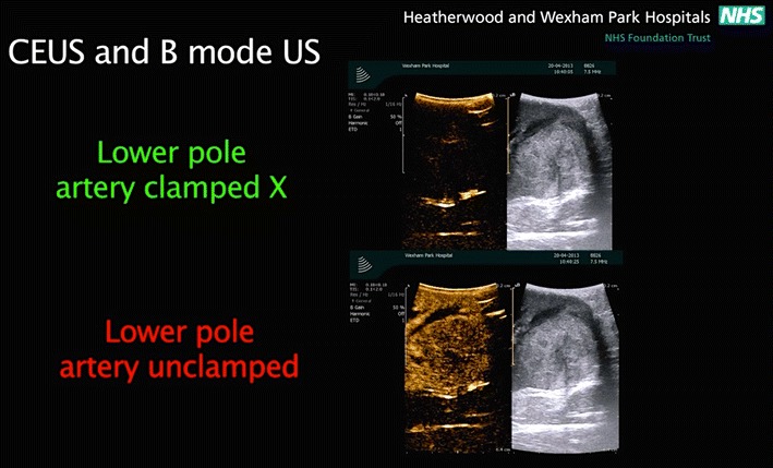
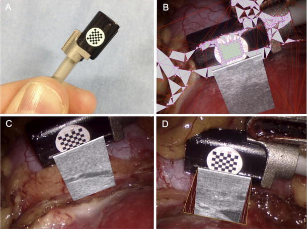
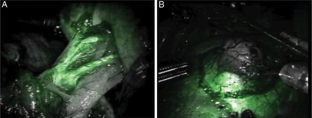
This review examines studies of intra-operative contrast-enhanced ultrasound (CEUS) and its emerging role and advantages in robotic-assisted nephron-sparing surgery. Contrast-enhanced ultrasound is a technology that combines the use of second-generation contrast agents consisting of microbubbles with existent ultrasound techniques. Until now, this novel technology has aided surgeons with procedures involving the liver. However, with recent advances in the CEUS technique and the introduction of robotics in nephron-sparing surgery, CEUS has proven to be efficacious in answering several clinical questions with respect to the kidneys. In addition, the introduction of the microbubble-based contrast agents has increased the image quality and signal uptake by the ultrasound probe. This has led to better, enhanced scanning of the macro and microvasculature of the kidneys, making CEUS a powerful diagnostic modality. This imaging method is capable of further lowering the learning curve and warm ischemia time (WIT) during robotic-assisted nephron-sparing surgery, with its increased level of capillary perfusion and imaging. CEUS has the potential to increase the sensitivity and specificity of intra-operative images, and can significantly improve the outcome of robotic-assisted nephron-sparing surgery by increasing the precision and diagnostic insight of the surgeon. The purpose of this article is to review the practical and potential uses of CEUS as an intra-operative imaging technique during robotic-assisted nephron-sparing surgery.

Keywords: CEUS; Contrast-enhanced ultrasound scan; Nephron sparing; Partial nephrectomy; Robotic assisted; SonoVue; Zero-ischemia.


Figures

Fig. 1
Fig. 1
Branches of the renal artery identified with different colored vessel loops for improved identification [5]
Fig. 2
Fig. 2
Using the robotic ProGrasp forceps the surgeon manipulates the robotic ultrasound probe over the kidney, to visualize the position of the tumor (B-mode ultrasound), and assess blood flow (using CEUS). The operating surgeon views the ultrasound images in the TilePro mode on the robot console. The B-mode ultrasound image is on the right, while the CEUS image is on the left of the TilePro image [5]
Fig. 3
Fig. 3
the difference between a conventional B-mode images on the right and the contrast-enhanced mode images on the left. SonoVue microbubble contrast agent was injected intravenously. The contrast-enhanced ultrasound mode in the monitor shows the circulation in the kidney and the segment containing the tumor in the lower pole. This facilitates and confirms selective ischemia to the desired renal segment
Fig. 4
Fig. 4
The ProART™ Robotic Transducer Type 8826 (courtesy of BK Medical)
Fig. 5
Fig. 5
a Ultrasound probe with attached chessboard pattern mounted in custom-made clip; b real-time automatic tracking and registration process; c superimposed ultrasound; d superimposed ultrasound with cutaway and 1-mm ruler (courtesy of Mayer et al.)
Fig. 6
Fig. 6
Fluorescence imaging during robotic partial nephrectomy using intravenously injected ICG illustrating on the left: a arterial phase enhancement of the primary, secondary, and tertiary arterial branches of the kidney and on the right: b hypo-fluorescent renal mass with surrounding normal fluorescent renal parenchyma (Courtesy of Silvers et al.) [15]
Fig. 7
Fig. 7
a Laparoscopic ultrasound probe being used for a right renal mass identification (a solid white arrow on the upper console image; solid black arrow on lower TilePro ultrasound image). The surgeon is trying to grab the laparoscopic probe with the robotic instrument to adjust the position (black arrow-head). b Robotic ultrasound probe being used to identify a right renal cystic renal cell carcinoma (solid white arrow). The robotic instrument is engaged with the notch on the probe (dashed black arrow), allowing the surgeon to independently maneuver the probe to identify tumor margins. The arrow head notes the scored resection margin of the far side of the tumor [8]

References

    1. Management of the clinical stage 1 renal mass: diagnosis and treatment recommendations. In: Guideline for management of the clinical stage1 renal mass pp 1–76. AUA Web site http://www.auanet.org/content/media/renalmass09.pdf. Updated 2009
    1. Ljungberg B, Cowan NC, Hanbury DC, Hora M, Kuczyk MA, Merseburger AS, Patard JJ, Mulders PF, Sinescu IC. European association of urology guideline group EAU guidelines on renal cell carcinoma: the 2010 update. Eur Urol. 2010;58:398–406. doi: 10.1016/j.eururo.2010.06.032. - DOI - PubMed
    1. Mottrie A, De Naeyer G, Schatteman P, Carpentier P, Sangalli M, Ficarra V. Impact of the learning curve on perioperative outcomes in patients who underwent robotic partial nephrectomy for parenchymal renal tumours”. Eur Urol. 2010;58:127–132. doi: 10.1016/j.eururo.2010.03.045. - DOI - PubMed
    1. Benway BM, Bhayani SB, Rogers CG, Dulabon LM, Patel MN, Lipkin M, et al. Robot assisted partial nephrectomy versus laparoscopic partial nephrectomy for renal tumors: a multi-institutional analysis of perioperative outcomes. J Urol. 2009;182:866–872. doi: 10.1016/j.juro.2009.05.037. - DOI - PubMed
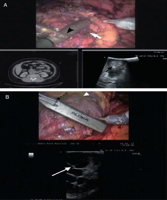
    1. Rao AR, Gray R, Mayer E, Motiwala H, Laniado M, Karim O. Occlusion Angiography Using Intraoperative Contrast-enhanced Ultrasound Scan (CEUS): A Novel Technique Demonstrating Segmental Renal Blood Supply to Assist Zero-ischaemia Robot-assisted Partial Nephrectomy. Eur Urol. 2013;63:913–919. doi: 10.1016/j.eururo.2012.10.034. - DOI - PubMed

MeSH terms