. 2015 Feb 26;160(5):893-903.
doi: 10.1016/j.cell.2015.01.031.

Mechanism of human antibody-mediated neutralization of Marburg virus

Affiliations

Mechanism of human antibody-mediated neutralization of Marburg virus

Andrew I Flyak et al. Cell. .

Abstract

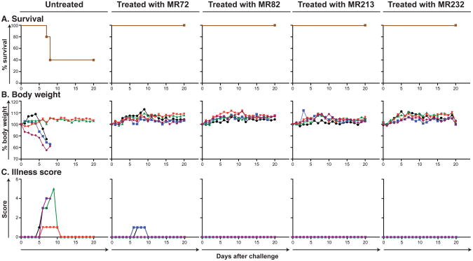
The mechanisms by which neutralizing antibodies inhibit Marburg virus (MARV) are not known. We isolated a panel of neutralizing antibodies from a human MARV survivor that bind to MARV glycoprotein (GP) and compete for binding to a single major antigenic site. Remarkably, several of the antibodies also bind to Ebola virus (EBOV) GP. Single-particle EM structures of antibody-GP complexes reveal that all of the neutralizing antibodies bind to MARV GP at or near the predicted region of the receptor-binding site. The presence of the glycan cap or mucin-like domain blocks binding of neutralizing antibodies to EBOV GP, but not to MARV GP. The data suggest that MARV-neutralizing antibodies inhibit virus by binding to infectious virions at the exposed MARV receptor-binding site, revealing a mechanism of filovirus inhibition.


Figures

Figure 1
Figure 1. MARV neutralizing mAbs display a unique binding pattern and target a distinct antigenic region on the GP surface
(A) Neutralization activity of MR77 (non-neutralizing antibody) or MR213 (neutralizing antibody) against VSV/GP-Uganda (red circles) or MARV-Uganda (black circles). Error bars represent the standard errors of the experiment performed in triplicate. (B) Binding of representative mAbs from four distinct Binding Groups to the MARV GP (blue squares) or MARV GPΔmuc (green squares). Dotted line indicates 0.5 μg/mL threshold for categorizing Group 3 antibodies as possessing low (3A) or high (3B) EC50 values. (C) Heatmap showing the neutralization potency of MARV GP-specific mAbs against VSV/GP-Uganda or MARV-Uganda. IC50 value for each virus-mAb combination is shown, with dark red, orange, yellow or white shading indicating high, intermediate, low or no potency. IC50 values greater than 1,000 μg/mL are indicated by >. Neutralization assays were performed in triplicate. (D) Data from competition binding assays using mAbs from Binding Groups 2, 3A or 3B. Numbers indicate the percent binding of the competing mAb in the presence of the first mAb, compared to binding of competing mAb alone. MAbs were judged to compete for the same site if maximum binding of the competing mAb was reduced to <30% of its un-competed binding (black boxes with white numbers). MAbs were considered non-competing if maximum binding of the competing mAb was >70% of its un-competed binding (white boxes with red numbers). Grey boxes with black numbers indicate an intermediate phenotype (between 30 and 70% of un-competed binding). See also Figures S1 to S5.
Figure 2
Figure 2. Neutralizing antibodies from a human survivor of MARV bind to the receptor-binding site of GP at two distinct angles of approach
(A) Representative reference-free 2D class averages of the MARV GPΔMuc:MR Fab complexes. (B) EM reconstructions of seven Fab fragments of neutralizing antibodies bound to MARV GPΔmuc (side views). All seven antibodies target a similar epitope on the top of GP. (C) These antibodies can be subdivided based on their angles of approach: i) those that bind toward the top and side of GP1 at a shallow angle relative to the central three-fold axis (MR72 in red, MR78 in orange, MR201 in yellow or MR82 in green) and ii) those that bind at a steeper angle toward the top of GP1 (MR191 in cyan, MR111 in blue or MR198 in purple). (D) The crystal structure of EBOV GPΔmuc (GP1 in white and GP2 in dark grey) is modeled into the MARV GP density (mesh) and the angles of approach of the neutralizing antibodies are indicated with arrows, colored as in (B). The footprint of the antibodies is indicated by a black circle targeting residues in the putative receptor-binding site (RBS) through a variety of approach angles.
Figure 3
Figure 3. Generation of escape mutants for MARV neutralizing antibodies
(A) VSV-MARV-72.5 (dotted lines) or VSV-MARV-78.1 (dashed line) escape mutations mapped onto the domain schematic of MARV GP. RBS = Receptor binding site; GLC = glycan cap; MUC = mucin-like domain. (B) Neutralization activity of antibodies from Binding Group 3B against wild-type VSV/GP-Uganda (circles, straight curves), VSV/GP-72.5 (squares, dotted curves) or VSV/GP-78.1 (triangles, dashed curves) escape mutant viruses.
Figure 4
Figure 4. Breadth of binding or neutralization of human MARV-specific mAbs for diverse filoviruses
(A) A heat map showing the binding in ELISA of neutralizing mAbs from Binding Group 3B to the MARV and EBOV GPs. EC50 value for each antigen-mAb combination is shown, with dark red shading indicating lower EC50 values and orange or yellow shading indicating intermediate or higher EC50 values. EC50 values greater than 1,000 μg/mL are indicated by >. (B) A heatmap showing the neutralization breadth of mAbs from Binding Group 3B. The IC50 value for each virus-mAb combination is shown, with dark red shading indicating increased potency and orange or yellow shading indicating intermediate or low potency. IC50 values greater than 1,000 μg/mL are indicated by >. Neutralization assays were performed in triplicate.
Figure 5
Figure 5. Survival and clinical overview of mice treated with MARV mAbs
Groups of mice at 5 animals per group were injected with individual mAbs by the intraperitoneal route twice: 1 h prior and 24 h after MARV challenge at 100 μg per treatment. Untreated animals served as controls. (A) Kaplan-Meier survival curves. (B) Body weight (C) Illness score

References

    1. Beniac DR, Melito PL, Devarennes SL, Hiebert SL, Rabb MJ, Lamboo LL, Jones SM, Booth TF. The organisation of Ebola virus reveals a capacity for extensive, modular polyploidy. PloS One. 2012;7:e29608. - PMC - PubMed
    1. Brauburger K, Hume AJ, Mühlberger E, Olejnik J. Forty-five years of Marburg virus research. Viruses. 2012;4:1878–1927. - PMC - PubMed
    1. Brochet X, Lefranc MP, Giudicelli V. IMGT/V-QUEST: the highly customized and integrated system for IG and TR standardized V-J and V-D-J sequence analysis. Nucleic Acids Res. 2008;36:W503–508. - PMC - PubMed
    1. Carette JE, Raaben M, Wong AC, Herbert AS, Obernosterer G, Mulherkar N, Kuehne AI, Kranzusch PJ, Griffin AM, Ruthel G, et al. Ebola virus entry requires the cholesterol transporter Niemann-Pick C1. Nature. 2011;477:340–343. - PMC - PubMed
    1. Carragher B, Kisseberth N, Kriegman D, Milligan RA, Potter CS, Pulokas J, Reilein A. Leginon: An automated system for acquisition of images from vitreous ice specimens. J Struct Biol. 2000;132:33–45. - PubMed

Publication types

MeSH terms