. 2015 Jul;144(1):87-91.
doi: 10.1007/s00418-015-1312-8. Epub 2015 Feb 28.

Hair follicle bulge cultures yield class III β-tubulin-positive melanoglial cells

Affiliations

Hair follicle bulge cultures yield class III β-tubulin-positive melanoglial cells

H Locher et al. Histochem Cell Biol. 2015 Jul.

Abstract

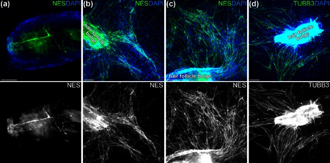
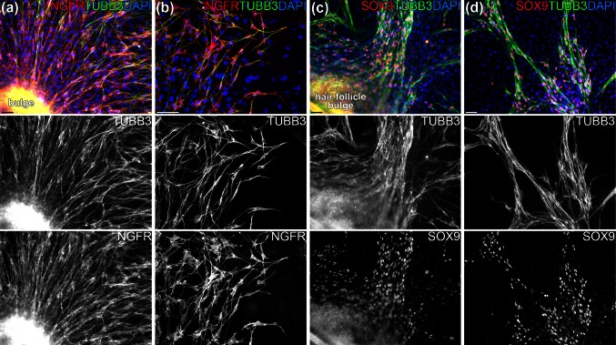
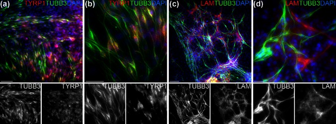
Class III β-tubulin (TUBB3)-positive cells from the hair follicle bulge are thought to be neuronal cells derived from a local neural crest stem cell. However, TUBB3 has recently been shown to be expressed in the melanocytic lineage. To evaluate the neural-crest-associated immunophenotype of TUBB3-positive cells from hair follicle bulge explants, we dissected hair follicle bulges out from mouse whisker pads and cultured for 1 month and assessed outgrowing cells by means of immunocytochemistry using the biomarkers TUBB3, nestin, NGFR, SOX9, TYRP1 and laminin. Large amounts of TUBB3-positive cells could be cultured that co-expressed nestin, NGFR, SOX9 and, to a lesser degree, TYRP1, matching a melanoglial phenotype. In addition, a small population of TUBB3-negative but laminin-positive cells was found, which presumably are of glial origin. It can be concluded that cells of melanoglial origin can easily be obtained from hair follicle bulge explants. These cells may be of use in experimental animal or human disease and wound healing models. Notably, the TUBB3-positive cells are of melanoglial rather than neuronal origin.


Figures

Fig. 1
Fig. 1
Nestin-positive and TUBB3-positive cells migrate from the hair follicle bulge. a Hair follicle bulge at the start of the culture, showing elongated nestin-positive (NES) cells (green) residing within the explant. b, c After 4 weeks in culture, numerous nestin-positive cells (green) are found growing in tracks extending from the bulge explant. d Hair follicle bulge explants after 4 four weeks of culture immunostain for TUBB3 in a pattern similar to that of nestin. Cell nuclei are stained blue with DAPI. Scalebars 100 μm (a) or 200 μm (bd)
Fig. 2
Fig. 2
TUBB3-positive hair follicle bulge cells co-express NGFR and SOX9. a, b Double immunostaining for TUBB3 (green) and NGRF (red) showing that both proteins are expressed in the same subgroup of cells. c, d Double immunostaining for TUBB3 (green) and SOX9 (red). The strong co-expression of SOX9 indicates that the TUBB3-positive cells are of melanoglial, rather than neuronal, origin. Cell nuclei are stained blue with DAPI. Scalebars 50 μm
Fig. 3
Fig. 3
Melanocyte and glial marker expression of TUBB3-positive cells. a Hair follicle bulge explant after 4 weeks of culture immunostained for TUBB3 (green) and TYRP1 (red), showing weak expression of TYRP1 in the TUBB3-positive cells. b Higher magnification of the lower left area in a. c, d Double immunostaining for TUBB3 (green) and laminin (red) shows a subpopulation of laminin-positive cells largely growing along the same migration patterns of the TUBB3-positive cells. Cell nuclei are stained blue with DAPI. Scalebars 200 μm (a, c), 100 μm (b) or 20 μm (d)

References

    1. Adameyko I, Lallemend F. Glial versus melanocyte cell fate choice: Schwann cell precursors as a cellular origin of melanocytes. Cell Mol Life Sci. 2010;67:3037–3055. doi: 10.1007/s00018-010-0390-y. - DOI - PMC - PubMed
    1. Adameyko I, Lallemend F, Furlan A, et al. Sox2 and Mitf cross-regulatory interactions consolidate progenitor and melanocyte lineages in the cranial neural crest. Development. 2012;139:397–410. doi: 10.1242/dev.065581. - DOI - PMC - PubMed
    1. Akasaka K, Maesawa C, Shibazaki M, et al. Loss of class III beta-tubulin induced by histone deacetylation is associated with chemosensitivity to paclitaxel in malignant melanoma cells. J Invest Dermatol. 2009;129:1516–1526. doi: 10.1038/jid.2008.406. - DOI - PubMed
    1. Amoh Y, Mii S, Aki R, et al. Multipotent nestin-expressing stem cells capable of forming neurons are located in the upper, middle and lower part of the vibrissa hair follicle. Cell Cycle. 2012;11:3513–3517. doi: 10.4161/cc.21803. - DOI - PMC - PubMed
    1. Djian-Zaouche J, Campagne C, Reyes-Gomez E, et al. Pax3(GFP), a new reporter for the melanocyte lineage, highlights novel aspects of PAX3 expression in the skin. Pigment Cell Melanoma Res. 2012;25:545–554. doi: 10.1111/j.1755-148X.2012.01024.x. - DOI - PubMed

Publication types

LinkOut - more resources