. 2015 Apr;61(3-4):369-78.
doi: 10.1007/s10858-015-9912-0. Epub 2015 Feb 28.

Transverse relaxation dispersion of the p7 membrane channel from hepatitis C virus reveals conformational breathing

Affiliations

Transverse relaxation dispersion of the p7 membrane channel from hepatitis C virus reveals conformational breathing

Jyoti Dev et al. J Biomol NMR. 2015 Apr.

Abstract

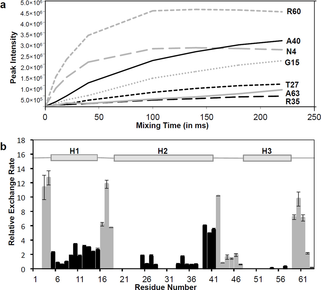
The p7 membrane protein encoded by hepatitis C virus (HCV) assembles into a homo-hexamer that selectively conducts cations. An earlier solution NMR structure of the hexameric complex revealed a funnel-like architecture and suggests that a ring of conserved asparagines near the narrow end of the funnel are important for cation interaction. NMR based drug-binding experiments also suggest that rimantadine can allosterically inhibit ion conduction via a molecular wedge mechanism. These results suggest the presence of dilation and contraction of the funnel tip that are important for channel activity and that the action of the drug is attenuating this motion. Here, we determined the conformational dynamics and solvent accessibility of the p7 channel. The proton exchange measurements show that the cavity-lining residues are largely water accessible, consistent with the overall funnel shape of the channel. Our relaxation dispersion data show that residues Val7 and Leu8 near the asparagine ring are subject to large chemical exchange, suggesting significant intrinsic channel breathing at the tip of the funnel. Moreover, the hinge regions connecting the narrow and wide regions of the funnel show strong relaxation dispersion and these regions are the binding sites for rimantadine. Presence of rimantadine decreases the conformational dynamics near the asparagine ring and the hinge area. Our data provide direct observation of μs-ms dynamics of the p7 channel and support the molecular wedge mechanism of rimantadine inhibition of the HCV p7 channel.


Figures

Fig. 1
Fig. 1. Purification and TROSY-HSQC spectra of p7
a. The p7 peptide was purified from a mixture of trpLE, p7, trpLE-p7 fusion protein by HPLC using a C18 column and a gradient of acetonitrile. b. FPLC elution profile after applying buffer containing 3 mM DPC, 100 mM NaCl, and 25 mM MES (pH 6.5) running through a Superdex 200 10/300 GL column (GE Healthcare) to remove excess detergent. c. SDS-PAGE of the FPLC elution fraction showing the purity of the p7 in the final NMR sample. d. Two-dimensional 15N TROSY-HSQC spectrum of 2H-15N- labeled p7 hexamer reconstituted in DPC micelles at pH 6.5 (recorded at 1H frequency of 600 MHz). The labels are residue-specific assignments of the amide resonances.
Fig. 2
Fig. 2. Peak intensities and proton exchange rate
a. Peak intensity of amide resonances as a function of mixing time (Tm) for selected residues of p7. Black lines represent residues in the helical regions and gray lines represent ones in the loop region. b. Water-amide proton exchange rates for each residue of the p7 channel (calculated from normalized peak intensities as described in Materials and Methods). Gray bars represent residues in the loop region and black bars represent residues in helical regions.
Fig. 3
Fig. 3. Solvent accessibility profile of the p7 channel
Mapping proton exchange rates for the three helices on the NMR structure of p7 (PDB: 2M6X). A gradient of gray to blue shows increasing exchange rate, with blue color indicating fastest exchange rate.
Fig. 4
Fig. 4. CPMG relaxation dispersion analysis of the p7 channel
a. Difference between R2,eff of CPMG frequency 100Hz and 900Hz (ΔR2) for each residue of the p7 channel recorded at two different magnetic fields, 600 MHz (red triangles) and 700 MHz (blue squares). Error bars were calculated based on uncertainty in repeat CPMG experiments. b. Mapping residues that show significant relaxation dispersion, Val7, Leu8 and Phe19 on the p7 structure (PDB: 2M6X). These three residues are marker by red spheres.
Fig. 5
Fig. 5. CPMG relaxation dispersion curves for p7 in the presence and absence of rimantadine
CPMG relaxation dispersion curves for three residues, Val7, Leu8 and Phe19 at two different magnetic fields, 600 MHz (red) and 700 MHz (blue) in the apo state (top panel) and in the presence of 5 mM rimantadine (bottom panel). Circles represent data points and the dotted line represents the fit for two-state model.
Fig. 6
Fig. 6. Proposed model for channel function and inhibition
The residues that show conformational exchange are shown with green arrows. The breathing of H1 helix (blue arrows), which is supported by our results, could facilitate opening and closing of the channel. In the open state, Asn9 is able to bind and release Ca2+ ions. Conformational dynamics at the hinge region between H1 and H2 could explain the mechanism of drug inhibition, as the drug binds at this region. Drug binding could act as a molecular wedge to prevent breathing of the complex, and therefore closing the channel.

References

    1. Agarkova I, Dunigan D, Gurnon J, Greiner T, Barres J, Thiel G, Van Etten JL. Chlorovirus-mediated membrane depolarization of Chlorella alters secondary active transport of solutes. J Virol. 2008;82:12181–12190. - PMC - PubMed
    1. Carrere-Kremer S, Montpellier-Pala C, Cocquerel L, Wychowski C, Penin F, Dubuisson J. Subcellular localization and topology of the p7 polypeptide of hepatitis C virus. Journal of Virology. 2002;76:3720–3730. - PMC - PubMed
    1. Carver JP, Richards RE. A general two-site solution for the chemical exchange produced dependence of T2 upon the carr-Purcell pulse separation. Journal of Magnetic Resonance. 1972;6:89–105.
    1. Cook GA, Opella SJ. Secondary structure, dynamics, and architecture of the p7 membrane protein from hepatitis C virus by NMR spectroscopy. Biochim Biophys Acta. 2011;1808:1448–1453. - PMC - PubMed
    1. Cuello LG, Jogini V, Cortes DM, Perozo E. Structural mechanism of C-type inactivation in K(+) channels. Nature. 2010;466:203–208. - PMC - PubMed

Publication types

MeSH terms

LinkOut - more resources