

Dynamic stiffening of poly(ethylene glycol)-based hydrogels to direct valvular interstitial cell phenotype in a three-dimensional environment
- PMID: 25725554
- PMCID: PMC4346780
- DOI: 10.1016/j.biomaterials.2015.01.047
Dynamic stiffening of poly(ethylene glycol)-based hydrogels to direct valvular interstitial cell phenotype in a three-dimensional environment
Abstract
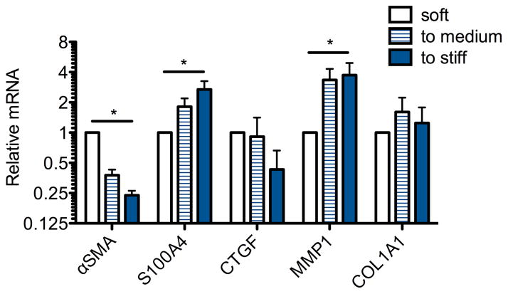
Valvular interstitial cells (VICs) are active regulators of valve homeostasis and disease, responsible for secreting and remodeling the valve tissue matrix. As a result of VIC activity, the valve modulus can substantially change during development, injury and repair, and disease progression. While two-dimensional biomaterial substrates have been used to study mechanosensing and its influence on VIC phenotype, less is known about how these cells respond to matrix modulus in a three-dimensional environment. Here, we synthesized MMP-degradable poly(ethylene glycol) (PEG) hydrogels with elastic moduli ranging from 0.24 kPa to 12 kPa and observed that cell morphology was constrained in stiffer gels. To vary gel stiffness without substantially changing cell morphology, cell-laden hydrogels were cultured in the 0.24 kPa gels for 3 days to allow VIC spreading, and then stiffened in situ via a second, photoinitiated thiol-ene polymerization such that the gel modulus increased from 0.24 kPa to 1.2 kPa or 13 kPa. VICs encapsulated within soft gels exhibited αSMA stress fibers (∼ 40%), a hallmark of the myofibroblast phenotype. Interestingly, in stiffened gels, VICs became deactivated to a quiescent fibroblast phenotype, suggesting that matrix stiffness directs VIC phenotype independent of morphology, but in a manner that depends on the dimensionality of the culture platform. Collectively, these studies present a versatile method for dynamic stiffening of hydrogels and demonstrate the significant effects of matrix modulus on VIC myofibroblast properties in three-dimensional environments.
Keywords: ECM; Elasticity; Heart valve; Hydrogel; Three-dimensional cell culture; Valvular interstitial cells.
Copyright © 2015 Elsevier Ltd. All rights reserved.
Figures


References
-
- Durbin AD, Gotlieb AI. Advances towards understanding heart valve response to injury. Cardiovasc Pathol. 2002;11:69–77. - PubMed
-
- Rajamannan NM, Evans FJ, Aikawa E, Grande-Allen KJ, Demer LL, Heistad DD, et al. Calcific Aortic Valve Disease: Not Simply a Degenerative Process: A Review and Agenda for Research From the National Heart and Lung and Blood Institute Aortic Stenosis Working Group. Executive Summary: Calcific Aortic Valve Disease - 2011 Update. Circulation. 2011;124:1783–91. - PMC - PubMed
-
- Fayet C, Bendeck MP, Gotlieb AI. Cardiac valve interstitial cells secrete fibronectin and form fibrillar adhesions in response to injury. Cardiovasc Pathol. 2007;16:203–11. - PubMed
-
- Liu AC, Gotlieb AI. Characterization of cell motility in single heart valve interstitial cells in vitro. Histol Histopathol. 2007;22:873–82. - PubMed
Publication types
MeSH terms
Substances
Grants and funding
LinkOut - more resources
Full Text Sources
Other Literature Sources
Research Materials
