. 2015 Feb 28;6(6):4345-56.
doi: 10.18632/oncotarget.2999.

GATA1 induces epithelial-mesenchymal transition in breast cancer cells through PAK5 oncogenic signaling

Affiliations

GATA1 induces epithelial-mesenchymal transition in breast cancer cells through PAK5 oncogenic signaling

Yang Li et al. Oncotarget. .

Abstract

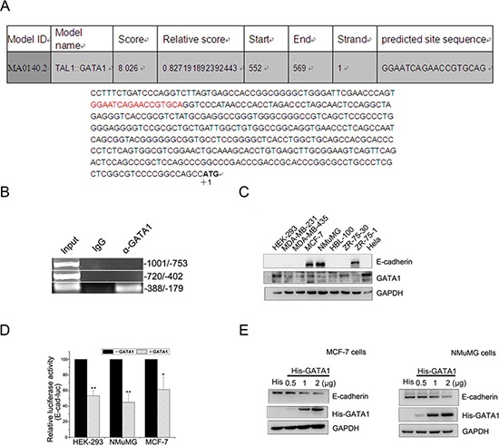
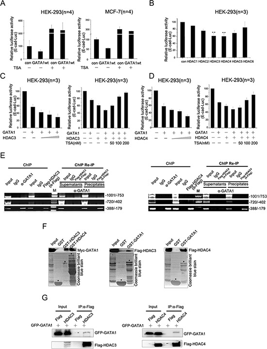
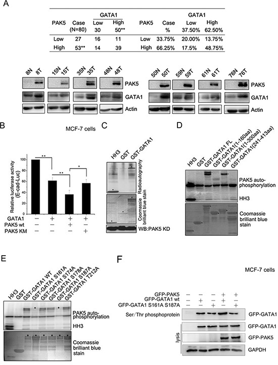
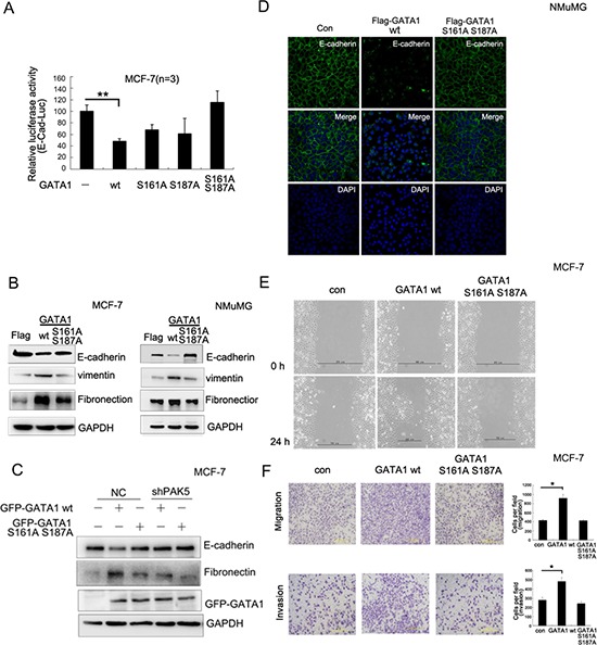
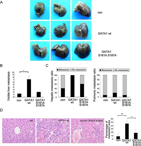
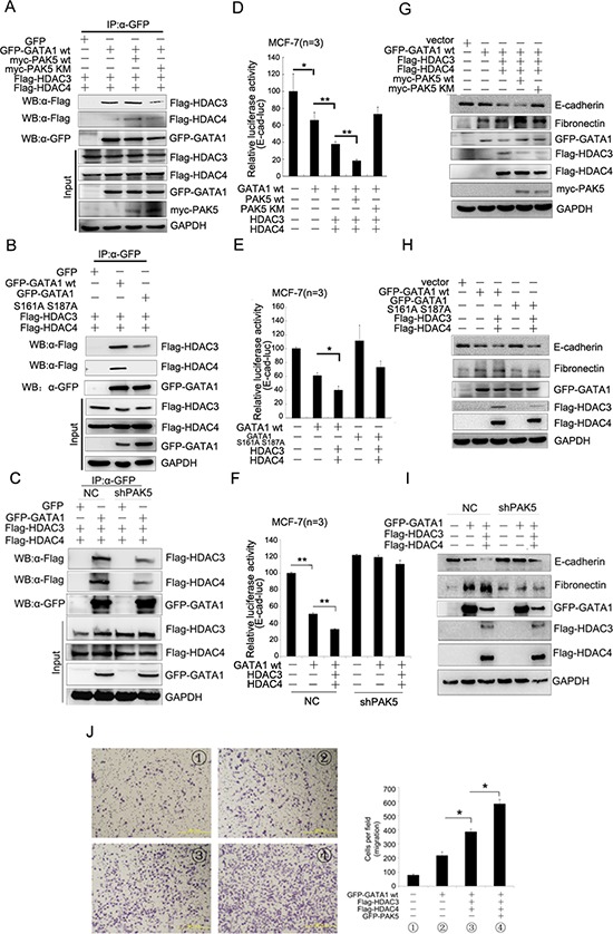
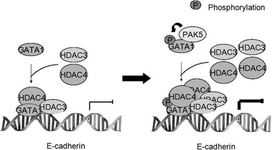
Epithelial-mesenchymal transition (EMT) is a key process in tumor metastatic cascade that is characterized by the loss of cell-cell junctions, resulting in the acquisition of migratory and invasive properties. E-cadherin is a major component of intercellular junctions and the reduction or loss of its expression is a hallmark of EMT. Transcription factor GATA1 has a critical anti-apoptotic role in breast cancer, but its function for metastasis has not been investigated. Here, we found that GATA1, as a novel E-cadherin repressor, promotes EMT in breast cancer cells. GATA1 binds to E-cadherin promoter, down-regulates E-cadherin expression, disrupts intercellular junction and promotes metastasis of breast cancer cell in vivo. Moreover, GATA1 is a new substrate of p21-activated kinase 5 (PAK5), which is phosphorylated on serine 161 and 187 (S161 and S187). GATA1 recruits HDAC3/4 to E-cadherin promoter, which is reduced by GATA1 S161A S187A mutant. These data indicate that phosphorylated GATA1 recruits more HDAC3/4 to promote transcriptional repression of E-cadherin, leading to the EMT of breast cancer cells. Our findings provide insights into the novel function of GATA1, contributing to a better understanding of the EMT, indicating that GATA1 and its phosphorylation may play an important role in the metastasis of breast cancer.


Conflict of interest statement

Conflict of interest

The authors declare no conflict of interest.

Figures

Figure 1
Figure 1. GATA1 binds to E-cadherin promoter and down-regulates E-cadherin
(A) Nucleotide sequence of the E-cadherin promoter was analyzed. Potential transcription factor binding motifs are red. ATG is indicated by +1. (B) GATA1 binds to E-cadherin promoter (–388/–179) detected by ChIP assays. (C) Protein expression levels of E-cadherin and GATA1 in mammary cell lines. (D) HEK-293, NMuMG and MCF-7 cell lines were transfected with pGL2-E-cad-luc, pRL-TK and pcDNA-GATA1 or control plasmid for luciferase assays. *p < 0.05, **p < 0.01. (E) MCF-7 and NMuMG cells were transfected with 0.5 μg, 1 μg, 2 μg His tagged-GATA1 plasmid, and western blot analysis was performed.
Figure 2
Figure 2. GATA1 recruits HDAC3/4 to down-regulate E-cadherin transcription
(A) pGL2-E-cad-luc and pRL-TK plasmids were co-transfected with pcDNA-GATA1 or control plasmid into HEK-293 cells and MCF7 cells. Then cells treated with or without TSA for luciferase assay. (B) HEK-293 cells were transfected with pGL2-E-cad-luc plasmid together with HDAC constructs expressing HDAC1–6, respectively. **p < 0.01. (C–D) HEK-293 cells were transfected with pGL2-E-cad-luc, pcDNA-GATA1 and increasing amounts of HDAC3/4 as indicated for Luciferase Assays. Simultaneously, increasing amounts of TSA was added to HEK-293 cells transfected with GATA1 and HDAC3/4 for Luciferase Assays. (E) MCF-7 cells were transfected with Flag-HDAC3/4 expression plasmids. ChIP assay was carried out using anti-GATA1 or anti-Flag antibody, followed by PCR with primers amplifying the E-cadherin promoter region (–1001/–753, –720/–402, –388/–179). ChIP Re-IP, soluble chromatin, prepared from MCF-7 cells transfected with Flag-HDAC3/4, was firstly immunoprecipitated with antibody against GATA1, then reimmunoprecipitated with anti-Flag antibody. (F) In vitro translated Myc-GATA1 or Flag-HDAC3/4 was incubated with GST-HDAC3/4 or GST-GATA1 fusion proteins for GST pull-down assay. (G) HEK-293 cells were transfected with GFP-GATA1 and Flag-HDAC3 or GFP-GATA1 and Flag-HDAC4. Lysates were immunoprecipitated with Flag antibodies and immunoblotted with Flag and GFP antibodies.
Figure 3
Figure 3. GATA1 is a physiological substrate of p21-activated kinase 5
(A) Western blot analysis demonstrates the protein level of PAK5 and GATA1 in breast cancer tissues and matched adjacent noncancerous tissues. N, matched adjacent noncancerous tissue; T, breast cancer tissue. **p < 0.01. (B) PAK5wt/KM and GATA1 were transfected into MCF-7 cells as indicated for Luciferase Assays. *p < 0.05, **p < 0.01. (C) HEK-293 cells transfected with Myc-PAK5KD (PAK5 kinase domain) were lysed for IP with anti-Myc antibody, the immunoprecipitated PAK5 kinase was incubated with GST or GST-GATA1 for in vitro kinase assay. Histone H3 (HH3) served as a positive control. (D) In vitro kinase assay using commercial PAK5 kinase and GST-GATA1FL (Full length, 1–413aa) or GST-GATA1 deletion mutants (1–160aa, 1–300aa and 241–413aa). Histone H3 (HH3) served as a positive control. (E) In vitro kinase assay using commercial PAK5 kinase and GST-GATA1 wt or GST-GATA1 single-site mutations as indicated. (F) MCF-7 cells transfected with GATA1 wt/S161A S187A and PAK5 wt were used for Ser/Thr phosphoprotein purification. Then concentrated protein was used for western blot. The top lane, the phosphorylated GATA1 from these cells lysates was used for immunoblotting using anti-GFP antibody. Total cells lysates were used for immunoblotting by anti-GFP, GAPDH antibodies.
Figure 4
Figure 4. PAK5-mediated GATA1 phosphorylation regulates EMT in breast cancer cells
(A) MCF-7 cells were transfected with wild type or different mutants of GATA1 for Luciferase assay. **p < 0.01. (B) Empty vector, Flag-GATA1 wt or Flag-GATA1 S161A S187A was stably expressed in MCF-7 and NMuMG cells through lentivirus. Cell lysates from these cells were used for immunoblotting using anti-Fibronectin, E-cadherin, vimentin, GAPDH antibodies. (C) Stably knocked down shPAK5 or NC (non-specific control) cells were transfected with GATA1 wt. Western blot analyzed the marker of EMT. (D) The same transfection as (B), NMuMG cells were cultured 24 hours and analyzed by immunofluorescence using anti-E-cadherin antibody followed by Alexa Flour 488 (green) antibody and nucleus was stained by DAPI (blue). (E–F) Effects of different types of GATA1 over-expression on the migration of cultured MCF-7 cells were examined by wound-healing (E) and transwell migration chambers (F) assays. Results are representative of three independent experiments. Migrated cells were plotted as the average number of cells per field of view. In the low lane, transwell migration chambers were treated with 10% matrigel, but not in the top lane. *p < 0.05.
Figure 5
Figure 5. Phosphorylation of GATA1 enhances breast cancer cell metastasis in vivo
1 × 10⁷ MCF-7 cells stably expressing empty vector, GATA1 wt or GATA1 S161A S187A were injected into nude mice through tail vein. (A) Livers were dissected after injection and macroscopically photographed. (B) Graphical representation of the number of liver metastases from each mouse (mean ± s.d.); *p < 0.05. (C) Effect of MCF7 cells with stable expression of control Flag, Flag GATA1 wt, Flag GATA1 S161A S187A on incidence of liver and lung metastasis. (D) Representative image of H&E-stained liver sections of mice and percentage of metastatic liver surface area relative to tatal live surface area. scale bar, 50 μm. 30 independent visions were counted. *p < 0.05, **p < 0.01.
Figure 6
Figure 6. The phosphorylated GATA1 recruits more HDAC3/4 to promote transcriptional repression of E-cadherin
(A) HEK-293 cells were transfected with GFP-GATA1, Myc-PAK5wt/KM, Flag-HDAC3/4 as indicated. Lysates were immunoprecipitated with anti-GFP antibody and immunoblotted with anti-Flag, anti-GFP and anti-myc antibody. (B) HEK-293 cells were co-transfected with different combination of plasmids as indicated. Lysates were immunoprecipitated with anti-GFP antibody and immunoblotted with anti-Flag and anti-GFP antibody. (C) MCF-7 cells stably infected with shPAK5 were co-transfected with different combination of plasmids as indicated. Lysates were immunoprecipitated with anti-GFP antibody and immunoblotted with anti-Flag and anti-GFP antibody. (D) PAK5wt/KM and GATA1, HDAC3/4 were transfected into MCF-7 cells as indicated for Luciferase Assays. *p < 0.05, **p < 0.01. (E) GATA1 wt and GATA1 S161A S187A, HDAC3/4 were transfected into MCF-7 cells as indicated for Luciferase Assays. *p < 0.05. (F) GATA1, HDAC3/4 were transfected into stably PAK5 knockdown MCF-7 cells as indicated for Luciferase Assays. **p < 0.01. (G–I) The same transfection as D, E, F cell lysates from these cells were used for immunoblotting using anti-Fibronectin, E-cadherin antibodies. (J) GATA1, HDAC3/4 and PAK5 were transfected into MCF-7 cells, then transwell assay was used. Results are representative of three independent experiments. Migrated cells were plotted as the average number of cells per field of view. *p < 0.05.
Figure 7
Figure 7. Proposed model showing the role of GATA1 and its phosphorylation in repressing E-cadherin transcription

References

    1. Polyak K, Weinberg RA. Transitions between epithelial and mesenchymal states: Acquisition of malignant and stem cell traits. Nat Rev Cancer. 2009;9:265–273. - PubMed
    1. Thiery JP, Acloque H, Huang RY, Nieto MA. Epithelial-mesenchymal transitions in development and disease. Cell. 2009;139:871–890. - PubMed
    1. Thiery JP, Sleeman JP. Complex networks orchestrate epithelial-mesenchymal transitions. Nat Rev Mol Cell Biol. 2006;7:131–142. - PubMed
    1. Yilmaz M, Christofori G. EMT, the cytoskeleton, and cancer cell invasion. Cancer Metastasis Rev. 2009;28:15–33. - PubMed
    1. Cowin P, Welch DR. Breast cancer progression: controversies and consensus in the molecular mechanisms of metastasis and EMT. J Mammary Gland Biol Neoplasia. 2007;12:99–102. - PMC - PubMed

Publication types

LinkOut - more resources