Coronavirus envelope (E) protein remains at the site of assembly
- PMID: 25726972
- PMCID: PMC4550588
- DOI: 10.1016/j.virol.2015.02.005
Coronavirus envelope (E) protein remains at the site of assembly
Abstract
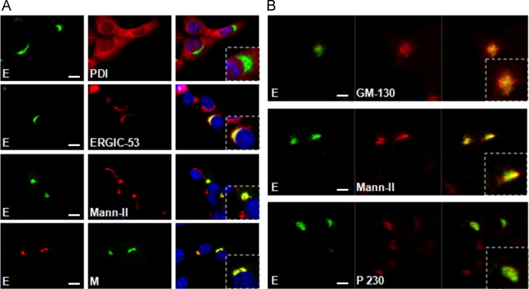
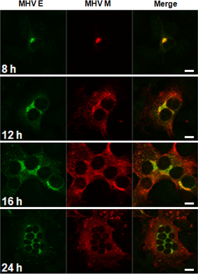
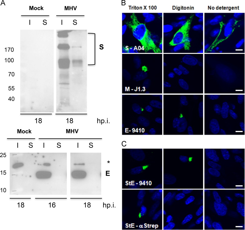
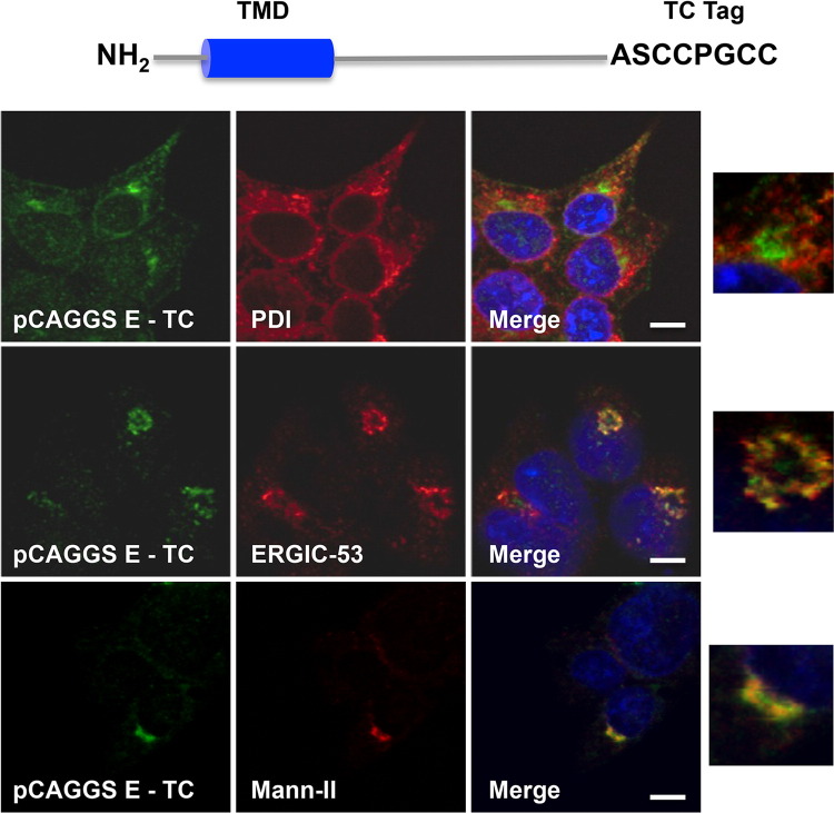
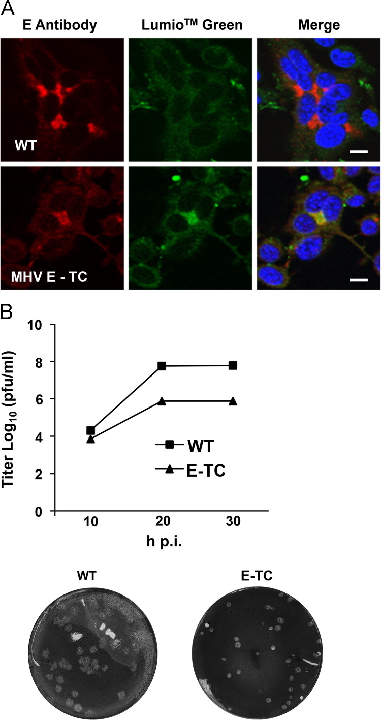
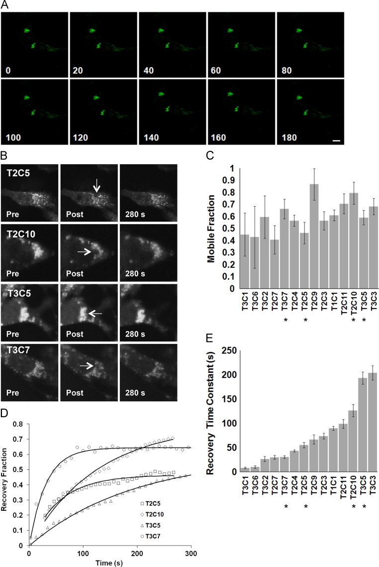
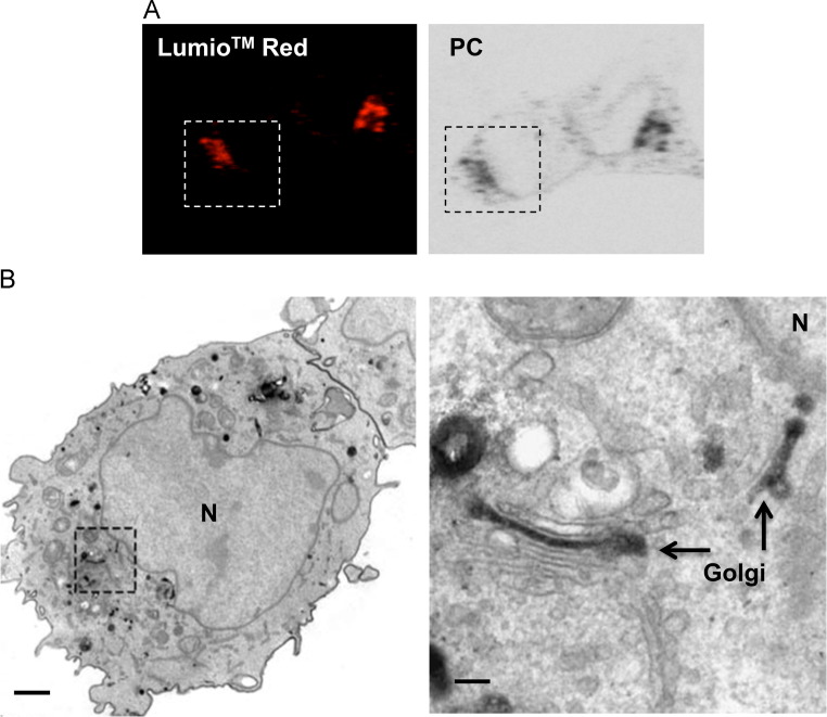
Coronaviruses (CoVs) assemble at endoplasmic reticulum Golgi intermediate compartment (ERGIC) membranes and egress from cells in cargo vesicles. Only a few molecules of the envelope (E) protein are assembled into virions. The role of E in morphogenesis is not fully understood. The cellular localization and dynamics of mouse hepatitis CoV A59 (MHV) E protein were investigated to further understanding of its role during infection. E protein localized in the ERGIC and Golgi with the amino and carboxy termini in the lumen and cytoplasm, respectively. E protein does not traffic to the cell surface. MHV was genetically engineered with a tetracysteine tag at the carboxy end of E. Fluorescence recovery after photobleaching (FRAP) showed that E is mobile in ERGIC/Golgi membranes. Correlative light electron microscopy (CLEM) confirmed the presence of E in Golgi cisternae. The results provide strong support that E proteins carry out their function(s) at the site of budding/assembly.
Keywords: CLEM; Coronavirus; Envelope protein; FRAP; Live-cell imaging; Protein transport/localization; Virus assembly.
Copyright © 2015 Elsevier Inc. All rights reserved.
Figures


Similar articles
-
Envelope glycoprotein interactions in coronavirus assembly.J Cell Biol. 1995 Oct;131(2):339-49. doi: 10.1083/jcb.131.2.339. J Cell Biol. 1995. PMID: 7593163 Free PMC article.
-
Hepatitis C Virus Lipoviroparticles Assemble in the Endoplasmic Reticulum (ER) and Bud off from the ER to the Golgi Compartment in COPII Vesicles.J Virol. 2017 Jul 12;91(15):e00499-17. doi: 10.1128/JVI.00499-17. Print 2017 Aug 1. J Virol. 2017. PMID: 28515296 Free PMC article.
-
Assembly and Cellular Exit of Coronaviruses: Hijacking an Unconventional Secretory Pathway from the Pre-Golgi Intermediate Compartment via the Golgi Ribbon to the Extracellular Space.Cells. 2021 Feb 26;10(3):503. doi: 10.3390/cells10030503. Cells. 2021. PMID: 33652973 Free PMC article. Review.
-
Characterization of the coronavirus mouse hepatitis virus strain A59 small membrane protein E.J Virol. 2000 Mar;74(5):2333-42. doi: 10.1128/jvi.74.5.2333-2342.2000. J Virol. 2000. PMID: 10666264 Free PMC article.
-
Incorporation of spike and membrane glycoproteins into coronavirus virions.Viruses. 2015 Apr 3;7(4):1700-25. doi: 10.3390/v7041700. Viruses. 2015. PMID: 25855243 Free PMC article. Review.
Cited by
-
Leaving no stone unturned: Allosteric targeting of SARS-CoV-2 spike protein at putative druggable sites disrupts human angiotensin-converting enzyme interactions at the receptor binding domain.Inform Med Unlocked. 2020;21:100451. doi: 10.1016/j.imu.2020.100451. Epub 2020 Oct 16. Inform Med Unlocked. 2020. PMID: 33083517 Free PMC article.
-
SARS-CoV-2 Evolutionary Adaptation toward Host Entry and Recognition of Receptor O-Acetyl Sialylation in Virus-Host Interaction.Int J Mol Sci. 2020 Jun 26;21(12):4549. doi: 10.3390/ijms21124549. Int J Mol Sci. 2020. PMID: 32604730 Free PMC article. Review.
-
Immunogenicity and Efficacy of Monovalent and Bivalent Formulations of a Virus-Like Particle Vaccine against SARS-CoV-2.Vaccines (Basel). 2022 Nov 24;10(12):1997. doi: 10.3390/vaccines10121997. Vaccines (Basel). 2022. PMID: 36560407 Free PMC article.
-
SARS-CoV-2 Envelope (E) Protein Binds and Activates TLR2 Pathway: A Novel Molecular Target for COVID-19 Interventions.Viruses. 2022 May 8;14(5):999. doi: 10.3390/v14050999. Viruses. 2022. PMID: 35632741 Free PMC article.
-
Design of a Multiepitope-Based Peptide Vaccine against the E Protein of Human COVID-19: An Immunoinformatics Approach.Biomed Res Int. 2020 May 11;2020:2683286. doi: 10.1155/2020/2683286. eCollection 2020. Biomed Res Int. 2020. PMID: 32461973 Free PMC article.
References
Publication types
MeSH terms
Substances
Grants and funding
LinkOut - more resources
Full Text Sources
Other Literature Sources
Miscellaneous