Review
. 2015 Mar 2;5(1):21-34.
doi: 10.1002/9780470942390.mo140166.

Overview of biological mechanisms and applications of three murine models of bone repair: closed fracture with intramedullary fixation, distraction osteogenesis, and marrow ablation by reaming

Affiliations
Review

Overview of biological mechanisms and applications of three murine models of bone repair: closed fracture with intramedullary fixation, distraction osteogenesis, and marrow ablation by reaming

Beth Bragdon et al. Curr Protoc Mouse Biol. .

Abstract

Fractures are one of the most common large-organ, traumatic injuries in humans, and osteoporosis-related fractures are the fastest growing health care problem of aging. Elective orthopedic surgeries of the bones and joints also represent some of most common forms of elective surgeries performed. Optimal repair of skeletal tissues is necessary for successful outcomes of these many different orthopedic surgical treatments. Research focused on post-natal skeletal repair is therefore of immense clinical importance and of particular relevance in situations in which bone tissue healing is compromised due to the extent of tissue trauma or specific medical co-morbidities. Three commonly used murine surgical models of bone healing, closed fracture with intramedullary fixation, distraction osteogenesis (DO), and marrow ablation by reaming, are presented. The biological aspects of these models are contrasted and the types of research questions that may be addressed with these models are presented.

Keywords: distraction osteogenesis; fracture; marrow ablation; murine models; orthopedic surgery.


Figures

Figure 1
Figure 1
Progression of Fracture Healing. All bone repair studies that are depicted in the figures were carried out in C57/BJ mice at 8 to 12 weeks. A) Radiographic progression of trauma induced simple transverse fracture healing fixed with an intramedullary pin. Post-operative days (POD) after trauma are denoted in the figure. The stages of the fracture healing are indicated in the figure with the black segment of each bar indicating the medium period when that stage of healing is maximal. B) The progression of changing tissue and material compositions across the time course of femur fracture healing. Transverse histological sections from day 10 and day 35 fracture calluses were stained with Safranin O and fast green. Cartilage is stained red and other tissues are shades of blue. Transverse cross-sections of day 14 and day 21 specimens are μCT reconstructions showing the distribution of tissue mineral, and translucent μCT reconstructions are composite rendering of consecutive reconstructions using contrast enhancement agent [CE] (Hayward et al., 2013) to distinguish cartilage from mineralized tissues. Images are pseudo-colored: yellow, original cortical bone; blue, cartilage; and green, new bone.
Figure 2
Figure 2
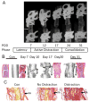
Progression of Bone Formation During Distraction Osteogenesis. All bone repair studies that are depicted in the figures were carried out in C57/BJ background mice at 8 to 12 weeks. Radiographic progression and the stages of bone formation induced by distraction osteogenesis are shown. Post-operative days (POD) after trauma are denoted in the figure. Arrow depicts osteotomy. B) The progression of changing tissue and material compositions across the time course of distraction osteogenesis. Days after surgery at which transverse histological sections and μCT were obtained from are denoted in the figure. Sections were stained with hematoxylin and eosin. Arrows denote the size of the gap. C) Comparisons of angiogenic response elicited by distraction osteogenesis. The vascular tissue beds within the muscle and bone compartments of the upper mouse leg of control (no surgery), osteotomy and osteotomy followed by distraction are depicted.
Figure 3
Figure 3
Progression of Bone Formation After Marrow Ablation by Reaming. A) Gross appearance of transverse section of the tibia at seven days after reaming. Large arrow denotes the entry point of the syringe needles that were used to ream the bone. Histological image depicts the extensive trabecular bone formation in repose to the reaming. B) Series of higher magnification images of the stages of intramedullary bone formation which peaks at day 7 POD followed by resorption seen with the extensive formation of multi- nucleated osteoclast (denoted with arrows) at day 14 POD. At day 21 POD the marrow space is returning to its pre reaming configuration however many more adipocytes are now seen in the marrow. C) The structural progression of changing material compositions across the time course of marrow ablation as characterized by μCT POD days 7 and 21.
Figure 4
Figure 4. Comparison of Representative Gene Expression during the three different Surgical Models
Two representative genes are shown for each biological process, bone (Osterix and Osteocalcin), cartilage (Sox9 and Aggrecan), and resorption (TRAP and RANKL) during fracture, DO, and marrow ablation. No detectible (ND) expression was observed for Aggrecan during marrow ablation.

References

    1. Abou-Khalil R, Colnot C. Cellular and molecular bases of skeletal regeneration: What can we learn from genetic mouse models? Bone. 2014;64:211–221. - PubMed
    1. Ai-Aql ZS, Alagl AS, Graves DT, Gerstenfeld LC, Einhorn TA. Molecular mechanisms controlling bone formation during fracture healing and distraction osteogenesis. Journal of dental research. 2008;87:107–118. - PMC - PubMed
    1. Aronson J, Good B, Stewart C, Harrison B, Harp J. Preliminary studies of mineralization during distraction osteogenesis. Clinical orthopaedics and related research. 1990;250:43–49. - PubMed
    1. Aronson J. Experimental and clinical experience with distraction osteogenesis. The Cleft palate-craniofacial journal. 1994;31:473–482. - PubMed
    1. Balogh ZJ, Reumann MK, Gruen RL, Mayer-Kuckuk P, Schuetz MA, Harris IA, Gabbe BJ, Bhandari M. Advances and future directions for management of trauma patients with musculoskeletal injuries. The Lancet. 2012;380:1109–1119. - PubMed

Publication types

LinkOut - more resources