Clinical validation of a gene expression signature that differentiates benign nevi from malignant melanoma
- PMID: 25727210
- PMCID: PMC6681167
- DOI: 10.1111/cup.12475
Clinical validation of a gene expression signature that differentiates benign nevi from malignant melanoma
Abstract
Background: Histopathologic examination is sometimes inadequate for accurate and reproducible diagnosis of certain melanocytic neoplasms. As a result, more sophisticated and objective methods have been sought. The goal of this study was to identify a gene expression signature that reliably differentiated benign and malignant melanocytic lesions and evaluate its potential clinical applicability. Herein, we describe the development of a gene expression signature and its clinical validation using multiple independent cohorts of melanocytic lesions representing a broad spectrum of histopathologic subtypes.
Methods: Using quantitative reverse-transcription polymerase chain reaction (PCR) on a selected set of 23 differentially expressed genes, and by applying a threshold value and weighting algorithm, we developed a gene expression signature that produced a score that differentiated benign nevi from malignant melanomas.
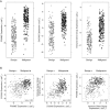
Results: The gene expression signature classified melanocytic lesions as benign or malignant with a sensitivity of 89% and a specificity of 93% in a training cohort of 464 samples. The signature was validated in an independent clinical cohort of 437 samples, with a sensitivity of 90% and specificity of 91%.
Conclusions: The performance, objectivity, reliability and minimal tissue requirements of this test suggest that it could have clinical application as an adjunct to histopathology in the diagnosis of melanocytic neoplasms.
Keywords: melanoma; molecular diagnositcs; pathology; real time PCR; validation studies.
© 2015 The Authors. Journal of Cutaneous Pathology published by John Wiley & Sons Ltd.
Figures


References
-
- Farmer ER, Gonin R, Hanna MP. Discordance in the histopathologic diagnosis of melanoma and melanocytic nevi between expert pathologists. Hum Pathol 1996; 27: 528. - PubMed
-
- McGinnis KS, Lessin SR, Elder DE, et al. Pathology review of cases presenting to a multidisciplinary pigmented lesion clinic. Arch Dermatol 2002; 138: 617. - PubMed
-
- Shoo BA, Sagebiel RW, Kashani‐Sabet M. Discordance in the histopathologic diagnosis of melanoma at a melanoma referral center. J Am Acad Dermatol 2010; 62: 751. - PubMed
-
- Veenhuizen KC, De Wit PE, Mooi WJ, et al. Quality assessment by expert opinion in melanoma pathology: experience of the pathology panel of the Dutch Melanoma Working Party. J Pathol 1997; 182: 266. - PubMed
-
- Barnhill RL, Argenyi ZB, From L, et al. Atypical Spitz nevi/tumors: lack of consensus for diagnosis, discrimination from melanoma, and prediction of outcome. Hum Pathol 1999; 30: 513. - PubMed
Publication types
MeSH terms
LinkOut - more resources
Full Text Sources
Other Literature Sources
Medical
Research Materials

