. 2015 Mar;41(5):686-98.
doi: 10.1111/ejn.12828.

Single-unit analysis of somatosensory processing in the core auditory cortex of hearing ferrets

Affiliations

Single-unit analysis of somatosensory processing in the core auditory cortex of hearing ferrets

M Alex Meredith et al. Eur J Neurosci. 2015 Mar.

Abstract

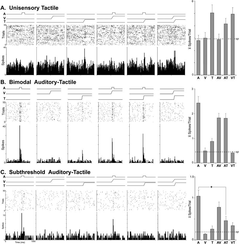
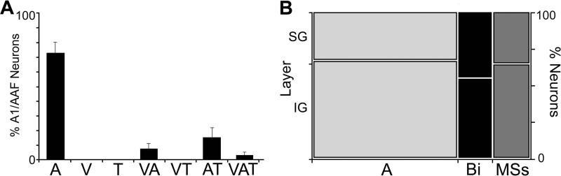
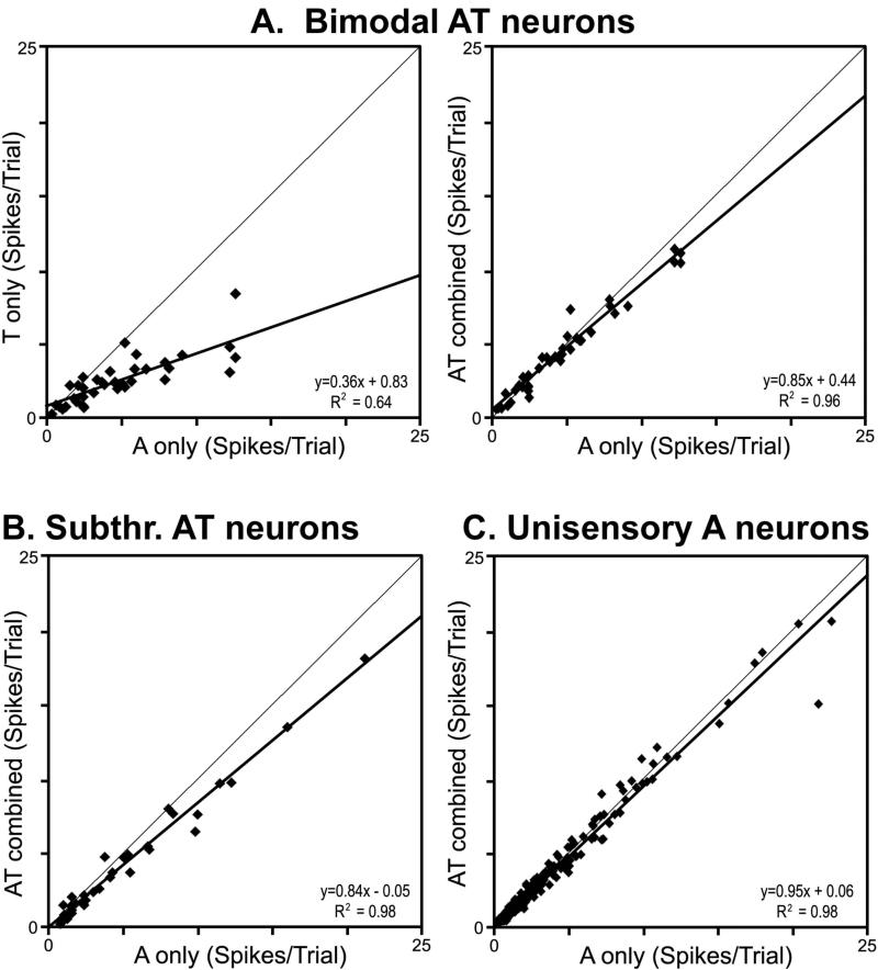
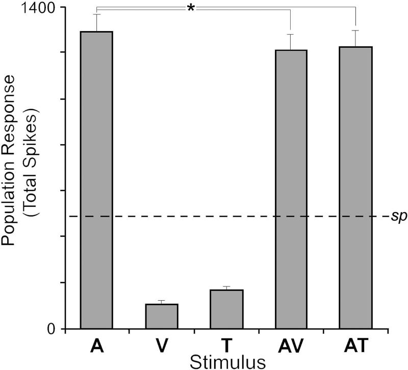
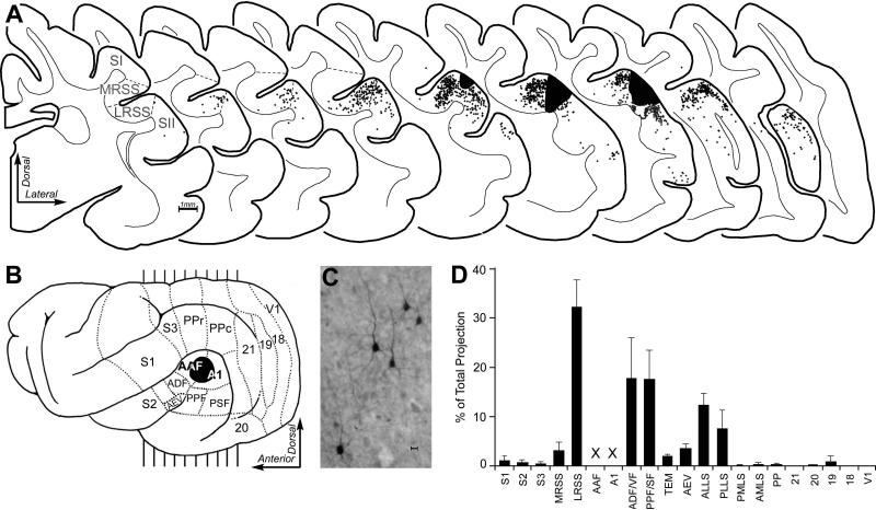
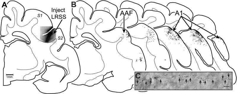
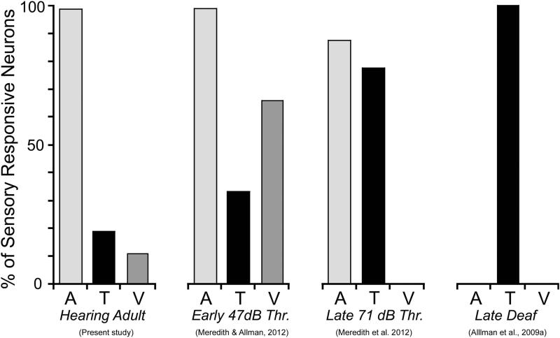
The recent findings in several species that the primary auditory cortex processes non-auditory information have largely overlooked the possibility of somatosensory effects. Therefore, the present investigation examined the core auditory cortices (anterior auditory field and primary auditory cortex) for tactile responsivity. Multiple single-unit recordings from anesthetised ferret cortex yielded histologically verified neurons (n = 311) tested with electronically controlled auditory, visual and tactile stimuli, and their combinations. Of the auditory neurons tested, a small proportion (17%) was influenced by visual cues, but a somewhat larger number (23%) was affected by tactile stimulation. Tactile effects rarely occurred alone and spiking responses were observed in bimodal auditory-tactile neurons. However, the broadest tactile effect that was observed, which occurred in all neuron types, was that of suppression of the response to a concurrent auditory cue. The presence of tactile effects in the core auditory cortices was supported by a substantial anatomical projection from the rostral suprasylvian sulcal somatosensory area. Collectively, these results demonstrate that crossmodal effects in the auditory cortex are not exclusively visual and that somatosensation plays a significant role in modulation of acoustic processing, and indicate that crossmodal plasticity following deafness may unmask these existing non-auditory functions.

Keywords: auditory; crossmodal; deaf; plasticity; tactile; vision.


Figures

Figure 1
Figure 1
A screen-shot of waveforms recorded over an extended epoch from one site in A1to demonstrate (top) the raw signals after thresholding, (bottom-left) cluster-cutting plot and (bottom-right) waveform discrimination for identification of single-unit activity (white=1 unit; grey=2ndunit).
Figure 2
Figure 2
Location of core auditory cortices of Anterior auditory field (AAF) and Primary auditory field (A1) on a lateral view of the ferret brain (inset; functional divisions indicated by dotted lines from Bizley et al., 2005) and the histological reconstruction of recording penetrations within those regions. Vertical lines on lateral view of cortex (inset) indicate approximate level from which the enlarged coronal sections were derived. The long straight lines indicate the location of an electrode shaft and the cross-hash or marks indicate the location of a recorded neuron. Each mark indicates one neuron and the Key indicates the sensory response type (A=auditory; AT=auditory-tactile; AV=auditory-visual; AVT= auditory-visual-tactile; not all neurons are plotted due to overlap. Recording penetrations that entered the banks of the suprasylvian sulcus (where visual and somatosensory representations have been identified) were excluded.
Figure 3
Figure 3
Single-unit recordings from neurons in core auditory cortex of hearing ferrets in response to auditory (A), visual (V), tactile (T) stimuli and their combinations (AV, AT, and VT). Each of the data panels indicate stimulus traces (top lines), rasters (1 dot=1 spike; 1 row= 1 presentation) and histograms (10ms time bin). In part A, this neuron was responsive only to the tactile stimulus and its effectiveness was not significantly influenced by the presence of an auditory or visual cue (a unisensory tactile neuron), as summarized in the bar graph (far right). In part B, the neuron was activated by an auditory stimulus and by a tactile stimulus presented alone (a bimodal neuron), and the activity levels were diminished when the two stimuli were combined, as summarized in the bar graph (far right). In part C, this neuron was activated by an auditory stimulus, but none of the other stimuli presented alone. However, when the auditory stimulus was combined with a tactile cue, the response was significantly (asterisk) diminished, as summarized in the bar graph (far right). In each graph, the dashed line indicates the level of spontaneous (sp) activity.
Figure 4
Figure 4
Part (A): The pattern and proportion (mean ± se) of sensory responsiveness for neurons from core auditory cortices of hearing ferrets. A=unimodal auditory; V=unimodal visual; T=unimodal tactile; VA=influenced by visual and auditory stimuli; VT=influenced by visual and tactile stimuli; AT=influenced by auditory and tactile stimuli; VAT=influenced by all three stimulus modalities. Part (B): The proportion of unisensory auditory neurons (A – light gray), bimodal (Bi – Black) and Subthreshold Multisensory (MSs- gray) that were identified in supragranular (SG) or infragranular (IG) laminar locations of core auditory cortex.
Figure 5
Figure 5
Activity levels evoked in the different types of neurons encountered within core auditory cortex show a consistent effect of non-auditory stimulation. For neurons identified as bimodal (activated by auditory and by tactile cues), their responses to tactile cues was consistently far lower than that elicited by auditory stimulation (part A-left) and the combination of AT stimulation almost always resulted in a lower response than evoked by auditory stimulation alone (part A-right). Similarly, for neurons identified as subthreshold (activation by auditory cues modulated by tactile) showed that the combination of AT stimulation resulted in a lower response than evoked by auditory stimulation by itself (part B). The same cross-modal suppressive effect was even observed for the population of unisensory auditory neurons (part C), where combined AT stimulation produced a slight but significant depression in response when compared (for the same neuron) to auditory stimulation alone.
Figure 6
Figure 6
Non-auditory effects on auditory cortex function are minimal when presented alone and are moderately suppressive when combined with auditory stimulation. Within the present sample, an auditory stimulus (A) elicited an average ~1300 spikes (± s.e.) from the population of activated neurons (sp=spontaneous level). When visual (V) or tactile (T) stimulation was presented alone, few neurons were activated and they responded with relatively few spikes. When combinations of auditory and non-auditory stimuli (AV, AT) were given, their responses averaged significantly (*, p<0.05) lower than auditory stimulation alone, but much higher than spontaneous levels or non-auditory stimulation alone. Hence, non-auditory stimuli largely modulate the output of core auditory cortex.
Fig 7
Fig 7
Anatomical tracer injected into core auditory cortex produced retrogradely labeled neurons in multiple sections through ipsilateral cortex, as illustrated in part A. In part B, the lateral view of ferret cortex shows its functional subdivisions (borders indicated by dashed lines), the location of the BDA-tracer deposit (blackened area), and indications (vertical lines) of the anterior-posterior level from which the coronal sections in part A were derived. The coronal sections are serially arranged (anterior=left) and span the somatosensory and auditory cortices. Tracer injection (large black area) into A1/AAF retrogradely labeled neurons (each dot=1 labeled neuron), examples of which are depicted in the micrograph labeled “C” (scale bar = 10μm). Note that areas of somatosensory cortex (S1; S2) are largely devoid of label, although neurons projecting to A1/AAF were consistently observed within the somatosensory LRSS region. No labeled neurons were identified in sections anterior or posterior to those depicted. In part “D,” the bar graph summarizes (mean ± s.e.; n=4 cases) the proportion of retrogradely labeled neurons found in each cortical functional subdivision. Note the largest single cortical projection source to the core auditory regions is the LRSS. Labeled neurons within A1/AAF were not included (x x) in this analysis of extrinsic inputs to those regions.
Figure 8
Figure 8
LRSS projects to auditory areas A1/AAF. Part (A) shows a coronal section through the cortical hempisphere containing somatosensory areas S1 and S2, with the suprasylvian sulcus located between the two regions. Within the lateral bank of that sulcus is located the LRSS, which was injected with BDA tracer (photomicrograph, at arrow labeled LRSS). Part (B) shows serial coronal sections through the auditory cortices A1/AAF (at black arrows) with the locations of terminal boutons plotted (1 dot = 1 labeled bouton) confirming that LRSS projections terminate within both A1 and AAF. Part (C) shows representative example of labeled axons and boutons (at the black arrows) photomicrographed from within A1 (1000x, oil).
Figure 9
Figure 9
A summary of the presence of non-auditory effects in core auditory cortex from normal hearing (left – present study) and for several published studies following hearing loss where hearing threshold (Thr.) averaged 47dB SPL (early-hearing impaired; Meredith & Allman, 2012), 71 dB SPL (late-deaf; Meredith et al., 2013) or showed profound (>90dB SPL; late deaf; Allman et al., 2009a) hearing loss. These results indicate that the proportion of crossmodal effects increases with increasing levels of hearing loss. A=auditory; T=tactile; V=visual.

References

    1. Aitkin LM, Kenyon CE, Philpott P. The representation of the auditory and somatosensory systems in the external nucleus of the cat inferior colliculus. J. Comp. Neurol. 1981;196:25–40. - PubMed
    1. Allman BL, Bittencourt-Navarrete RE, Keniston LP, Medina A, Wang MY, Meredith MA. Do cross-modal projections always result in multisensory integration? Cereb. Cortex. 2008;18:2066–2076. - PMC - PubMed
    1. Allman BL, Keniston LP, Meredith MA. Adult-deafness induces somatosensory conversion of ferret auditory cortex. Proc. Natl. Acad. Sci. USA. 2009a;106:5925–5930. - PMC - PubMed
    1. Allman BL, Keniston LP, Meredith MA. Not just for bimodal neurons anymore: The contribution of unimodal neurons to cortical multisensory processing. Brain Topogr. 2009b;21:157–167. - PMC - PubMed
    1. Auer ET, Jr., Bernstein LE, Sungkarat W, Singh M. Vibrotactile activation of the auditory cortices in deaf versus hearing adults. Neuroreport. 2007;18:645–648. - PMC - PubMed

Publication types

LinkOut - more resources