Review
. 2015 May:479-480:619-26.
doi: 10.1016/j.virol.2015.02.003. Epub 2015 Feb 26.

Poxvirus membrane biogenesis

Affiliations
Review

Poxvirus membrane biogenesis

Bernard Moss. Virology. 2015 May.

Abstract

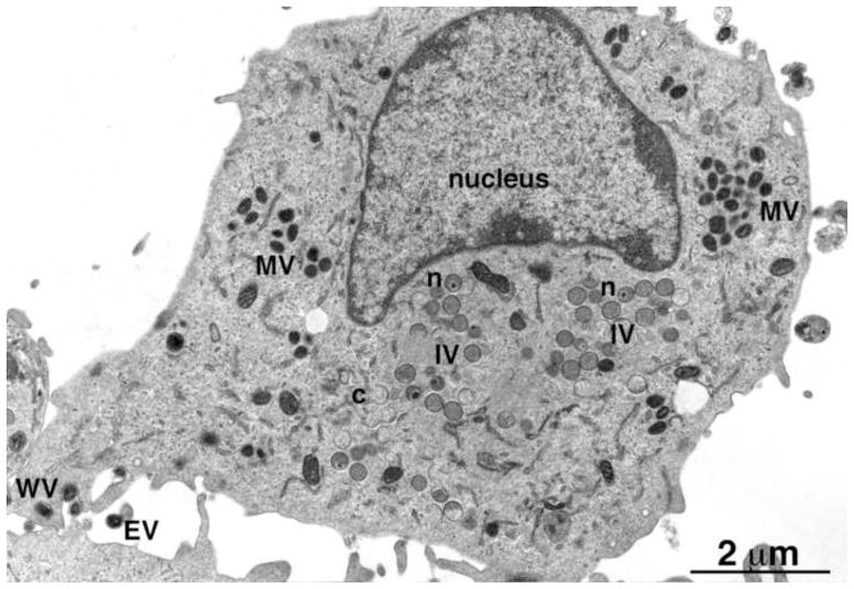
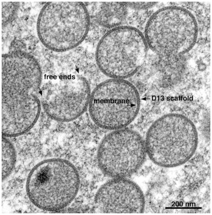
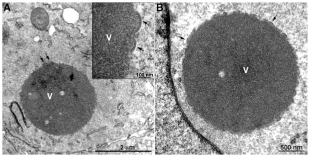
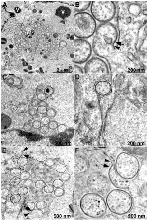
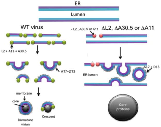
Poxviruses differ from most DNA viruses by replicating entirely within the cytoplasm. The first discernible viral structures are crescents and spherical immature virions containing a single lipoprotein membrane bilayer with an external honeycomb lattice. Because this viral membrane displays no obvious continuity with a cellular organelle, a de novo origin was suggested. Nevertheless, transient connections between viral and cellular membranes could be difficult to resolve. Despite the absence of direct evidence, the intermediate compartment (ERGIC) between the endoplasmic reticulum (ER) and Golgi apparatus and the ER itself were considered possible sources of crescent membranes. A break-through in understanding poxvirus membrane biogenesis has come from recent studies of the abortive replication of several vaccinia virus null mutants. Novel images showing continuity between viral crescents and the ER and the accumulation of immature virions in the expanded ER lumen provide the first direct evidence for a cellular origin of this poxvirus membrane.

Keywords: Endoplasmic reticulum; Vaccinia virus; Viral membrane proteins; Virus morphogenesis.


Figures

Fig. 1
Fig. 1
Transmission electron microscopic image of a HeLa cell infected with VACV. Abbreviations: MV, mature virion; IV, immature virion; WV, wrapped virion; EV, extracellular enveloped virion; n, IV with nucleoid. Scale bar at bottom. Provided by A. Weisberg.
Fig. 2
Fig. 2
Transmission electron microscopic image of a cell infected with VACV showing IVs forming within a virus factory. Free ends, lipoprotein membrane and D13 scaffold are labeled with arrows. Scale bar at bottom. Provided by A. Weisberg.
Fig. 3
Fig. 3
Transmission electron microscopic images of a BS-C-1 cell infected with a VACV L2 deletion mutant showing dense inclusions. Arrows point to short crescents. V, dense inclusion of viroplasm. Inset, high magnification of portion of inclusion. Scale bars shown. Adapted from (Maruri-Avidal et al., 2013a)
Fig. 4
Fig. 4
Transmission electron microscopic images of cells infected with a VACV L2 and A30.5 deletion mutants showing association of ER and IV-like structures. (A, B) VACV L2 deletion mutant infection of BS-C-1 cells. (C–F) VACV A30.5 deletion mutant infection of RK-13 cells. Arrows, membrane with spicule layer; Arrowheads, smooth membrane. Scale bars shown. Adapted from (Maruri-Avidal et al., 2013c).
Fig. 5
Fig. 5
Model for formation of viral membranes during infection with wild-type (WT) and mutant VACV. L2, A30.5, A11, A17, A14 and D13 refer to VACV proteins; ΔL2, ΔA30.5 and ΔA11 refer to VACV deletion mutants. Adapted from (Maruri-Avidal et al., 2013c).

References

    1. Ansarah-Sobrinho C, Moss B. Role of the I7 protein in proteolytic processing of vaccinia virus membrane and core components. J Virol. 2004;78:6335–6343. - PMC - PubMed
    1. Aridor M, Fish KN, Bannykh S, Weissman J, Roberts TH, Lippincott-Schwartz J, Balch WE. The Sar1 GTPase coordinates biosynthetic cargo selection with endoplasmic reticulum export site assembly. J Cell Biol. 2001;152:213–229. - PMC - PubMed
    1. Bahar MW, Graham SC, Stuart DI, Grimes JM. Insights into the evolution of a complex virus from the crystal structure of vaccinia virus D13. Structure. 2011;19:1011–1020. - PMC - PubMed
    1. Baldick CJ, Moss B. Resistance of vaccinia virus to rifampicin conferred by a single nucleotide substitution near the predicted NH2 terminus of a gene encoding an Mr 62,000 polypeptide. Virology. 1987;156:138–145. - PubMed
    1. Betakova T, Moss B. Disulfide bonds and membrane topology of the vaccinia virus A17L envelope protein. J Virol. 2000;74:2438–2442. - PMC - PubMed

Publication types

MeSH terms

LinkOut - more resources