. 2015 Jun;27(6):1186-97.
doi: 10.1016/j.cellsig.2015.02.024. Epub 2015 Feb 26.

Participation of proteasome-ubiquitin protein degradation in autophagy and the activation of AMP-activated protein kinase

Affiliations

Participation of proteasome-ubiquitin protein degradation in autophagy and the activation of AMP-activated protein kinase

Shaoning Jiang et al. Cell Signal. 2015 Jun.

Abstract

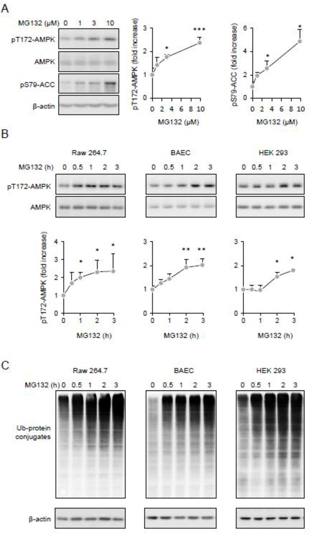
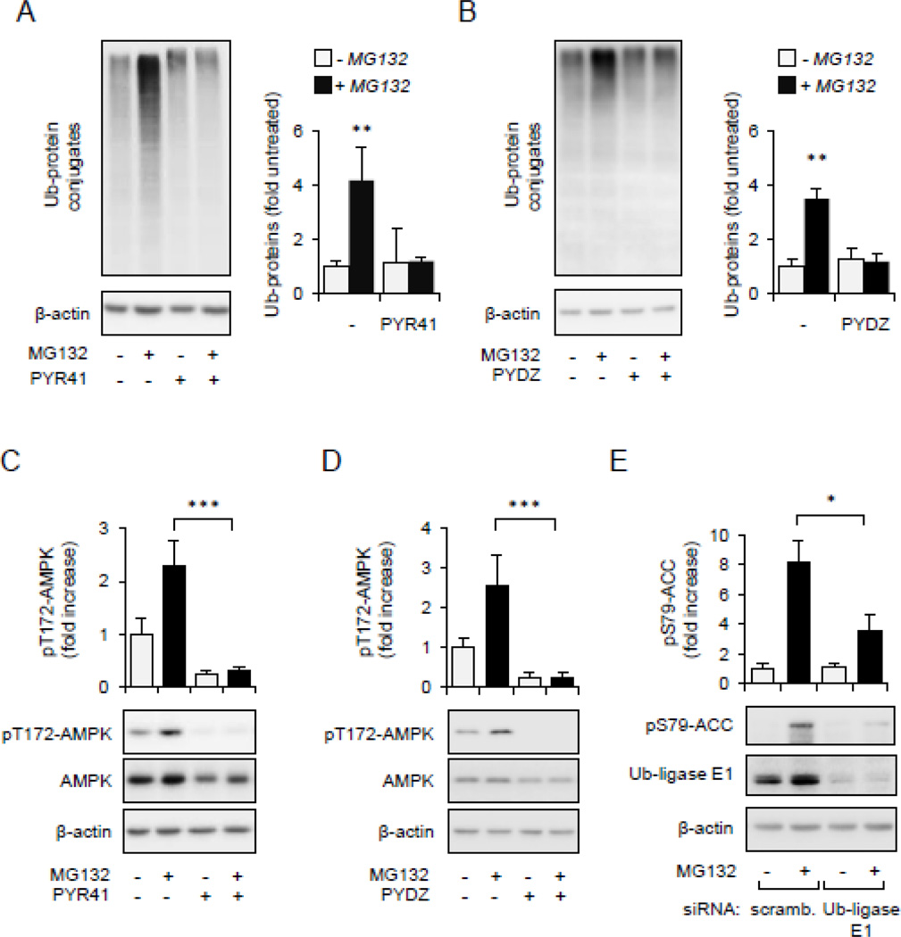
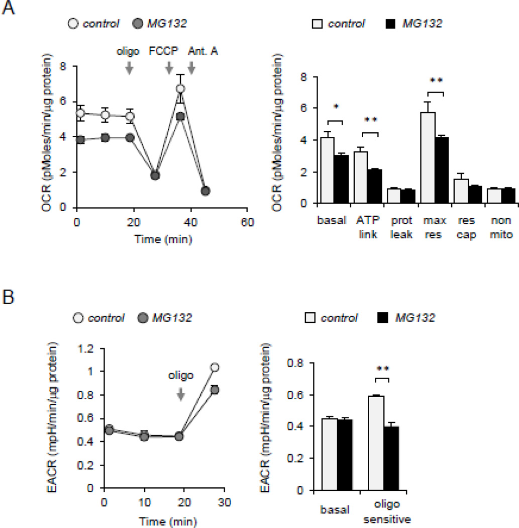
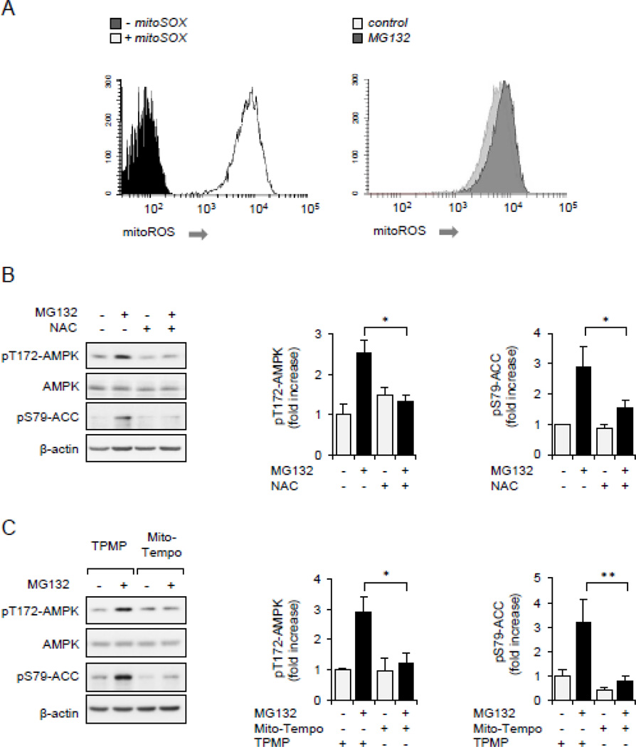
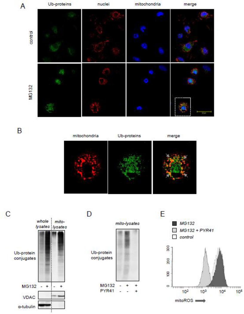
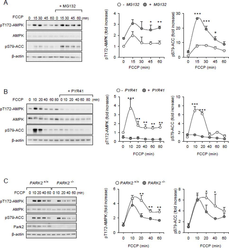
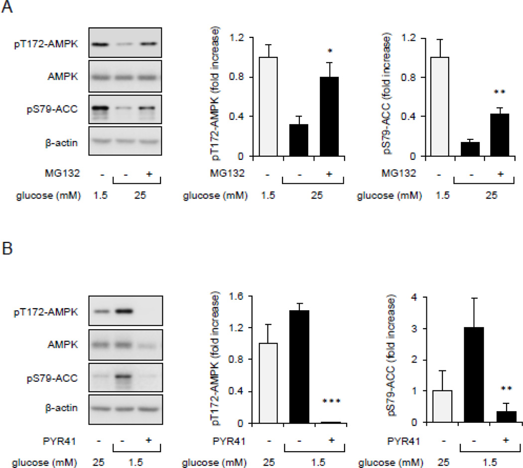
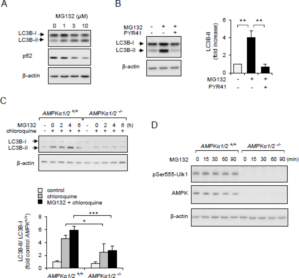
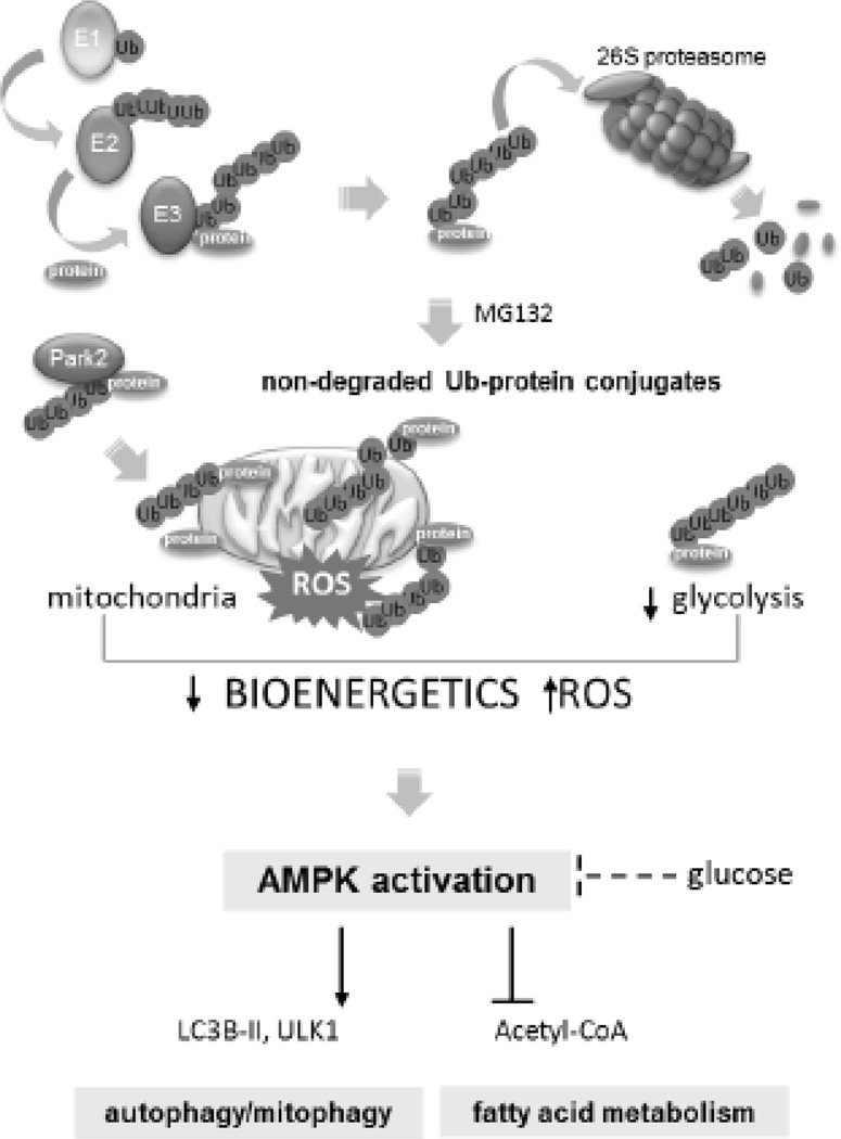
Although activation of the AMP-activated protein kinase (AMPK) as well as of ubiquitin/proteasome degradative pathways play an essential role in the preservation of metabolic homeostasis, little is known concerning interactions between protein turnover and AMPK activity. In the present studies, we found that inhibition of the 26S proteasome resulted in rapid activation of AMPK in macrophages, epithelial and endothelial cells. This was associated with increased levels of non-degraded Ub-protein conjugates, in both cytosolic and mitochondrial fractions. Selective inhibitors of ubiquitination or siRNA-dependent knockdown of Ub-ligase E1 diminished AMPK activation in cells treated with MG132, a 26S proteasome inhibitor. In addition to inhibition of AMPK activation by Ub-ligase E1 inhibitors, deficiency in Park2 mitochondria-associated Ub-ligase E3 also reduced AMPK activation upon dissipation of mitochondrial membrane potential (Δψm). Accumulation of Ub-proteins was correlated with decreases in cellular bioenergetics, including mitochondria oxidative phosphorylation, and an increase in ROS formation. Antioxidants, such as N-acetyl-L-cysteine or mitochondria-targeted MitoTEMPO, effectively diminished MG132-induced AMPK activation. Glucose-dependent regulation of AMPK or AMPK-mediated autophagy was modulated by alterations in intracellular levels of Ub-protein conjugates. Our results indicate that accumulation of ubiquitinated proteins alter cellular bioenergetics and redox status, leading to AMPK activation.

Keywords: AMP kinase; Autophagy; Bioenergetics; PARK2; Proteasome; Ubiquitination.


Conflict of interest statement

Conflict of interest

All authors declare no conflict of interest

Figures

Figure 1
Figure 1
Inhibition 26S proteasome and accumulation of Ub-protein conjugates is associated with AMPK activation. (A). Representative Western blots show the amount of pThr172-AMPK or pSer79ACC, total AMPK and β-actin in Raw 264.7 cells treated with MG132 (0, 1, 3, or 10 µM) for 60 minutes. Quantitative data of optical bend densitometry are shown. Mean ± SD, n = 3, * P < 0.05, ** P < 0.01. (B and C). Raw 264.7 cells, BAEC or HEK 293 cells were treated with MG132 (10 µM) for indicated time. Representative Western blots (B) and quantitative data (C) show the extent of pThr172-AMPK, total AMPK, Ub-protein conjugates and β-actin Mean ± SD, n = 3, * P < 0.05, ** P < 0.01.
Figure 2
Figure 2
AMPK activity is dependent on accumulation of ubiquitinated proteins. Raw 264.7 cells were pre-treated with Ub-ligase E1 inhibitor PYR41 (0 or 50 µM) or PYZD4409 (0 or 50 µM) for 30 minutes followed by incubation with MG132 (0 or 10 µM) for additional 60 minutes. (A). Representative Western blots show the amount of Ub-protein conjugates and β–actin or (B) the extent of AMPK phosphorylation. Mean ± SD, n = 3, *** P < 0.001.
Figure 3
Figure 3
Cellular bioenergetics is implicated in AMPK activation. (A). Raw 264.7 cells were treated with MG132 (0 or 10 µM) for 60 minutes and then bioenergetic status determined using Seahorse Extracellular Flux Analyzer. The OCR was monitored over time and after subsequent injection of oligomycin (1 µg/ml), carbonyl cyanide 4-(trifluoromethoxy) phenylhydrazone (0.5 µM), and antimycin A (ant. A) (10 µM). Bar graph shows the indices of mitochondrial respiratory function, including basal OCR, ATP-linked OCR (ATP link), protein leak (prot leak), maximal respiration (max res), and non-mitochondrial respiration (non-mito) in control or MG132-treated Raw 264.7 macrophages. (B) ECAR was determined in control and MG132-treated cells. Measurements were performed before and after inclusion of oligomycin. Mean ± SEM, n = 3 – 8, * P < 0.05; ** P < 0.01.
Figure 4
Figure 4
ROS is associated with AMPK activation. (A).Raw 264.7 cells were incubated with fluorogenic probe mitoSOX (0 or 5 µM) for 60 minutes (left panel), or mitoSOX loaded cells were treated with MG132 (0, 10 µM) for 45 minutes and fluorescence determined using flow cytometry (right panel). (B and C).Raw 264.7 cells were pretreated with NAC (0 or 20 mM) for 30 minutes, TPMP or MitoTempo (0 or 1 µM) 1 for 15 minutes followed by inclusion of MG132 (0 or 10 µM) for an additional 60 minutes. Representative Western blots and quantitative data of pThr172-AMPK and pSer79-ACC are shown. Mean ± SEM, n = 3 – 8, * P < 0.05; ** P < 0.01.
Figure 5
Figure 5
Accumulation of Ub-protein conjugates is associated with mitoROS-dependent activation of AMPK. Peritoneal macrophages were incubated with or without MG312 (10 µM) for 2 hours followed by staining cells for Ub-protein and GRP75, a mitochondrial marker. (A and B) Representative images show mitochondria (red), Ub-protein conjugates (green) and nuclei (blue). Area of interest (dotted lines in A) is magnified and shown in panel (B). Arrows indicate overlap between mitochondria and Ub-protein conjugates. (C) The amount of Ub-protein conjugates, VDAC, and α-tubulin was determined using Western blot analysis of whole cell or mitochondrial extracts obtained from Raw 264.7 that were treated with MG132 (0 or 10 µM) for 60 minutes. (D). Raw 264.7 cells were pretreated with PYR41 (0 or 50 µM) for 30 minutes followed by exposure to MG132 (0 or 10 µM) for additional 60 minutes. Ub-protein conjugates obtained from mitochondrial fractions are shown. (E) The extent of ROS production was determined in Raw 264.7 macrophages pre-treated with PYR41 (0 or 50 µM) for 30 minutes followed by inclusion of MG132 (0 or 10 µM) for additional 60 minutes. MitoSOX fluorescence intensity was determined using flow cytometry.
Figure 6
Figure 6
FCCP-dependent activation of AMPK is diminished upon inclusion of E1 inhibitors. (A and B) Raw 264.7 were subsequently (A) MG132 (0 or 10 µM) or (B) PYR41 (0 or 50 µM) for 30 minutes followed by inclusion of FCCP (500 nM) for indicated time points. Representative Western blots and quantitative data show the amounts of pThr172-AMPK, pSer79-ACC (mean ± SD, n = 3, * P < 0.05; ** P < 0.01 or *** P < 0.001. (C). Wild type (PARK+/+) and Park2 deficient (PARK2−/−) peritoneal macrophages were incubated with FCCP (500 nM) for indicate time followed by Western Blot analysis of phospho- or total AMPK and ACC. Mean ± SD, n = 3, * P < 0.05; ** P < 0.01.
Figure 7
Figure 7
The effects of protein ubiquitination on glucose and nutrients-dependent regulation of AMPK activity. (A). Raw 264.7 cells were cultured in low glucose/serum medium for 60 minutes followed by inclusion of glucose (25 mM) and MG132 (0 or 10 µM) for additional 60 minutes. (B). Cells cultured in 25 mM glucose medium were exposed to low glucose (1.5 mM) in the presence or absence of PYR-41 (50 µM) for 60 minutes. Western blot and quantitative data in panels A and B show the extent of Thr172-AMPK and Ser79-ACC phosphorylation. Mean ± SEM, n = 3 – 7, * P < 0.05 or ** P < 0.01.
Figure 8
Figure 8
Non-degraded Ub-protein conjugates are involved in AMPK-dependent autophagy/mitophagy. Panel (A). HEK 293 cells were dose-dependently treated with MG132 for 4 hours. Western blots of LC3B-I, LC3B-II, p62 β-actin is shown. (B). HEK293 cells were first incubated with PYR41 (0 or 50 µM) followed by exposure to MG132 (0 or 10 µM) for 4 hours. Western blots and quantitative data show the extent of LC3B-I, LC3B-II and β-actin. Mean ± SEM, n = 3, * P < 0.05 or ** P < 0.01. (C). MEFs, wild type (AMPKα1/2+/+) or AMPK deficient fibroblasts (AMPKα1/2−/−), were time dependently treated with MG132 (10 µM) followed by Western Blot analysis of LC3B-I, LC3B-II and β-actin. Chloroquine was applied for 30 minutes, as indicated. (D).Western blot analysis quantitative data show the extent of pSer555-Ulk1, AMPKα and β-actin in MG132-treated MEF AMPKα1/2+/+ or AMPKα1/2−/−. Mean ± SEM, n = 3, * P < 0.05 or *** P < 0.001.
Figure 9
Figure 9
Proteasome/ubiquitin protein degradative pathway controls AMPK function. Proteasome inhibition and accumulation of non-degraded Ub-protein conjugates is associated with alterations in mitochondrial bioenergetics and ROS formation followed by activation of AMPK. Protein ubiquitination can also affect glucose-dependent regulation of AMPK activity. Once activated AMPK can influence bioenergetic and degradative pathways, including autophagy, mitochondrial quality control mitophagy and promotes fatty acids oxidation.

Similar articles

Cited by

References

    1. Glickman MH, Ciechanover A. Physiol Rev. 2002;82:373–428. - PubMed
    1. Hershko A. Cell Death Differ. 2005;12:1191–1197. - PubMed
    1. Ravid T, Hochstrasser M. Nat Rev Mol Cell Biol. 2008;9:679–690. - PMC - PubMed
    1. Gallastegui N, Groll M. Trends Biochem Sci. 2010;35:634–642. - PubMed
    1. Hershko A, Heller H, Elias S, Ciechanover A. J Biol Chem. 1983;258:8206–8214. - PubMed

Publication types

MeSH terms

LinkOut - more resources