. 2015 Oct;230(10):2447-2460.
doi: 10.1002/jcp.24975.

A SMYD3 Small-Molecule Inhibitor Impairing Cancer Cell Growth

Affiliations

A SMYD3 Small-Molecule Inhibitor Impairing Cancer Cell Growth

Alessia Peserico et al. J Cell Physiol. 2015 Oct.

Abstract

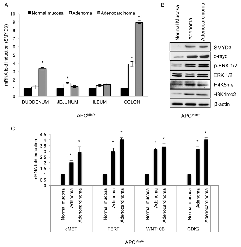
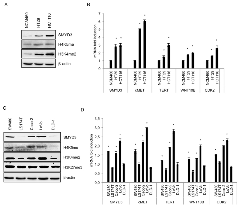
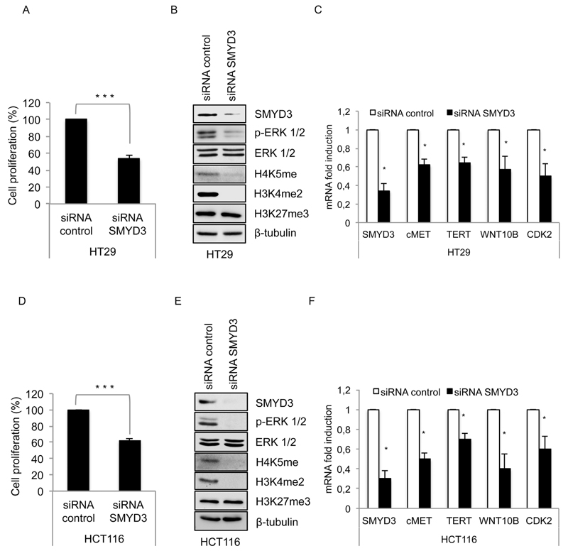
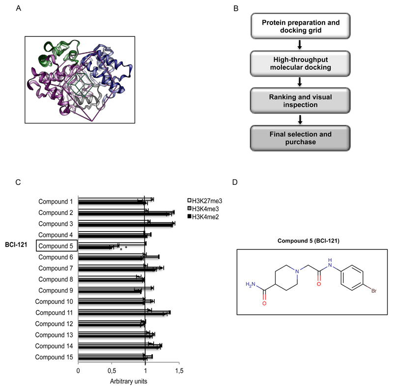
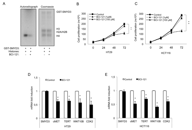
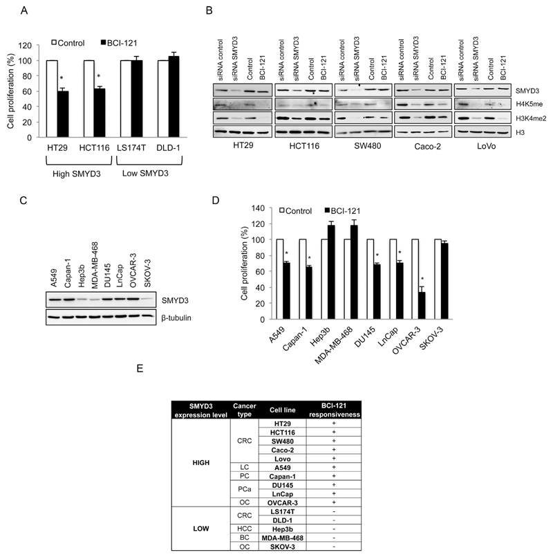
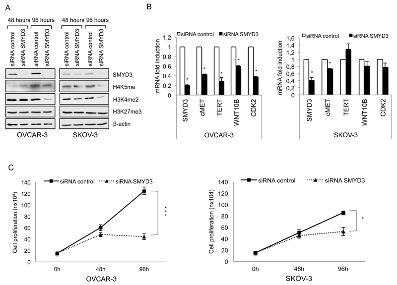
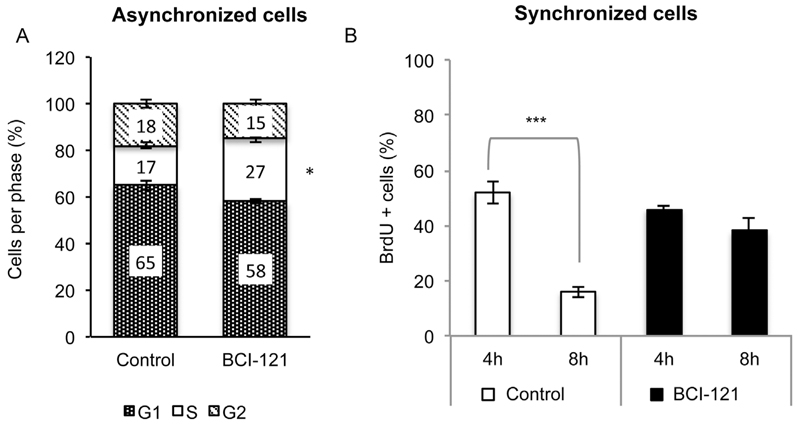
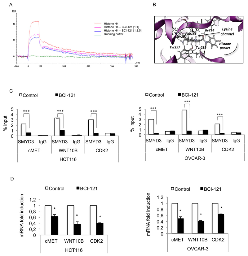
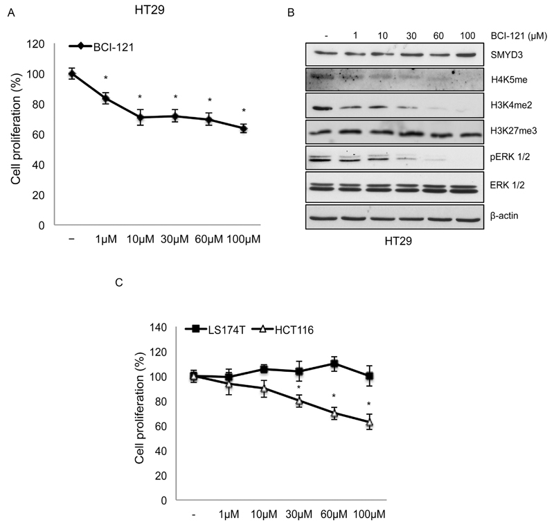
SMYD3 is a histone lysine methyltransferase that plays an important role in transcriptional activation as a member of an RNA polymerase complex, and its oncogenic role has been described in different cancer types. We studied the expression and activity of SMYD3 in a preclinical model of colorectal cancer (CRC) and found that it is strongly upregulated throughout tumorigenesis both at the mRNA and protein level. Our results also showed that RNAi-mediated SMYD3 ablation impairs CRC cell proliferation indicating that SMYD3 is required for proper cancer cell growth. These data, together with the importance of lysine methyltransferases as a target for drug discovery, prompted us to carry out a virtual screening to identify new SMYD3 inhibitors by testing several candidate small molecules. Here we report that one of these compounds (BCI-121) induces a significant reduction in SMYD3 activity both in vitro and in CRC cells, as suggested by the analysis of global H3K4me2/3 and H4K5me levels. Of note, the extent of cell growth inhibition by BCI-121 was similar to that observed upon SMYD3 genetic ablation. Most of the results described above were obtained in CRC; however, when we extended our observations to tumor cell lines of different origin, we found that SMYD3 inhibitors are also effective in other cancer types, such as lung, pancreatic, prostate, and ovarian. These results represent the proof of principle that SMYD3 is a druggable target and suggest that new compounds capable of inhibiting its activity may prove useful as novel therapeutic agents in cancer treatment.


Conflict of interest statement

Conflict of interest: The authors have no conflict of interest to declare.

Figures

Fig. 1
Fig. 1
SMYD3 is upregulated during colorectal tumorigenesis (A) SMYD3 mRNA expression increases during transition from normal to tumor colon mucosa in APCMin/+ mice. Tissue samples were obtained from control APCMin/+ mice (n = 10) and APCMin/+ mice treated with 12 mg/kg of azoxymethane (AOM) (n = 10). (B) Colorectal adenomas and carcinomas of APCMin/+ mice overexpress SMYD3 protein and show increased global levels of H4K5me and H3K4me2. cMYC and pERK expression was assessed as an internal control of the experiment. (C) SMYD3 expression profile correlates with mRNA expression of its target genes. β-actin was used as a loading control for immunoblotting and for real-time PCR data normalization. Statistical analysis was performed using Student’s t-tail test; *P < 0.05 was considered statistically significant.
Fig. 2
Fig. 2
SMYD3 shows high levels of expression in CRC cell lines. SMYD3 protein (A, C) and mRNA (B, D) expression is enhanced in several human CRC cell lines. (A, C) SMYD3 protein expression correlates with the global level of targeted histone methyl marks [H3K4me2 and H4K5me] and (B, D) with the transcriptional activation of its target genes in the indicated cell lines. The levels of a SMYD3 non-targeted methyl mark [H3K27me3] are not modulated. In B and D, fold induction is normalized to mRNA levels observed in NCM460 cells (arbitrarily set as 1). β-actin was used as a loading control for immunoblotting and for real-time PCR data normalization. Statistical analysis was performed using Student’s t-tail test; *P < 0.05 was considered statistically significant.
Fig. 3
Fig. 3
SMYD3 is required for proper cancer cell growth. In HT29 (A, B, C) and HCT116 (D, E, F) CRC cell lines, SMYD3 inhibition by RNAi impairs cell proliferation (A, D), decreases the level of targeted histone methyl marks [H4K5me, H3K4me2] and ERK 1/2 activation (B, E), and reduces the expression level of SMYD3 target genes (C, F). A non-targeted methyl mark [H3K27me3] was not affected by RNAi-mediated SMYD3 ablation. HT29 and HCT116 CRC cell lines were transfected with control siRNAs or SMYD3-specific siRNAs for 48 h. Cell proliferation was calculated using the WST-1 assay. β-tubulin was used as a loading control for immunoblotting and β-actin was used for real-time PCR data normalization. Statistical analysis was performed using Student’s t-tail test; *P < 0.05, **P < 0.01, and ***P < 0.001 were considered statistically significant.
Fig. 4
Fig. 4
Virtual screening procedure allowing identification of BCI-121 as a promising candidate for SMYD3 inhibition. (A) Structure of full-length SMYD3 highlighting the N-terminal SET domain (purple), the MYND domain (yellow), the post-SET domain (gray), and the C-terminal region (blue). The boxes define the area of the protein - centered on the histone binding site - that was considered to perform the docking screening calculation. The green box identifies the area containing at least one atom of the putative ligand, while the purple box identifies the area where all atoms of the ligands should lie. (B) Schematic representation of the virtual screening procedure. (C) Compound 5 [BCI-121] is the best candidate small molecule for SMYD3 inhibition among the initial set of 15 compounds selected through the virtual screening procedure. The global level of SMYD3 targeted [H3K4me2 and H3K4me3] and non-targeted [H3K27me3] histone methyl marks was measured by immunoblot in nuclear enriched fractions of CRC cells (HT29) treated with each compound (100 μM). Values shown correspond to histone methyl mark levels quantified by densitometric analysis and normalized to the loading control Lamin A/C (arbitrary units, untreated control at 48h = 1). (D) Structural formula of the selected compound BCI-121 (Figure generated with MarvinSketch v6.0.0 http://www.chemaxon.com). Statistical analysis was performed using Student’s t-tail test; *P < 0.05 was considered statistically significant.
Fig. 5
Fig. 5
BCI-121 inhibits SMYD3 activity in vitro and in CRC cell models and affects cell proliferation. (A) In vitro methylation assay using the indicated recombinant SMYD3 protein on a mixture of calf thymus histones in the presence and/or absence of BCI-121 showing a significant decrease in H4 methylation. Autoradiograph and Coomassie stained (loading control) gels are shown. BCI-121 inhibits cell proliferation in HT29 (B) and HCT116 (C) cells in a dose- and time-dependent manner. Cell proliferation was assessed by cell counting. The data presented are the mean values obtained for each analyzed time point (n = 4). (D, E) BCI-121 100 μM decreases the expression levels of SMYD3 target genes in both cell lines (the 48 h time point was evaluated). β-actin was used for normalization of real-time PCR data. Statistical analysis was performed using Student’s t-tail test; *P < 0.05, **P < 0.01, and ***P < 0.001 were considered statistically significant.
Fig. 6
Fig. 6
Basal SMYD3 expression levels predict BCI-121 treatment response. (A) Administration of BCI-121 affects proliferation of CRC cell lines expressing high levels of SMYD3. (B) BCI-121 treatment reduces targeted histone methyl marks [H4K5me and H3K4me2] to an extent comparable to that observed with RNAi. CRC cells were treated with BCI-121 and/or SMYD3-specific siRNAs for 48 h and H4K5me and H3K4me2 global levels were evaluated by immunoblot. H3 was used as a loading control. (C) SMYD3 protein is highly expressed in several cell lines derived from different types of cancer (A549 = lung cancer; Capan-1 = pancreatic cancer; Hep3b = hepatocellular carcinoma; MDA-MB-468 = breast cancer; DU145 and LnCap = prostate cancer; OVCAR-3 and SKOV-3 = ovarian cancer). β-tubulin was used as a loading control. (D) BCI-121 treatment impaired proliferation of cancer cells with high expression levels of SMYD3, while cancer cells expressing low levels of SMYD3 were not affected. (E) SMYD3 protein levels in cell lines that proved responsive to BCI-121 treatment. Table summarizing the data obtained in the cell lines derived from the different types of cancer tested in this study [CRC, colorectal cancer; LC, lung cancer; PC pancreatic cancer; PCa, prostate cancer; OC, ovarian cancer; BC, breast cancer; HCC, hepatocellular carcinoma]. Cancer cell lines were treated with BCI-121 (100 μM) for 72 h (A) and 96 h (D) and cell proliferation was calculated using the WST-1 assay. Statistical analysis was performed using Student’s t-tail test; *P < 0.05 was considered statistically significant.
Fig. 7
Fig. 7
SMYD3 is required for proper ovarian cancer cell growth. (A) SMYD3 genetic ablation in ovarian cancer cell models decreases the global level of targeted histone methyl marks [H3K4me2 and H4K5me] without affecting a non-targeted methyl mark [H3K27me3]. (B) SMYD3 genetic ablation levels correlate with deregulated mRNA expression of its target genes and (C) with decreased cell proliferation rate. OVCAR-3 and SKOV-3 cells were transfected with control or SMYD3-specific siRNAs for 48 h and 96 h. Levels of methyl marks were determined by immunoblot; expression of target genes was analyzed by real-time PCR (the 48 h time point was evaluated). Cell proliferation was assessed by cell counting. The number of cells for each indicated time point is plotted. β-actin was used as a loading control for immunoblotting and for normalization of real-time PCR data. Statistical analysis was performed using Student’s t-tail test; *P < 0.05, **P < 0.01, and ***P < 0.001 were considered statistically significant.
Fig. 8
Fig. 8
SMYD3 regulates S/G2 transition in cancer cells. (A) SMYD3 inhibition affects cell cycle progression in HT29 cells with a significant increase in the S-phase fraction. HT29 cells were treated with BCI-121 [100 μM] for 48 h and FACS analysis was performed following propidium iodide staining. (B) HCT116 synchronized cells treated with BCI-121 failed to exit S phase. Synchronization by double thymidine block was performed during treatment with BCI-121 (48 h). The 4 h (expected phase = S) and 8 h (expected phase = G2) post-release time points were analyzed through a BrdU incorporation assay to quantify BrdU-positive cells. Statistical analysis was performed using Student’s t-tail test; *P < 0.05, **P < 0.01, and ***P < 0.001 were considered statistically significant.
Fig. 9
Fig. 9
BCI-121 competes with target histones for SMYD3 binding in vitro and in cell line models. (A) Sensorgrams of H4 histone binding to SMYD3 in the presence and in the absence of BCI-121. (B) Predicted binding mode of BCI-121 to the histone binding site. (C) SMYD3 binding to the promoter of its target genes is abolished by the presence of BCI-121 in cancer cells. ChIP was performed in HCT116 and OVCAR-3 cells treated or not with BCI-121 (100 μM) for 72 h. Cells were cross-linked and immunoprecipitated with anti-SMYD3 and anti-IgG antibodies. The precipitated DNA was subjected to real-time PCR with specific primers, which amplify SMYD3 binding site elements of human target gene promoters (cMET, WNT10B, and CDK2). IgGs were used as an immunoprecipitation control. (D) Transcriptional activation of target genes correlates with impaired binding of SMYD3. β-actin was used for real-time PCR data normalization. Statistical analysis was performed using Student’s t-tail test; *P < 0.05 was considered statistically significant.
Fig. 10
Fig. 10
Dose-dependence of BCI-121 treatment in CRC cell lines. (A) BCI-121 treatment for 48 h induces a concentration-dependent reduction of CRC cell proliferation and (B) decreases the global levels of targeted histone methyl marks [H4K5me, H3K4me2] and ERK 1/2 activation. A non-targeted methyl mark [H3K27me3] was not affected. β-actin was used as a loading control for immunoblotting. (C) The dose-dependent effect of BCI-121 treatment (72 h) on cell growth is observed in cells expressing high levels of SMYD3 (HCT116), but not in cells with low levels of SMYD3 (LS174T). Cell proliferation was calculated using the WST-1 assay. Statistical analysis was performed using Student’s t-tail test; *P < 0.05, **P < 0.01, and ***P < 0.001 were considered statistically significant.

References

    1. Biggar KK, Li SSC. Non-histone protein methylation as a regulator of cellular signalling and function. Nat Rev Mol Cell Biol. 2015;16:5–17. - PubMed
    1. Chi P, Allis CD, Wang GG. Covalent histone modifications-miswritten, misinterpreted, and mis-erased in human cancers. Nat Rev Cancer. 2010;10:457–469. - PMC - PubMed
    1. Chiacchiera F, Matrone A, Ferrari E, Ingravallo G, Lo Sasso, Petruzzelli S, Salvatore M, Moschetta L, Simone A. P38alpha blockade inhibits colorectal cancer growth in vivo by inducing a switch from HIF1alpha- to FoxO-dependent transcription. Cell Death Differ. 2009;16:1203–1214. - PubMed
    1. Cock-Rada AM, Medjkane S, Janski N, Yousfi N, Perichon M, Chaussepied M, Chluba J, Langsley G, Weitzman JB. SMYD3 promotes cancer invasion by epigenetic upregulation of the metalloproteinase MMP-9. Cancer Res. 2012;72:810–820. - PMC - PubMed
    1. Copeland RA, Solomon ME, Richon VM. Protein methyltransferases as a target class for drug discovery. Nat Rev Drug Discov. 2009;8:724–732. - PubMed

Publication types

MeSH terms

Substances