

A variant extensor indicis muscle and the branching pattern of the deep radial nerve could explain hand functionality and clinical symptoms in the living patient
- PMID: 25729087
- PMCID: PMC4319448
A variant extensor indicis muscle and the branching pattern of the deep radial nerve could explain hand functionality and clinical symptoms in the living patient
Abstract
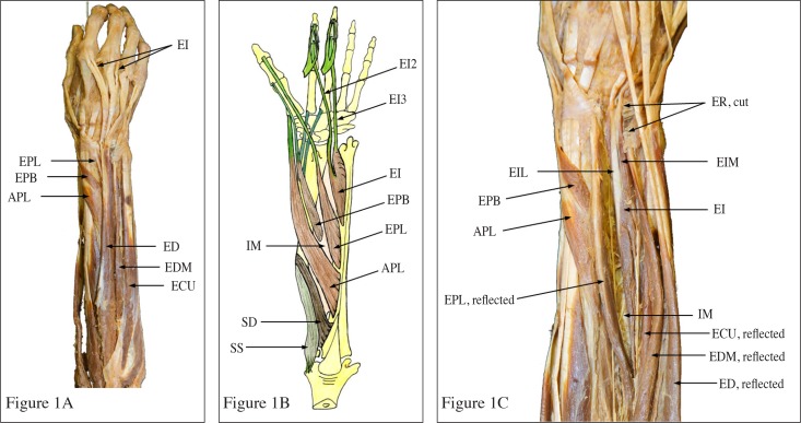
The purpose of this study is to document the topographic anatomy of an extensor indicis (EI) muscle with a double tendon and the associated distribution of the deep branch of the radial nerve (DBRN). Both EI tendons were positioned deep to the tendons of the extensor digitorum as they traversed the dorsal osseofibrous tunnel. They then joined the medial slips of the extensor expansion of the second and third digits. In all other dissected forearms, a tendon of the EI muscle joined the medial slip of the extensor expansion to the index finger. The DBRN provided short branches to the superficial extensor muscles, long branches to the abductor pollicis longus and extensor pollicis brevis muscles, and terminated as the posterior interosseous nerve. Descending deep to the extensor pollicis longus muscle, the posterior interosseous nerve sent branches to the extensor pollicis brevis and EI muscles. Understanding of the topographic anatomy of an EI with a double tendon, and the associated distribution of the DBRN, may contribute to accurate diagnosis and treatment of hand lesions.
L’objectif de cette étude est de documenter l’anatomie topographique du musculus extensor indicis (MEI), ou muscle extenseur de l’index, avec un tendon double et la distribution de la grosse branche du nerf radial (DGBNR) qui lui est associée. Les deux tendons du MEI sont positionnés profondément dans les tendons du extensor digitorum alors qu’ils traversent le tunnel ossofibreux dorsal. Ils joignent ensuite les portions internes de l’expansion de l’extenseur au niveau du deuxième et troisième doigt. Dans tous les autres avant-bras disséqués, un tendon du MEI joint la portion interne de l’expansion de l’extenseur à l’index. La DGBNR fournit de petites branches aux muscles d’extenseur superficiels, de grosses branches aux muscles du long abducteur du pouce et du court extenseur du pouce, pour finir en tant que nerf interosseux postérieur. Le nerf interosseux postérieur descend profondément dans le muscle du long extenseur du pouce et envoie des branches au court extenseur du pouce et au MEI. Une bonne compréhension de l’anatomie topographique d’un MEI avec double tendon, et la DGBNR associée, peut favoriser des diagnostics précis et améliorer le traitement de lésions aux mains.
Keywords: extensor indicis; neuropathy; radial nerve; variation; wrist pain.
Figures


References
-
- Jones BV, Ipswich RN. An anomalous extensor indicis muscle. J Bone Joint Surg Br. 1959;41-B(4):763–765. - PubMed
-
- Schenck RR. Variations of the extensor tendons of the fingers. Surgical significance. J Bone Joint Surg Am. 1964;46:103–110. - PubMed
-
- Bergman RA, Thompson SA, Afifi AK. Catalog of Human Variation. Baltimore-Munich: Urban & Schwazzenberg; 1984. pp. 40–155.
-
- Von Schroeder HP, Botte MJ. The extensor medii proprius and anomalous extensor tendons to the long finger. J Hand Surg Am. 1991;16(6):1141–1145. - PubMed
-
- Godwin Y, Ellis H. Distribution of the extensor tendons on the dorsum of the hand. Clin Anat. 1992;5:394–403.
LinkOut - more resources
Full Text Sources