. 2014 Apr 1;57(4):490-500.
doi: 10.1007/s11426-014-5086-y.

Synthetic biodegradable functional polymers for tissue engineering: a brief review

Affiliations

Synthetic biodegradable functional polymers for tissue engineering: a brief review

Guo BaoLin et al. Sci China Chem. .

Abstract

Scaffolds play a crucial role in tissue engineering. Biodegradable polymers with great processing flexibility are the predominant scaffolding materials. Synthetic biodegradable polymers with well-defined structure and without immunological concerns associated with naturally derived polymers are widely used in tissue engineering. The synthetic biodegradable polymers that are widely used in tissue engineering, including polyesters, polyanhydrides, polyphosphazenes, polyurethane, and poly (glycerol sebacate) are summarized in this article. New developments in conducting polymers, photoresponsive polymers, amino-acid-based polymers, enzymatically degradable polymers, and peptide-activated polymers are also discussed. In addition to chemical functionalization, the scaffold designs that mimic the nano and micro features of the extracellular matrix (ECM) are presented as well, and composite and nanocomposite scaffolds are also reviewed.

Keywords: functional polymers; scaffolds; synthetic biodegradable polymers; tissue engineering.


Figures

Figure 1
Figure 1
Polymers frequently used as scaffolds for tissue regeneration.
Figure 2
Figure 2
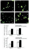
Representative fluorescence images of electrically stimulated PC-12 cells: (a) polypyrrole-PLGA random fibers (RF) at 0 mV/cm (unstimulated); (b) polypyrrole-PLGA aligned fibers (AF) at 0 mV/cm; (c) polypyrrole-PLGA random fibers at 10 mV/cm; (d) polypyrrole-PLGA aligned fibers at 10 mV/cm. Scale bars are 50 μm. (e) Median neurite lengths and (f) percentages of neurite-bearing PC12 cells when unstimulated and when electrically stimulated (10 mV/cm) on random and aligned polypyrrole-PLGA fibers [56]. Reprinted with the permission of Elsevier.
Figure 3
Figure 3
Degradable conducting polymers with different architectures.
Figure 4
Figure 4
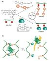
(a) Synthesis of azobenzene modified dextran (AB-Dex) and cyclodextrin modified dextran (CD-Dex) through the thiol-maleimide reaction. (b) Schematic representation of photoresponsive protein release system. Upon the UV-light irradiation, azobenzene moieties isomerise from trans to cis configurations that leads to the dissociation of crosslinking points and allows the entrapped protein to be released. Reproduced from ref. [76] with the permission of the Royal Society of Chemistry, 2010.
Figure 5
Figure 5
Synthesis of degradable hydrogels by click reaction of 4-arm PEG-N3 with dialkyne modified peptide [49]. Reprinted with the permission of John Wiley and Sons.
Figure 6
Figure 6
Formation of peptide-incorporated hydrogels using HRP and H2O2 [83]. Reprinted with the permission of Springer Verlag.
Figure 7
Figure 7
SEM micrographs of nanofibrous-gelatin scaffold, × 50 (left); pore-wall morphology of nanofibrous-gelatin scaffold, × l000 (middle); high magnification of nanofibrous-gelatin scaffold, × 10000 (right) [91]. Reprinted with the permission of Elsevier.
Figure 8
Figure 8
A schematic synthesis of star-shaped-PLLA and the fabrication of nanofibrous hollow microspheres. (a) PAMAM (G2) as an initiator to synthesize star-shaped-PLLA; (b) the synthesis of star-shaped-PLLA. Red coils represent the PLLA chains. (c) Fabrication of SS-PLLA microspheres using a surfactant-free emulsification process. (d) Nanofibrous hollow microspheres were prepared via phase separation, solvent extraction and freeze-drying [92]. With the permission of Nature Publishing Group.
Figure 9
Figure 9
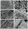
SEM micrographs of mineralized PLLA matrices. (a) Electrodeposition at 3 V, 60 °C for 60 min, (b) the magnified image of (a) and (c) mineralized in 1.5 × SBF for 12 days, (d) the magnified image of (c) and (e) mineralized in 1.5 × SBF for 30 days, (f) the magnified image of (e) [107]. Reprinted with the permission of Elsevier.

References

    1. Couch N, Wtlson R, Hager E, Murray J. Transplantatton of cadaver kidneys experience with 21 cases. Surgery. 1966;59:183–188. - PubMed
    1. Langer R, Vacanti JP. Tissue engineering. Science. 1993;260:920–926. - PubMed
    1. Guo BL, Glavas L, Albertsson AC. Biodegradable and electrically conducting polymers for biomedical applications. Prog Polym Sci. 2013;38:1263–1286.
    1. Ma PX. Biomimetic materials for tissue engineering. Adv Drug Delivery Rev. 2008;60:184–198. - PMC - PubMed
    1. Liu XH, Holzwarth JM, Ma PX. Functionalized synthetic biodegradable polymer scaffolds for tissue engineering. Macromol Biosci. 2012;12:911–919. - PubMed

LinkOut - more resources