. 2015 Jan;1(1):75-86.
doi: 10.1016/j.jcmgh.2014.11.002.

Acinar Cell Production of Leukotriene B4 Contributes to Development of Neurogenic Pancreatitis in Mice

Affiliations

Acinar Cell Production of Leukotriene B4 Contributes to Development of Neurogenic Pancreatitis in Mice

Rafiq A Shahid et al. Cell Mol Gastroenterol Hepatol. 2015 Jan.

Abstract

Background & aims: In the pancreas, activation of primary sensory nerves through the transient receptor potential ion channel TRPV1 contributes to the early stages of development of pancreatitis. Little is known about the mechanism by which this occurs. We investigated whether leukotriene B4 (LTB4) is an endogenous agonist of TRPV1 and mediates pancreatitis.

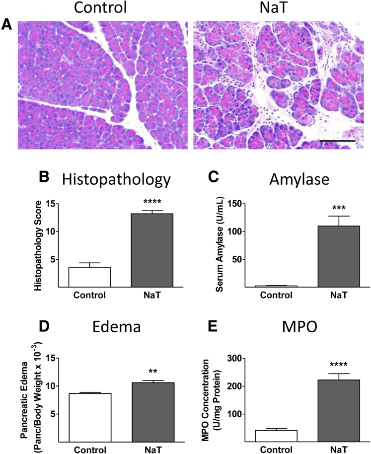
Methods: Acute inflammation was induced in the pancreata of Trpv1-/- mice and their wild-type littermates by retrograde infusion of the main pancreatic duct with 2% sodium taurocholate (NaT) or intraperitoneal injections of caerulein. Mice were also given injections of resiniferatoxin (an excitotoxin that desensitizes TRPV1) or MK886 (a drug that inhibits LTB4 biosynthesis). Pancreatic tissues and plasma were collected and analyzed.

Results: Retrograde perfusion of the main pancreatic ducts of wild-type mice with NaT caused severe acute pancreatitis; severity was reduced by co-administration of resiniferatoxin. Trpv1-/- mice developed a less severe pancreatitis following NaT administration than controls. Administration of MK886 before perfusion with NaT also significantly reduced the severity of pancreatitis in wild-type mice. Pancreatic tissues from mice given NaT had a marked increase in the level of 5-lipoxygenase immunoreactivity specifically in acinar cells. Bile acid and caerulein induced secretion of LTB4 by cultured pancreatic acinar cells; MK886 inhibited this process.

Conclusions: Administration of caerulein or intraductal bile acids in mice causes production of LTB4 by pancreatic acinar cells. This activates TRPV1 on primary sensory nerves to induce acute pancreatitis.

Keywords: gallstone pancreatitis; mouse model; neurogenic inflammation; secretagogue; vanilloid.


Figures

Figure 1
Figure 1
Intrapancreatic duct infusion of bile acids causes pancreatitis. The effects of pancreatic duct infusion of 50 μL of 2% NaT on pancreatic histopathology, serum amylase levels, pancreatic edema, and pancreatic MPO levels are shown (n = 6). (A) The effects of pancreatic duct infusion with 2% NaT on pancreatic histology. (B) The effects of NaT on histopathology score (∗∗∗∗P < .0001 vs control). (C) The effects of NaT on serum amylase levels (∗∗∗P < .001 vs control). (D) The effects of NaT on pancreatic edema (∗∗P < .01 vs control). (E) The effects of NaT on pancreatic MPO levels (∗∗∗∗P < .0001 vs control). Scale bar = 100 μm.
Figure 2
Figure 2
The effects of RTX desensitization of TRPV1 in pancreatitis induced by bile acid. NaT with or without 14 μg/mL RTX was infused into the pancreatic duct of wild-type mice (n = 6). (A) The effects of pancreatic duct infusion with 2% NaT on pancreatic histology and the protective effect of coinfusion of RTX. (B) The effects of NaT and NaT + RTX on histopathology score, expressed as a percentage of the control value (∗∗∗P < .001 vs NaT). (C) The effects of NaT and NaT + RTX on serum amylase, expressed as a percentage of the control value (∗P < .05 vs NaT). (D) The effects of NaT and NaT + RTX on pancreatic edema, expressed as a percentage of the control value (∗∗P < .01 vs NaT). (E) The effects of NaT and NaT + RTX on pancreatic MPO concentration, expressed as a percentage of the control value (∗∗∗∗P < .0001 vs NaT). Scale bar = 100 μm.
Figure 3
Figure 3
The effects of intraductal NaT in Trpv1−/−mice. NaT was infused into the pancreatic duct of Trpv1−/− mice (N = 6). (A) The effects of pancreatic duct infusion with 2% NaT on pancreatic histology in wild-type and Trpv1−/− mice. (B) The effects of NaT on histopathology score in wild-type and Trpv1−/− mice, expressed as a percentage of the control value (∗∗∗P < .001 vs NaT) (C) The effects of NaT on serum amylase in wild-type and Trpv1−/− mice, expressed as a percentage of the control value (∗P < .05 vs NaT). (D) The effects of NaT on pancreatic edema in wild-type and Trpv1−/− mice, expressed as a percentage of the control value. (E) The effects of NaT on pancreatic MPO concentration in wild-type and Trpv1−/− mice, expressed as a percentage of the control value (∗∗∗P < .001 vs NaT). Scale bar = 100 μm.
Figure 4
Figure 4
Bile acids stimulate LTB4formation in the pancreas. (A) Effect of pancreatic duct infusion of NaT on LTB4 concentrations in the head of the pancreas in vivo and inhibition of the NaT-stimulated increase in pancreatic LTB4 levels by pretreatment with 1 μmol/L MK886, a FLAP inhibitor (n = 6; ∗∗∗P < .001 vs control; ##P < .01 vs NaT). Inset: The effect of caerulein hyperstimulation on LTB4 concentrations in the head of the pancreas in vivo (n = 6; ∗∗∗P < .001 vs control). (B) Effect of TLCS on LTB4 secretion from wild-type mouse pancreatic acini in vitro. TLCS (500 μmol/L) significantly stimulated LTB4 secretion from pancreatic acini (n = 5–9; ∗∗P < .01 vs control) and this effect was significantly inhibited by 1 μmol/L MK886 (#P < .05 vs TLCS alone). Inset: The effect of caerulein hyperstimulation (100 pmol/L) on LTB4 secretion from pancreatic acini (n = 6; ∗∗P < .01 vs control). LTB4 levels were normalized to pancreatic protein concentrations and expressed as a percentage of the control values.
Figure 5
Figure 5
Bile acids regulate 5-lipoxygenase immunoreactivity in the mouse pancreas. (A) Photomicrographs of sections taken from the head of the mouse pancreas and stained immunohistochemically for 5-LO. Pancreatic duct infusion with vehicle (control) in wild-type mice resulted in little 5-LO immunoreactivity. Pancreatic duct infusion with NaT in wild-type mice (magnification 200×) elicited increased 5-LO immunoreactivity seen mainly in acinar cells localized mainly in the basal poles of the cells as seen at magnification 400×. Pancreatic duct infusion with NaT in Trpv1−/− mice caused a similar pattern of 5-LO expression as seen in wild-type mice. Pancreatic duct infusion with NaT after pretreatment with MK886 in wild-type mice resulted in the virtual abolition of 5-LO immunoreactivity. (B) The effects of NaT on 5-LO mRNA. The pancreatic ducts of wild-type mice were retrogradely infused with vehicle (control) or NaT, and 24 hours later portions of the head of the pancreas were separately collected. RNA was purified from one portion and reverse transcribed to cDNA, and the Alox5 gene (5-LO) was assessed by quantitative real-time PCR. There was no significant difference in the levels of 5-LO RNA in control versus NaT-treated sample. (C) Western blots showed that NaT did not affect the level of 5-LO protein expression in the head of the pancreas, and (D) densitometric analysis of the immunoblot confirmed the lack of significant differences. (E and F) The effect of acute pancreatitis on 5-LO immunoreactivity in the human pancreas: (E) control and (F) acute pancreatitis. Human acute pancreatitis patients showed a pattern of increased 5-LO immunoreactivity similar to that seen in mice. Scale bars = 50 μm.
Figure 6
Figure 6
Bile acid–induced pancreatitis requires 5-LO activation. Wild-type mice were pretreated with the FLAP inhibitor MK886 before intrapancreatic duct infusion of NaT (n = 6–7). (A) The effects of pancreatic duct infusion with 2% NaT on pancreatic histology and the protective effects of pretreatment with 10 mg/kg MK886 on pancreatic histology. (B) The effects of NaT and NaT + MK886 on histopathology score, expressed as a percentage of the control value (∗∗∗∗P < .0001 vs NaT). (C) The effects of NaT and NaT + MK886 on serum amylase, expressed as a percentage of the control value. (D) The effects of NaT and NaT + MK886 on pancreatic edema, expressed as a percentage of the control value (∗P < .05 vs NaT). (E) The effects of NaT and NaT + MK886 on pancreatic MPO concentration, expressed as a percentage of the control value (∗∗∗P < .001 vs NaT).
Figure 7
Figure 7
A model depicting the mechanisms by which intraductal bile acids cause acute pancreatitis in the mouse. Bile acids bind to the bile acid receptor Gpbar1 expressed on the apical plasma membranes of acinar cells. This interaction causes increased 5-LO activity in the acinar cell, resulting subsequently in LTB4 synthesis and secretion from the cell. Increased pancreatic LTB4 concentrations can then bind to TRPV1 receptors expressed by a subclass of primary sensory afferent nerves innervating the pancreas, resulting in the peripheral release of proinflammatory neurotransmitters such as substance P, which subsequently causes neutrophil recruitment, edema and necrosis, and acute pancreatitis.

References

    1. Aho H.J., Koskensalo S.M., Nevalainen T.J. Experimental pancreatitis in the rat: sodium taurocholate-induced acute haemorrhagic pancreatitis. Scand J Gastroenterol. 1980;15:411–416. - PubMed
    1. Wittel U.A., Wiech T., Chakraborty S. Taurocholate-induced pancreatitis: a model of severe necrotizing pancreatitis in mice. Pancreas. 2008;36:e9–21. - PubMed
    1. Laukkarinen J.M., Van Acker G.J., Weiss E.R. A mouse model of acute biliary pancreatitis induced by retrograde pancreatic duct infusion of Na-taurocholate. Gut. 2007;56:1590–1598. - PMC - PubMed
    1. Perides G., van Acker G.J., Laukkarinen J.M. Experimental acute biliary pancreatitis induced by retrograde infusion of bile acids into the mouse pancreatic duct. Nat Protoc. 2010;5:335–341. - PubMed
    1. Perides G., Laukkarinen J.M., Vassileva G. Biliary acute pancreatitis in mice is mediated by the G-protein-coupled cell surface bile acid receptor Gpbar1. Gastroenterology. 2010;138:715–725. - PMC - PubMed

LinkOut - more resources