. 2015 Apr;47(4):367-372.
doi: 10.1038/ng.3221. Epub 2015 Mar 2.

Analysis of the genetic phylogeny of multifocal prostate cancer identifies multiple independent clonal expansions in neoplastic and morphologically normal prostate tissue

Colin S Cooper #  1   2   3 Rosalind Eeles #  1   4 David C Wedge #  5 Peter Van Loo #  5   6   7 Gunes Gundem  5 Ludmil B Alexandrov  5 Barbara Kremeyer  5 Adam Butler  5 Andrew G Lynch  8 Niedzica Camacho  1 Charlie E Massie  9 Jonathan Kay  9 Hayley J Luxton  9 Sandra Edwards  1 ZSofia Kote-Jarai  1 Nening Dennis  4 Sue Merson  1 Daniel Leongamornlert  1 Jorge Zamora  5 Cathy Corbishley  10 Sarah Thomas  4 Serena Nik-Zainal  5 Sarah O'Meara  5 Lucy Matthews  1 Jeremy Clark  3 Rachel Hurst  3 Richard Mithen  11 Robert G Bristow  12   13   14 Paul C Boutros  12   15   16 Michael Fraser  13   14 Susanna Cooke  5 Keiran Raine  5 David Jones  5 Andrew Menzies  5 Lucy Stebbings  5 Jon Hinton  5 Jon Teague  5 Stuart McLaren  5 Laura Mudie  5 Claire Hardy  5 Elizabeth Anderson  5 Olivia Joseph  5 Victoria Goody  5 Ben Robinson  5 Mark Maddison  5 Stephen Gamble  5 Christopher Greenman  17 Dan Berney  18 Steven Hazell  4 Naomi Livni  4 ICGC Prostate GroupCyril Fisher  4 Christopher Ogden  4 Pardeep Kumar  4 Alan Thompson  4 Christopher Woodhouse  4 David Nicol  4 Erik Mayer  4 Tim Dudderidge  4 Nimish C Shah  9 Vincent Gnanapragasam  9 Thierry Voet  19 Peter Campbell  5 Andrew Futreal  5 Douglas Easton  20 Anne Y Warren #  21 Christopher S Foster #  22   23 Michael R Stratton  5 Hayley C Whitaker #  9 Ultan McDermott #  5 Daniel S Brewer #  1   3   24 David E Neal #  9   25
Collaborators, Affiliations

Analysis of the genetic phylogeny of multifocal prostate cancer identifies multiple independent clonal expansions in neoplastic and morphologically normal prostate tissue

Colin S Cooper et al. Nat Genet. 2015 Apr.

Erratum in

  • Corrigendum: analysis of the genetic phylogeny of multifocal prostate cancer identifies multiple independent clonal expansions in neoplastic and morphologically normal prostate tissue.
    Cooper CS, Eeles R, Wedge DC, Van Loo P, Gundem G, Alexandrov LB, Kremeyer B, Butler A, Lynch AG, Camacho N, Massie CE, Kay J, Luxton HJ, Edwards S, Kote-Jarai Z, Dennis N, Merson S, Leongamornlert D, Zamora J, Corbishley C, Thomas S, Nik-Zainal S, Ramakrishna M, O'Meara S, Matthews L, Clark J, Hurst R, Mithen R, Bristow RG, Boutros PC, Fraser M, Cooke S, Raine K, Jones D, Menzies A, Stebbings L, Hinton J, Teague J, McLaren S, Mudie L, Hardy C, Anderson E, Joseph O, Goody V, Robinson B, Maddison M, Gamble S, Greenman C, Berney D, Hazell S, Livni N; ICGC Prostate Group; Fisher C, Ogden C, Kumar P, Thompson A, Woodhouse C, Nicol D, Mayer E, Dudderidge T, Shah NC, Gnanapragasam V, Voet T, Campbell P, Futreal A, Easton D, Warren AY, Foster CS, Stratton MR, Whitaker HC, McDermott U, Brewer DS, Neal DE. Cooper CS, et al. Nat Genet. 2015 Jun;47(6):689. doi: 10.1038/ng0615-689b. Nat Genet. 2015. PMID: 26018901 No abstract available.

Abstract

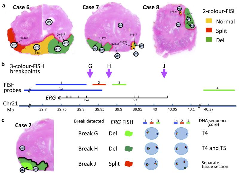
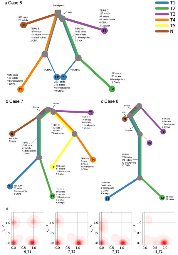
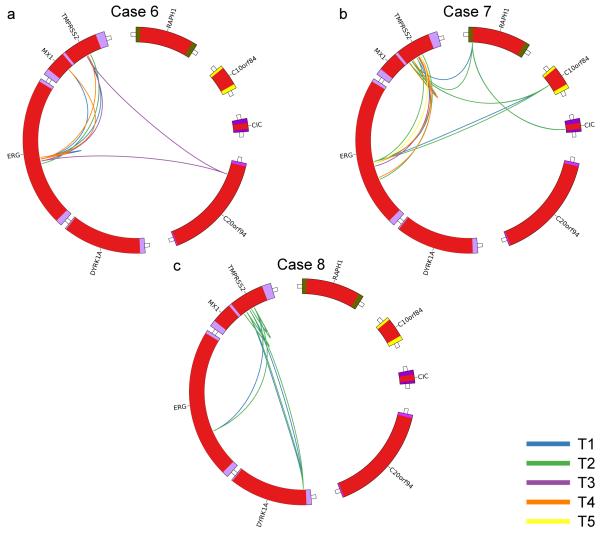
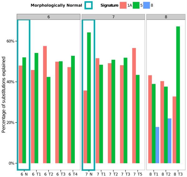
Genome-wide DNA sequencing was used to decrypt the phylogeny of multiple samples from distinct areas of cancer and morphologically normal tissue taken from the prostates of three men. Mutations were present at high levels in morphologically normal tissue distant from the cancer, reflecting clonal expansions, and the underlying mutational processes at work in morphologically normal tissue were also at work in cancer. Our observations demonstrate the existence of ongoing abnormal mutational processes, consistent with field effects, underlying carcinogenesis. This mechanism gives rise to extensive branching evolution and cancer clone mixing, as exemplified by the coexistence of multiple cancer lineages harboring distinct ERG fusions within a single cancer nodule. Subsets of mutations were shared either by morphologically normal and malignant tissues or between different ERG lineages, indicating earlier or separate clonal cell expansions. Our observations inform on the origin of multifocal disease and have implications for prostate cancer therapy in individual cases.


Figures

Figure 1
Figure 1
Prostate samples chosen for whole-genome sequencing. a, ERG rearrangements determined by fluorescence in situ hybridization (FISH). Case 7 is a multifocal cancer containing two separate foci (T1/T2/T4/T5 and T3). Case 8 is also designated as a multifocal cancer,(nodules T1/T2, and T3). Yellow: un-rearranged normal ERG gene; Red, ERG gene split but both 3′ and 5′ ends retained; Green, ERG gene rearranged but only its 3′ end retained. Panels b and c: 3-colour FISH used to distinguish different ERG-locus translocation breakpoints in Case 7. b, Position of the three FISH probes: probe 1 (blue, BAC RP11-164E1, and probe 1a, BACs RP11-95G19, RP11-720N21, CTD-2511E13) was labeled in Aqua (Kreatech 415 Platinum Bright): probe 2 (red, fosmid G248P80319F5 37Kb) labeled with Cy3; and Probe 3 (green, fosmid G248P86592E2 38.5k, and probe 4, BACs RP11-372O17, RP11-115E14, RP11-729O4) labeled with FITC. The purple arrows represent the positions of ERG breakpoints detected in these experiments. For the precise position of the ERG breakpoints G and H see Table 2. c, Left: Tumor areas with ERG locus breaks G and H are indicated as light and dark green respectively. Break J was found in an adjacent prostate section not show in this figure. Right: representations of the ERG FISH patterns. Original FISH images are show in Supplementary Fig. 1. “Split” denotes that 5′ and 3′ ERG signals were separated but retained in the cell. “Del” indicates that 5′ ERG signals were lost from the cell, while 3′ ERG signals were retained.
Figure 2
Figure 2
Phylogenies of multi-focal prostate cancers. a-c, Phylogenies revealing the relationships between sample clones for each case. Each line is associated with a clone from a particular sample. The length of each line is proportional to the weighted quantity of variations on a logarithmic scale. The thickness of a line indicates the proportion the clone makes up of that sample i.e. 48%/52% for 6_T1 and 12%/88% for 8_T3. The minor clone of 8_T3b has no detected unique variants. 8_T3 contained 43 mutations present as a 12% subclone (T3a) shared with 8_T1/8_T2. In validation experiments 8_T3 did not contain any of the five ERG and TMPRSS2 rearrangements present in 8_T1/8_T2 (Table 2)) or mutations that were unique to 8_T1/8_T2 (10,000 depth) indicating that it represents an earlier clone of 8_T1/8_T2 seeded into tissue sample 8_T3. The various TMPRSS2-ERG translocations are indicated by their TERG ID (Table 2). d, Example 2D density plots showing the posterior distribution of the fraction of cells bearing a mutation in two samples. The fraction of cells is modeled using a Bayesian Dirichlet processes. These plots illustrate samples that have shared clonal mutations (6_T1/6_T2), and branched (unrelated) mutations (7_T2/T_T3). There are two examples of samples with a subclone. 7_T2/7_T5 has a peak at (0,0.72), which represents subclonal mutations in 72% of cells in 7_T5 that have occurred only in this sample, after divergence from the other samples. Similarly, 8_T1/8_T3 has a peak at (0.54,0), representing subclonal mutations in 54% of cells in T1 only.
Figure 3
Figure 3
Patterns of ERG alterations. a-c, Circos plots highlighting ERG rearrangements present in each prostate. Each color represents a different cancer sample as indicated.
Figure 4
Figure 4
Relative contributions of mutational signatures to the total mutation burden of each sample. The mutational spectra, as defined by the triplets of nucleotides around each substitution, of each sample were deconvoluted into mutational processes using 22 distinct signatures determined from 7,042 cancers as described previously,. The signature designations (1a, 5, 8) match those reported previously. For sample 7_T4 and 8_N there were too few mutations to be able to accurately identify the contributions of the mutational signatures.

Comment in

References

REFERENCE LIST

    1. Andreoiu M, Cheng L. Multifocal prostate cancer: biologic, prognostic, and therapeutic implications. Hum. Pathol. 2010;41:781–793. - PubMed
    1. Cheng L, et al. Evidence of independent origin of multiple tumors from patients with prostate cancer. J. Natl. Cancer Inst. 1998;90:233–237. - PubMed
    1. Kobayashi M, et al. Molecular analysis of multifocal prostate cancer by comparative genomic hybridization. Prostate. 2008;68:1715–1724. - PubMed
    1. Boyd LK, et al. High-resolution genome-wide copy-number analysis suggests a monoclonal origin of multifocal prostate cancer. Genes Chromosomes Cancer. 2012;51:579–589. - PubMed
    1. Lindberg J, et al. Exome sequencing of prostate cancer supports the hypothesis of independent tumour origins. Eur. Urol. 2013;63:347–353. - PubMed

METHODS-ONLY REFERENCES

    1. Warren AY, et al. Method for sampling tissue for research which preserves pathological data in radical prostatectomy. Prostate. 2013;73:194–202. - PubMed
    1. Li H, Durbin R. Fast and accurate short read alignment with Burrows-Wheeler transform. Bioinformatics. 2009;25:1754–1760. - PMC - PubMed
    1. Ye K, Schulz MH, Long Q, Apweiler R, Ning Z. Pindel: a pattern growth approach to detect break points of large deletions and medium sized insertions from paired-end short reads. Bioinformatics. 2009;25:2865–2871. - PMC - PubMed
    1. Campbell PJ, et al. Identification of somatically acquired rearrangements in cancer using genome-wide massively parallel paired-end sequencing. Nat. Genet. 2008;40:722–729. - PMC - PubMed
    1. Zerbino DR, Birney E. Velvet: algorithms for de novo short read assembly using de Bruijn graphs. Genome Res. 2008;18:821–829. - PMC - PubMed

Publication types