

In vivo NAD assay reveals the intracellular NAD contents and redox state in healthy human brain and their age dependences
- PMID: 25730862
- PMCID: PMC4352772
- DOI: 10.1073/pnas.1417921112
In vivo NAD assay reveals the intracellular NAD contents and redox state in healthy human brain and their age dependences
Abstract
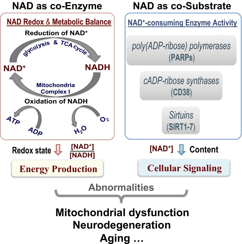
NAD is an essential metabolite that exists in NAD(+) or NADH form in all living cells. Despite its critical roles in regulating mitochondrial energy production through the NAD(+)/NADH redox state and modulating cellular signaling processes through the activity of the NAD(+)-dependent enzymes, the method for quantifying intracellular NAD contents and redox state is limited to a few in vitro or ex vivo assays, which are not suitable for studying a living brain or organ. Here, we present a magnetic resonance (MR) -based in vivo NAD assay that uses the high-field MR scanner and is capable of noninvasively assessing NAD(+) and NADH contents and the NAD(+)/NADH redox state in intact human brain. The results of this study provide the first insight, to our knowledge, into the cellular NAD concentrations and redox state in the brains of healthy volunteers. Furthermore, an age-dependent increase of intracellular NADH and age-dependent reductions in NAD(+), total NAD contents, and NAD(+)/NADH redox potential of the healthy human brain were revealed in this study. The overall findings not only provide direct evidence of declined mitochondrial functions and altered NAD homeostasis that accompany the normal aging process but also, elucidate the merits and potentials of this new NAD assay for noninvasively studying the intracellular NAD metabolism and redox state in normal and diseased human brain or other organs in situ.
Keywords: NAD; aging; human brain; in vivo 31P MR spectroscopy; redox state.
Conflict of interest statement
The authors declare no conflict of interest.
Figures


References
-
- Harden A, Young WJ. The alcoholic ferment of yeast-juice. Proc R Soc Lond B Biol Sci. 1906;77(519):405–420.
-
- Chance B, Ito T. Control of endogenous adenosine triphosphatase activity by energy-linked pyridine nucleotide reduction in mitochondria. Nature. 1962;195:150–153. - PubMed
-
- Belenky P, Bogan KL, Brenner C. NAD+ metabolism in health and disease. Trends Biochem Sci. 2007;32(1):12–19. - PubMed
-
- Lin SJ, Guarente L. Nicotinamide adenine dinucleotide, a metabolic regulator of transcription, longevity and disease. Curr Opin Cell Biol. 2003;15(2):241–246. - PubMed
Publication types
MeSH terms
Substances
Grants and funding
LinkOut - more resources
Full Text Sources
Other Literature Sources
Medical
