. 2015 Mar 3;112(9):2829-34.
doi: 10.1073/pnas.1424400112. Epub 2015 Feb 17.

Mutant IDH is sufficient to initiate enchondromatosis in mice

Affiliations

Mutant IDH is sufficient to initiate enchondromatosis in mice

Makoto Hirata et al. Proc Natl Acad Sci U S A. .

Abstract

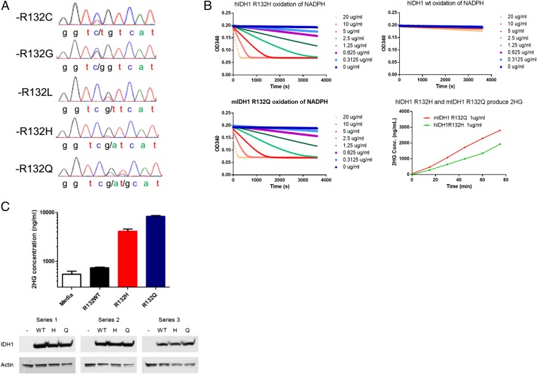
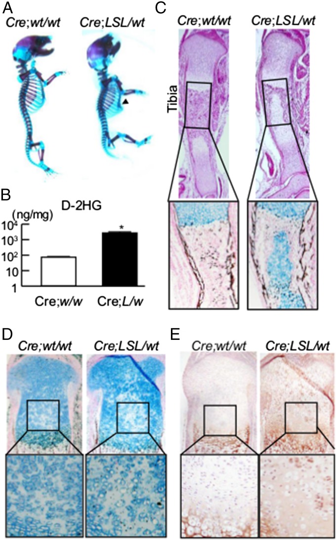
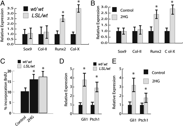
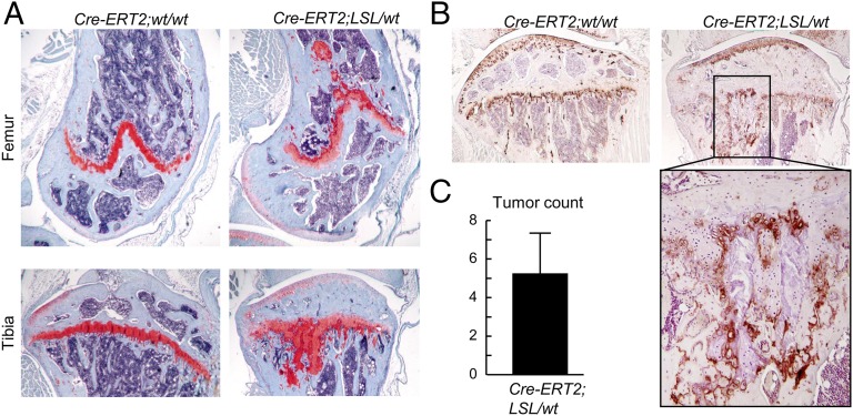
Enchondromas are benign cartilage tumors and precursors to malignant chondrosarcomas. Somatic mutations in the isocitrate dehydrogenase genes (IDH1 and IDH2) are present in the majority of these tumor types. How these mutations cause enchondromas is unclear. Here, we identified the spectrum of IDH mutations in human enchondromas and chondrosarcomas and studied their effects in mice. A broad range of mutations was identified, including the previously unreported IDH1-R132Q mutation. These mutations harbored enzymatic activity to catalyze α-ketoglutarate to d-2-hydroxyglutarate (d-2HG). Mice expressing Idh1-R132Q in one allele in cells expressing type 2 collagen showed a disordered growth plate, with persistence of type X-expressing chondrocytes. Chondrocyte cell cultures from these animals or controls showed that there was an increase in proliferation and expression of genes characteristic of hypertrophic chondrocytes with expression of Idh1-R132Q or 2HG treatment. Col2a1-Cre;Idh1-R132Q mutant knock-in mice (mutant allele expressed in chondrocytes) did not survive after the neonatal stage. Col2a1-Cre/ERT2;Idh1-R132 mutant conditional knock-in mice, in which Cre was induced by tamoxifen after weaning, developed multiple enchondroma-like lesions. Taken together, these data show that mutant IDH or d-2HG causes persistence of chondrocytes, giving rise to rests of growth-plate cells that persist in the bone as enchondromas.

Keywords: cartilage tumor; hedgehog; isocitrate dehydrogenase.


Conflict of interest statement

The authors declare no conflict of interest.

Figures

Fig. 1.
Fig. 1.
IDH1 R132Q mutations confer an enzymatic activity that converts α-KG to d-2HG. (A) Chromatograms generated by Sanger sequencing at the locus coding IDH1-R132 in human chondrosarcoma samples. (B) NADPH consumption and d-2HG production by IDH1 mutant proteins. Recombinant R132Q, R132H, and WT IDH1 proteins were assayed for NADPH consumption activity at different protein concentrations. This neoactivity is acquired by known IDH1 driver mutations. MS identified the production of d-2HG by R132Q and R132H mutant proteins. Liquid chromatography (LC) /MS was used to identify and measure the concentration of d-2HG in enzymatic assays of recombinant protein by comparing spectra with a d-2HG standard. Time-dependent d-2HG production was detected in the reactions catalyzed by IDH1 R132Q and IDH1 R132Q proteins but not WT protein. (C) Cellular production of d-2HG by R132Q and R132H mutant IDH1. HEK293 cells were transiently transfected with expression plasmids containing WT IDH1 or R132H or R132Q mutations. After 48 h, d-2HG concentration in the culture media was measured by LC/MS and compared with a media alone control, and protein expression was assessed by Western blot.
Fig. 2.
Fig. 2.
Col2a1-Cre;Idh1-KI mice showed impaired chondrocyte differentiation. (A) Whole-mount skeletal staining with alcian blue and alizarin red in Col2a1-Cre;Idh1-KI mutant and WT littermates. Arrowhead shows pectus excavatum in the mutant mice. (B) d-2HG levels determined by liquid chromatography/MS in 10 mg limbs harvested from WT and Idh1 mutant littermates at E16.5. (C) Representative (Upper) H&E and (Lower) alcian blue and von Kossa staining of tibias at E16.5 from both genotypes. (D and E) Representative alcian blue and von Kossa staining and Col10a1 staining in proximal tibias at postnatal day 0 from both genotypes.
Fig. 3.
Fig. 3.
Idh1-R132Q and 2HG increase the expression of hypertrophic chondrocyte markers and increase cell proliferation. (A) Expression of markers of hypertrophic chondrocytes is significantly higher in chondrocytes from mice expressing Idh1-R132Q. Relative expression is shown, with levels in WT cells arbitrarily set to one. (B) Treatment of WT cells with 2HG has a similar effect on gene expression as observed in cells expressing Idh1-R132Q. (C) Percentage of cells incorporating BrdU in mutant and WT cells treated with 2-HD or AGI-5198. (D and E) Cells expressing the mutant Idh or cells from mice expressing WT Idh treated with 2HG up-regulate expression of Hh target genes. Data are given as means, and error bars represent SDs. *P < 0.05 vs. WT control.
Fig. 4.
Fig. 4.
Col2a1-Cre/ERT2;Idh1-KI mice develop multiple enchondroma-like cartilage lesions with dysregulated chondrocyte differentiation. (A and B) Safranin O staining and Col10 immunohistochemistry in (A, Upper) femurs and (A, Lower) tibias of WT and Col2-Cre/ERT2;Idh1-KI littermates. Multiple cartilage lesions were detected adjacent to growth plates in mutant mice. Col10 immunohistochemistry in the high-power field showed patchy expression of Col10 within the cartilage lesions. (C) Tumor counts in Idh1 mutant mice (n = 5). Data are given as means, and error bars represent SDs.

References

    1. Hong ED, Carrino JA, Weber KL, Fayad LM. Prevalence of shoulder enchondromas on routine MR imaging. Clin Imaging. 2011;35(5):378–384. - PubMed
    1. Walden MJ, Murphey MD, Vidal JA. Incidental enchondromas of the knee. AJR Am J Roentgenol. 2008;190(6):1611–1615. - PubMed
    1. Bovee JV, Hogendoorn PC, Wunder JS, Alman BA. Cartilage tumours and bone development: Molecular pathology and possible therapeutic targets. Nature Rev Cancer. 2010;10(7):481–488. - PubMed
    1. Levesque J, Bell RS, Marx R, Wunder JS. University of Toronto Division of Orthopaedic Surgery . A Guide to Orthopaedic Tumors. Division of Orthopaedic Surgery, Department of Surgery, University of Toronto; Toronto: 1996. p. 132.
    1. Schwartz HS, et al. The malignant potential of enchondromatosis. J Bone Joint Surg Am. 1987;69(2):269–274. - PubMed

Publication types