. 2015 Mar 5;519(7541):92-6.
doi: 10.1038/nature14232. Epub 2015 Feb 25.

Dietary emulsifiers impact the mouse gut microbiota promoting colitis and metabolic syndrome

Affiliations

Dietary emulsifiers impact the mouse gut microbiota promoting colitis and metabolic syndrome

Benoit Chassaing et al. Nature. .

Erratum in

Abstract

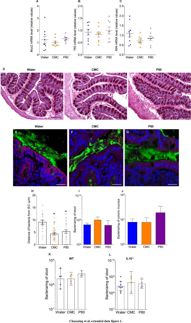
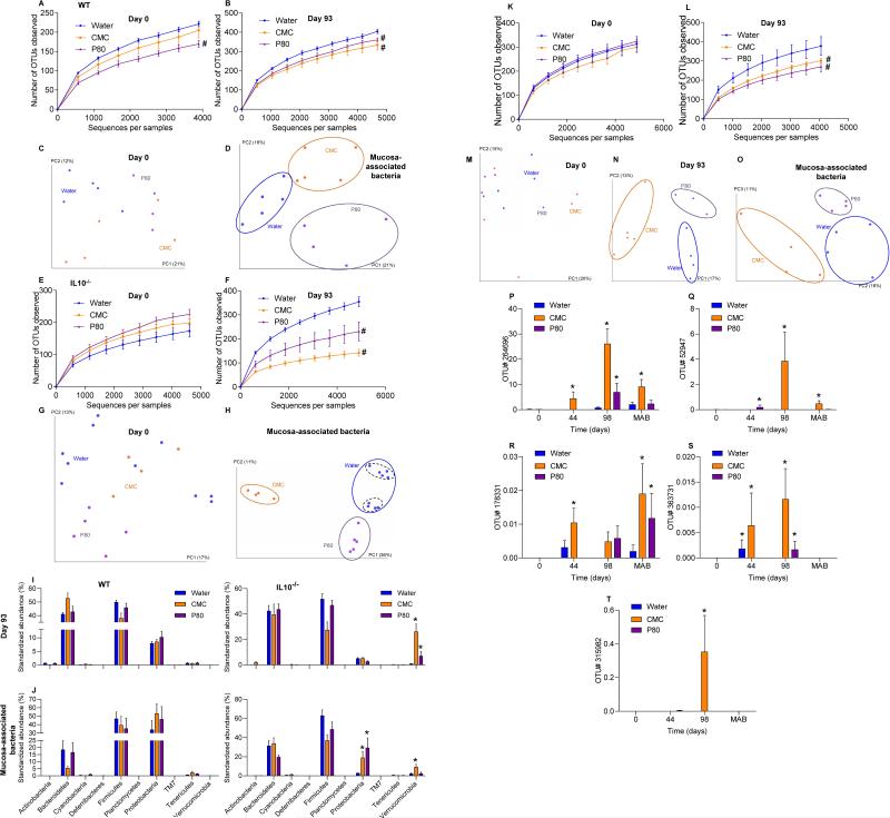
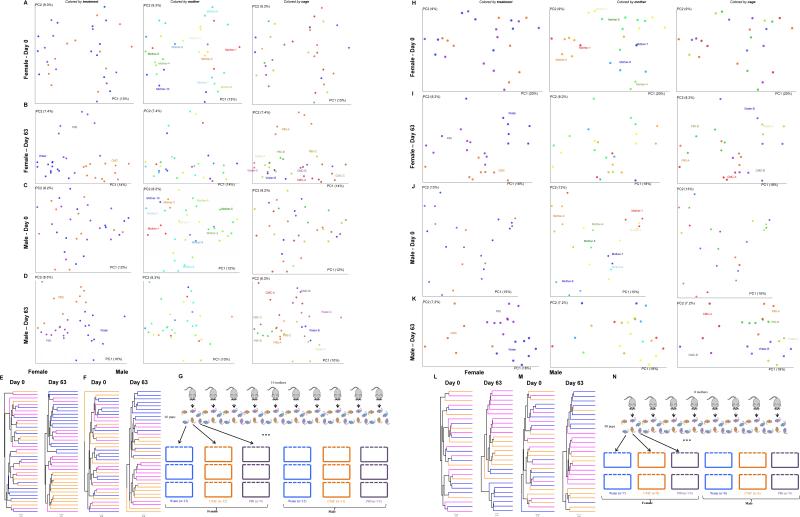
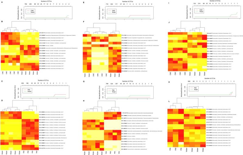
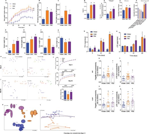
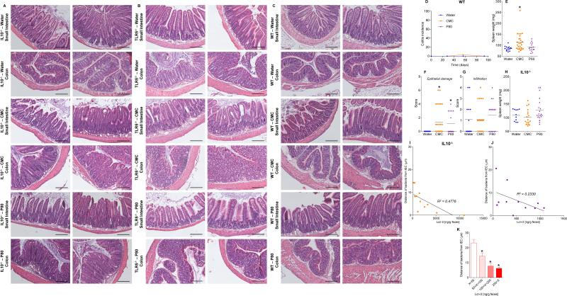
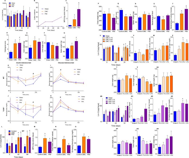
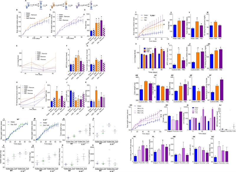
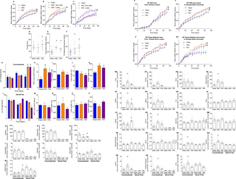
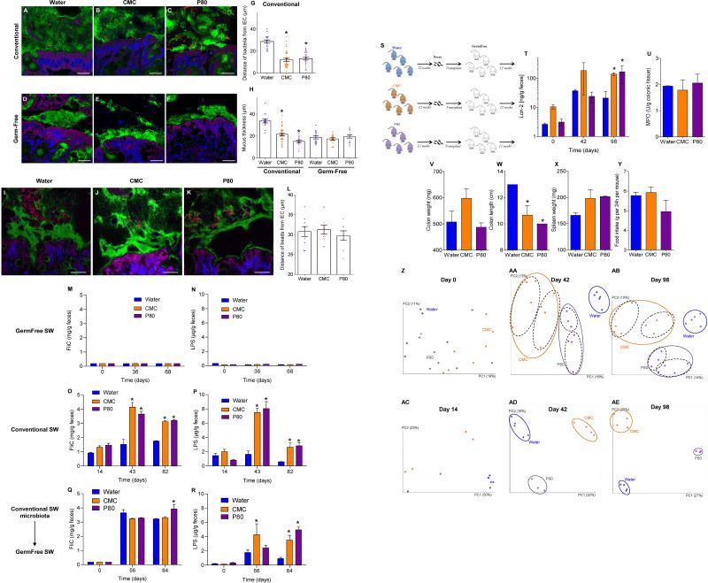
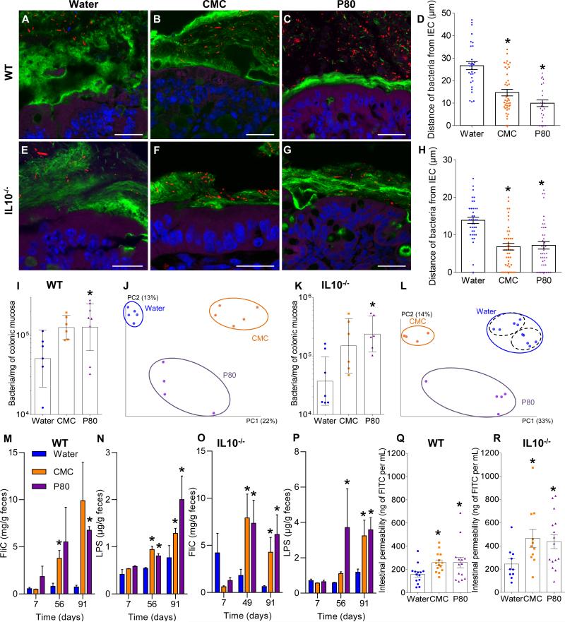
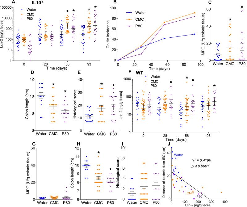
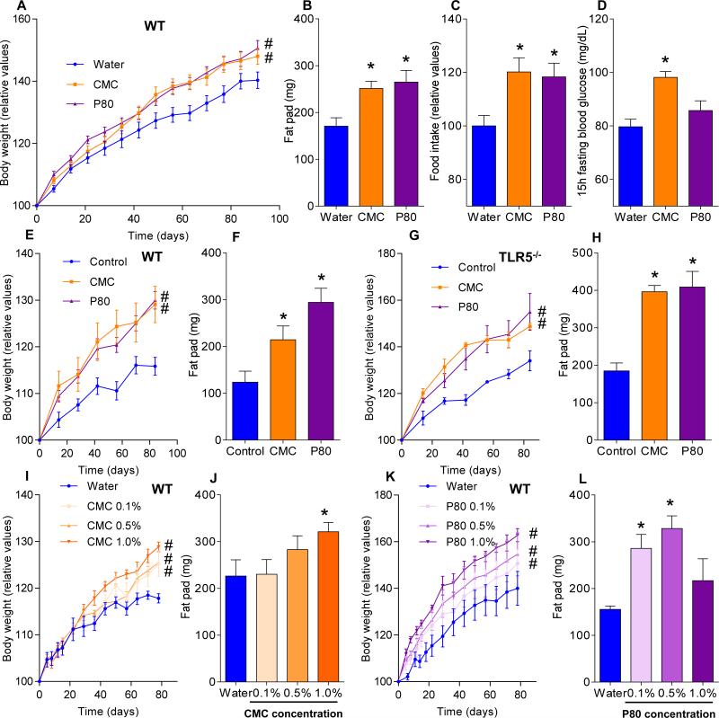
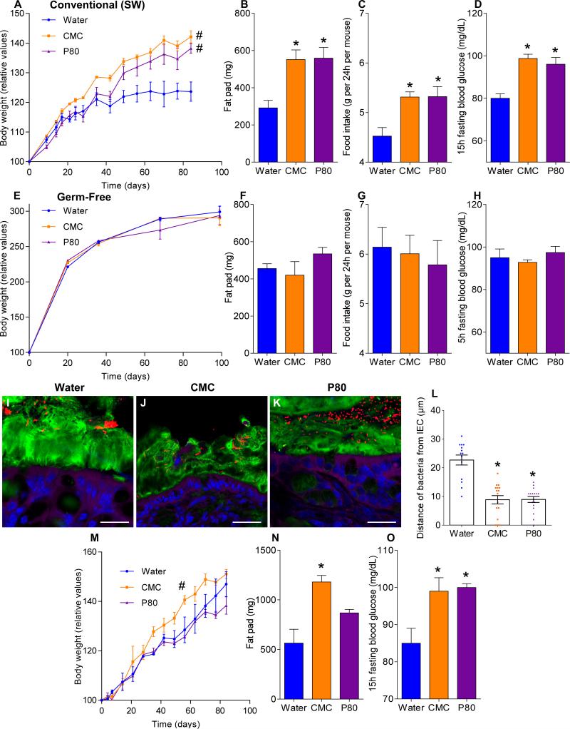
The intestinal tract is inhabited by a large and diverse community of microbes collectively referred to as the gut microbiota. While the gut microbiota provides important benefits to its host, especially in metabolism and immune development, disturbance of the microbiota-host relationship is associated with numerous chronic inflammatory diseases, including inflammatory bowel disease and the group of obesity-associated diseases collectively referred to as metabolic syndrome. A primary means by which the intestine is protected from its microbiota is via multi-layered mucus structures that cover the intestinal surface, thereby allowing the vast majority of gut bacteria to be kept at a safe distance from epithelial cells that line the intestine. Thus, agents that disrupt mucus-bacterial interactions might have the potential to promote diseases associated with gut inflammation. Consequently, it has been hypothesized that emulsifiers, detergent-like molecules that are a ubiquitous component of processed foods and that can increase bacterial translocation across epithelia in vitro, might be promoting the increase in inflammatory bowel disease observed since the mid-twentieth century. Here we report that, in mice, relatively low concentrations of two commonly used emulsifiers, namely carboxymethylcellulose and polysorbate-80, induced low-grade inflammation and obesity/metabolic syndrome in wild-type hosts and promoted robust colitis in mice predisposed to this disorder. Emulsifier-induced metabolic syndrome was associated with microbiota encroachment, altered species composition and increased pro-inflammatory potential. Use of germ-free mice and faecal transplants indicated that such changes in microbiota were necessary and sufficient for both low-grade inflammation and metabolic syndrome. These results support the emerging concept that perturbed host-microbiota interactions resulting in low-grade inflammation can promote adiposity and its associated metabolic effects. Moreover, they suggest that the broad use of emulsifying agents might be contributing to an increased societal incidence of obesity/metabolic syndrome and other chronic inflammatory diseases.


Figures

Extended data figure 1
(A-D) Dietary emulsifiers did not affect mucus and mucus-related genes expression in WT mice. WT mice were exposed to drinking water containing CMC or P80 (1.0%) for 12 weeks. (A-C) mRNA expression analysis by q-RT-PCR of Muc2 (A), Tff3 (B) and Klf4 (C) genes in the colonic mucosa. Points are from individual mice, bar represent the mean +/- S.E.M, (n=9). (D) Colons were stained using Periodic Acid Schiff. Bar = 200μm.Pictures are representatives of 10 biological replicates. (E-J) Dietary emulsifiers alter microbiota localization, composition, and pro-inflammatory potential in TLR5−/− mice. TLR5 −/− mice were exposed to drinking water containing CMC or P80 (1.0%) for 12 weeks. (E-G) Confocal microscopy analysis of microbiota localization; Muc2 (green), actin (purple), bacteria (red), and DNA (Blue). Bar = 20μm. (H) Distances of closest bacteria to intestinal epithelial cells per condition over 5 high-powered fields per mouse. Pictures are representatives of 5 biological replicates. (I-J) PCR-based quantification of total bacterial load (I) and bacterial load adhered to colonic mucosa (J). Data are the means +/- S.E.M., (n=5). Significance was determined using one-way ANOVA corrected for multiple comparisons with Sidak test, * indicates p<0.05 compared to water-treated group. (K-L) Dietary emulsifiers do not modify total bacterial load in WT and IL10−/− mice. WT (K) and IL10−/− (L) mice were exposed to drinking water containing CMC or P80 (1.0%) for 12 weeks. (K) Total bacterial load in stool of WT and (L) IL10−/− mice. Points are from individual mice. Data are geometric means with 95% confidence interval (n=5 for K except n=4 for CMC and P80-treated groups; for L, n=8, 4 and 6 for water, CMC and P80-treated groups, respectively).
Extended data figure 2
(A-H) Dietary emulsifiers induce profound alterations in gut microbiota composition in WT and IL10−/− mice. WT and IL10−/− mice were exposed to drinking water containing CMC or P80 (1.0%) for 12 weeks. (A, E) Day 0 and (B, F) day 93 microbiota richness and diversity in WT (A, B) and IL10−/− (E, F). (C-D, G-H) Principal coordinates analysis (PCoA) of the unweighted UniFrac distance matrix of fecal microbiota (C, G) and mucosa-associated bacteria (D, H) in WT (C-D) and IL10−/− (G-H) mice. Treatment of each mouse is indicated by point color and matching colored circles represent clustering by treatment (blue = water; orange = CMC; purple = P80). Black dashed circles represent mice sharing a cage. Data are the means +/- S.E.M. (n=5, except n=4 for WT mice P80-treated, n=4 for IL10−/− mice CMC-treated and n=9 for IL10−/− mice water-treated). Significance was determined using 2-way group ANOVA (# indicates p<0.05) compared to water-treated group. (I-J) Phylum characterization of emulsifier-induced alteration of gut microbiota composition in WT and IL10−/− mice. WT and IL10−/− mice were exposed to drinking water containing CMC or P80 (1.0%) for 12 weeks. Relative abundance of phyla are represented for fecal microbiota at day 93 (I) and for colonic mucosa associated bacteria (J). Data are the means +/- S.E.M. (n=5). Significance was determined using two-way ANOVA corrected for multiple comparisons with Bonferroni test, * indicates p<0.05 compared to water-treated group. (K-O)Dietary emulsifiers induce profound alterations in gut microbiota composition in TLR5−/− mice. TLR5−/− mice were exposed to drinking water containing CMC or P80 (1.0%) for 12 weeks. (K) Day 0 and (L) day 93 microbiota richness and diversity (n=5). (M-O) PCoA of the unweighted UniFrac distance matrix of fecal microbiota at day 0 (M), day 93 (N) and of mucosa-associated bacteria (O). Data are the means +/- S.E.M. (for M, n=4, 5 and 5 for water, CMC and P80-treated groups, respectively; for N, n=4, 5 and 3 for water, CMC and P80-treated groups, respectively; for O, n=4). Significance was determined using 2-way group ANOVA (# indicates p<0.05) compared to water-treated group. (P-T) Prevalence analysis of OTUs related to mucolytic bacteria in IL10−/− mice treated with dietary emulsifier. IL10−/− mice were exposed to drinking water containing CMC or P80 (1.0%) for 12 weeks. OTUs (P) Prok_MSA # 52947 (related to Clostridium perfringens), (Q) 264696 (related to Akkermansia muciniphila), (R) 315982 (related to Clostridium perfringens), (S) 363731 (related to Akkermansia muciniphila), and (T) 178331(related to Akkermansia muciniphila) were analyzed. Data are expressed as % of the total sequences analyzed and are the means +/- S.E.M. (n=6, except n=9 for IL10−/− mice water-treated). Significance was determined using two-way ANOVA corrected for multiple comparisons with Bonferroni test, * indicates p<0.05 compared to water-treated group.
Extended data figure 3
(A-G) Dietary emulsifiers alter gut microbiota composition in WT mice relative to littermate controls. All the female (n=33, A-B) and male (n=33, C-D) mice from 10 different litters were placed into cages in a manner such that each litter was split equally amongst the 3 experimental groups that were to receive water, CMC or P80 (3 cages per sex per condition). Mice were exposed to drinking water containing CMC or P80 (1.0%) for 8 weeks. (A, C) Day 0 and (B, D) day 63 PCoA of the unweighted UniFrac distance matrix of fecal microbiota in WT mice. Treatment of each mouse is indicated by point color (blue = water; orange = CMC; purple = P80). Mother of each mouse is indicated by point color. Cage of each mouse is indicated by point color. (E-F) Mice were clustered using UPGMA (Unweighted Pair Group Method with Arithmetic mean) method. Treatment of each mouse is indicated by line color (blue = water; orange = CMC; purple = P80). (G) Schematic representation of the above experimental design. (H-N) Dietary emulsifiers alter gut microbiota composition in TLR5−/− mice relative to littermate controls. All the female (n=25, H-I) and male (n=23, J-K) mice from 8 different litters were placed into cages in a manner such that each litter was split equally amongst the 3 experimental groups that were to receive water, CMC or P80 (2 cages per sex per condition). Mice were exposed to drinking water containing CMC or P80 (1.0%) for 8 weeks. (H, J) Day 0 and (I, K) day 63 PCoA of the unweighted UniFrac distance matrix of fecal microbiota in TLR5−/− mice. Treatment of each mouse is indicated by point color (blue = water; orange = CMC; purple = P80). Mother of each mouse is indicated by point color. Cage of each mouse is indicated by point color. (L-M) Mice were clustered using UPGMA (Unweighted Pair Group Method with Arithmetic mean) method. Treatment of each mouse is indicated by line color (blue = water; orange = CMC; purple = P80). (N) Schematic representation of the above experimental design.
Extended data figure 4
(A-D) Misclassification error rate and heatmap representation of the 15 most significantly altered OTUs in WT mice treated with dietary emulsifier. WT mice were exposed to drinking water containing CMC (A-B) or P80 (C-D) (1.0%) for 12 weeks. (A, C) Misclassification error rate showing that 15 OTUs were sufficient to successfully discriminate microbiota from each experimental group (error rate = 0). (B, D) Heatmap representation of the 15 most significantly altered OTUs in WT mice treated with dietary emulsifier. Colors represent relative expression (white and red for underrepresented and overrepresented, respectively). The 15 OTUs were listed on the right part using their Greengenes Prok_MSA IDs, and assigned taxonomy were labeled starting phylum, then class, order, family, and genus. Dendrogram on the upper part represents sample clustering. (E-H) Misclassification error rate and heatmap representation of the 15 most significantly altered OTUs in IL10−/− mice treated with dietary emulsifier. IL10−/− mice were exposed to drinking water containing CMC (E-F) or P80 (G-H) (1.0%) for 12 weeks. See legend of A-D for more details. (I-L)Misclassification error rate and heatmap representation of the 15 most significantly altered OTUs in TLR5−/− mice treated with dietary emulsifier. TLR5−/− mice were exposed to drinking water containing CMC (I-J) or P80 (K-L) (1.0%) for 12 weeks. See legend of A-D for more details (for A-B, n=5; for C-D, n=5 and 4 for water and P80-treated groups, respectively; for E-F, n=5 and 4 for water and CMC-treated groups, respectively; for G-H, n=5 and 4 for water and P80-treated groups, respectively; for I-J, n=4; for K-L, n=4 and 3 for water and P80-treated groups, respectively).
Extended data figure 5
(A-G) Dietary emulsifiers promote metabolic syndrome in adult WT mice. 4 months-old male WT mice were exposed to drinking water containing CMC or P80 (1.0%) for 8 weeks (2 cages per condition). (A) Body weight over time, (B) 15-hours fasting blood glucose concentration, (C) food intake measurement, (D) spleen weights, (E) fat-pad weights, (F) colon weights and (G) colon lengths. Data are the means +/- S.E.M. (n=10). Significance was determined using one-way ANOVA corrected for multiple comparisons with Sidak test (* indicates p<0.05 compared to water-treated group) or 2-way group ANOVA (# indicates p<0.05 compared to water-treated group). (H-P) Dietary emulsifiers alter gut microbiota composition in adult WT mice. 4 months-old male WT mice were exposed to drinking water containing CMC or P80 (1.0%) for 8 weeks (2 cages per condition). (H) Day 0 and (I) day 63 PCoA of the unweighted UniFrac distance matrix of fecal microbiota in WT mice. Treatment of each mouse is indicated by point color (blue = water; orange = CMC; purple = P80). Cage of each mouse is indicated by point color (for H, n=7, 8 and 8 for water, CMC and P80-treated groups, respectively). (J) Day 0 and day 63 microbiota richness and diversity. (K) Day 63 Jackknifed PCoA of the unweighted UniFrac distance matrix of fecal microbiota in WT mice. Treatment of each mouse is indicated by point color (blue = water; orange = CMC; purple = P80) (n=8). (L) After clustering of mouse fecal microbiota using PCoA of the unweighted UniFrac distance matrix, a representative mouse has been used to illustrate the time point evolution of the microbiota (n=1). (M) After clustering of mouse fecal microbiota using PCoA of the unweighted UniFrac distance matrix, evolution of the principal component 1 between day 0 and day 63 has been calculated for each mouse (n=10). (N) Average of the UniFrac unweighted distance for each group (water, CMC and P80) between day 0 and day 63 has been calculated (n=10). (O) Average of the UniFrac unweighted distance within group (water, CMC and P80) has been calculated (n=10). (P) Average of the UniFrac unweighted distance between group (cages, water, CMC and P80) or within group (water) at day 63 has been calculated (n=10). Significance was determined using one-way ANOVA corrected for multiple comparisons with Sidak test (* indicates p<0.05 compared to water-treated group) or 2-way group ANOVA (# indicates p<0.05 compared to water-treated group). (Q-R) Dietary emulsifiers increase pro-inflammatory potential of intestinal microbiota in TLR5−/− mice. TLR5−/− mice were exposed to drinking water containing CMC or P80 (1.0%) for 12 weeks. Bioactive levels of fecal flagellin (Q) and LPS (R) assayed with TLR5 and TLR4 reporter cells. Data are the means +/- S.E.M. (n=10). Significance was determined using two-way ANOVA corrected for multiple comparisons with Bonferroni test, * indicates p<0.05 compared to water-treated group. (S-V)Dietary emulsifiers increase serum immune reactivity. WT (S, T) and IL10−/− (U, V) mice were exposed to drinking water containing CMC or P80 (1.0%) for 12 weeks. (S, U) Serum immune reactivity to flagellin and (T, V) LPS in WT (S, T) and IL10−/− (U, V) mice. Points are from individual mice. Data are the means +/- S.E.M. (for S, n=18, 30 and 16 for water, CMC and P80-treated groups, respectively; for T, n=18, 31 and 17 for water, CMC and P80-treated groups, respectively; for U, n=21, 20 and 23 for water, CMC and P80-treated groups, respectively; for V, n=27, 20 and 25 for water, CMC and P80-treated groups, respectively). Significance was determined using one-way ANOVA corrected for multiple comparisons with Sidak test, * indicates p<0.05 compared to water-treated group.
Extended data figure 6
A-Dietary emulsifiers induce histopathologically robust inflammation in IL10−/− mice. IL10−/− mice were exposed to drinking water containing CMC or P80 (1.0%) for 12 weeks. Colon and small intestine were H&E stained. Bar = 200μm. Pictures are representatives of 15 biological replicates. B-Dietary emulsifiers induce histopathologically robust inflammation in TLR5−/− mice. TLR5−/− mice were exposed to drinking water containing CMC or P80 (1.0%) for 12 weeks. Colon and small intestine were H&E stained. Bar = 200μm. Pictures are representatives of 5 biological replicates. CHistopathology of emulsifier-treated WT mice. WT mice were exposed to drinking water containing CMC or P80 (1.0%) for 12 weeks. Colon and small intestine were H&E stained. Bar = 200μm. Pictures are representatives of 15 biological replicates. (D-H) Dietary emulsifiers elicit low-grade intestinal inflammation in WT and splenomegaly in IL10−/− mice. WT (D-G) and IL10-/- (H) mice were exposed to drinking water containing CMC or P80 (1.0%) for 12 weeks. (D) Colitis incidence over time, (E, H) spleen weights, (F) epithelial damage and (G) infiltration scores. Points are from individual mice, bar represent the mean. (for E-G, n=14, 27 and 16 for water, CMC and P80-treated groups, respectively; for H, n=11, 18 and 20 for water, CMC and P80-treated groups, respectively). Significance was determined using one-way ANOVA corrected for multiple comparisons with Sidak test, * indicates p<0.05 compared to water-treated group. (I-K) Extent of intestinal inflammation correlates with perturbation in microbiota localization in WT and IL10−/− mice. IL10−/− mice were exposed to drinking water containing (I) CMC or (J) P80 (1.0%) for 12 weeks. Fecal levels of the inflammatory marker Lcn2 as well as confocal microscopy analysis of microbiota localization and estimation of the distances of the closest bacteria to intestinal epithelial cells were determined, and plotted in X and Y axis, respectively. Linear regression line was draft and R2 was determined. (n=11). (K) Analysis of bacterial-epithelial distance upon stratification of levels of gut inflammatory marker fecal Lcn2, using both WT and IL10-/- mice exposed to drinking water containing CMC or P80 (1.0%) for 12 weeks. Mice were grouped according to their fecal Lcn2 levels and bacterial-epithelial distances were then plotted (mean +/- S.E.M.). Significance was determined using oneway ANOVA corrected for multiple comparisons with Sidak test (* indicates p<0.05 compared to X<50 ng/g group).
Extended data figure 7
(A-G) Dietary emulsifiers promote intestinal inflammation in TLR5−/− mice. TLR5−/− mice were exposed to drinking water containing CMC or P80 (1.0%) for 12 weeks. (A) Fecal levels of the inflammatory marker Lcn2 over time, (B) colitis incidence over time, (C) MPO levels, (D) histological score, (E) colon weights, (F) colon lengths and (G) spleen weights. Data are the means +/- S.E.M. or geometric means with 95% confidence interval (for A) (n=5). Significance was determined using one-way ANOVA corrected for multiple comparisons with Sidak test or using two-way ANOVA corrected for multiple comparisons with Bonferroni test, * indicates p<0.05 compared to water-treated group. (H-K) Dietary emulsifiers induce metabolic syndrome in WT and TLR5-/- mice. (H-I) WT and (J-K) TLR5−/− mice were exposed to drinking water containing CMC or P80 (1.0%) for 12 weeks. (H, J) Glucose tolerance and (I, K) insulin sensitivity were analyzed. Data are the means +/- S.E.M. (n=5).Significance was determined using 2-way group ANOVA (# indicates p<0.05) compared to water-treated group. (L-T) Emulsifier-supplemented chow elicits low-grade intestinal inflammation in WT mice. WT mice were given mouse chow containing CMC or P80 (1.0%) for 12 weeks. (L) Fecal levels of the inflammatory marker Lcn2 over time, (M) MPO levels, (N) food intake measurement, (O) 15-hours fasting blood glucose concentration, (P) colon weights, (Q) colon lengths, (R) spleen weights, and PCR-based quantification of (S) total bacterial load and (T) bacterial load adhered to colonic mucosa. Data are the means +/- S.E.M. or geometric means with 95% confidence interval (for A) (n=5). Significance was determined using one-way ANOVA corrected for multiple comparisons with Sidak test or using two-way ANOVA corrected for multiple comparisons with Bonferroni test, * indicates p<0.05 compared to control group. (U-AH) Dose response characterization of dietary emulsifiers on intestinal inflammation. WT mice were exposed to drinking water containing 0.1-1.0% CMC (U-AA) or P80 (AB-AH) for 12 weeks. (U, AB) Fecal levels of the inflammatory marker Lcn2 over time, (V, AC) MPO levels, (W, AD) food intake measurement, (X, AE) 15-hours fasting blood glucose concentration, (Y, AF) colon weights, (Z, AG) colon lengths and (AA, AH) spleen weights. Data are the means +/- S.E.M. (n=5). Significance was determined using one-way ANOVA corrected for multiple comparisons with Sidak test or using two-way ANOVA corrected for multiple comparisons with Bonferroni test, * indicates p<0.05 compared to water-treated group.
Extended data figure 8
(A-K) Emulsifiers-induced metabolic syndrome is partially reversible by 6 weeks post-emulsifier treatment. (A) Schematic representation of the experiment. (B, C) Body weight over time, (D) fat-pad weight, (E) fecal levels of the inflammatory marker Lcn2 over time, (F) MPO levels, (G) food intake measurement, (H) 15-hours fasting blood glucose concentration, (I) colon weights, (J) colon lengths and (K) spleen weights. Data are the means +/- S.E.M. (n=5). Significance was determined using one-way ANOVA corrected for multiple comparisons with Sidak test (* indicates p<0.05 compared to water-treated group) or 2-way group ANOVA (# indicates p<0.05 compared to water-treated group). (L-S) Sodium sulfite did not induce robust or low-grade intestinal inflammation. WT and IL10−/− mice were exposed to drinking water containing sodium sulfite (1.0%) for 12 weeks. (L, M) Body weight over time, (N) colon weights, (O) colon lengths, (P) spleen weights, (Q) fat-pad weight, (R) MPO levels and (S) serum levels of the inflammatory marker Lcn2. Data are the means +/- S.E.M. (n=5). Points are from individual mice. Significance was determined using one-way ANOVA corrected for multiple comparisons with Sidak test. (T-W) Dietary emulsifiers promote metabolic syndrome in TLR5−/− mice. TLR5−/− mice were exposed to drinking water containing CMC or P80 (1.0%) for 12 weeks. (T) Body weight over time, (U) fatpad weight, (V) food intake measurement, and (W) 15-hours fasting blood glucose concentration. Data are the means +/- S.E.M. (n=5). Significance was determined using one-way ANOVA corrected for multiple comparisons with Sidak test (* indicates p<0.05 compared to water-treated group) or 2-way group ANOVA (# indicates p<0.05 compared to water-treated group). (X-AF) Emulsifier-supplemented chow promotes intestinal inflammation in TLR5−/− mice. TLR5−/− mice were given mouse chow containing CMC or P80 (1.0%) for 12 weeks. (X) Fecal levels of the inflammatory marker Lcn2 over time, (Y) MPO levels, (Z) food intake measurement, (AA) 15-hours fasting blood glucose concentration, (AB) colon weights, (AC) colon lengths, (AD) spleen weights, and PCR-based quantification of (AE) total bacterial load and (AF) bacterial load adhered to colonic mucosa. Data are the means +/- S.E.M. (n=5). Significance was determined using one-way ANOVA corrected for multiple comparisons with Sidak test or using two-way ANOVA corrected for multiple comparisons with Bonferroni test, * indicates p<0.05 compared to control group. (AG-AO) Dose response characterization of dietary emulsifiers on intestinal inflammation in TLR5−/− mice. TLR5−/− mice were exposed to drinking water containing 0.1-1.0% P80 for 12 weeks. (AG) Body weight over time, (AH) fat pad weight, (AI) fecal levels of the inflammatory marker Lcn2 over time, (AJ) MPO levels, (AK) food intake measurement, (AL) 15-hours fasting blood glucose concentration, (AM) colon weights, (AN) colon lengths, and (AO) spleen weights. Data are the means +/- S.E.M. (n=3). Points in D are from individual mice. Significance was determined using one-way ANOVA corrected for multiple comparisons with Sidak test (* indicates p<0.05 compared to water-treated group) or 2-way group ANOVA (# indicates p<0.05 compared to water-treated group).
Extended data figure 9
(A-F) Dietary emulsifiers promotes metabolic syndrome in IL10−/− mice. IL10−/− mice were exposed to drinking water containing CMC or P80 (1.0%) for 12 weeks. (A-C) Body weight over time, (D) fat-pad weight, (E) food intake measurement, and (F) 15-hours fasting blood glucose concentration. Data are the means +/- S.E.M. (for A, n=24, 18 and 21 for water, CMC and P80-treated groups, respectively; for B, n=14, 11 and 8 for water, CMC and P80-treated groups, respectively; for C, n=14, 9 and 9 for water, CMC and P80-treated groups, respectively; for D, n=14, 18 and 20 for water, CMC and P80-treated groups, respectively; for E, n=15, 11 and 12 for water, CMC and P80-treated groups, respectively; for F, n=21, 17 and 20 for water, CMC and P80-treated groups, respectively). Significance was determined using one-way ANOVA corrected for multiple comparisons with Sidak test (* indicates p<0.05 compared to water-treated group) or 2-way group ANOVA (# indicates p<0.05 compared to water-treated group). Points are from individual mice and red points in F represent mice with overt colitis. (G-P) Emulsifiers-induced low-grade intestinal inflammation was abolished under germ-free conditions. (G-K) Conventionally-housed and germ-free (L-P) Swiss-Webster mice were exposed to drinking water containing CMC or P80 (1.0%) for 12 weeks. (G, L) Fecal levels of the inflammatory marker Lcn2 over time, (H, M) MPO levels, (I, N) colon weights, (J, O) colon lengths and (K, P) spleen weights. Data are the means +/- S.E.M. (n=8 for conventionally-housed mice and n=4 for germ-free mice). Significance was determined using one-way ANOVA corrected for multiple comparisons with Sidak test or using two-way ANOVA corrected for multiple comparisons with Bonferroni test, * indicates p<0.05 compared to control group. (Q-W) Dietary emulsifiers induce perturbations in fecal short-chain fatty acid composition. Fecal short-chain fatty acids composition was analyzed at the Metabolomics Core of the University of Michigan. (Q) Acetate, (R) propionate, (S) butyrate, (T) isovalerate, (U) valerate (V) hexanoate and (W) heptanoate were analyzed. Data are the means +/- S.E.M. (n=5, 8 and 7 for water, CMC and P80 conventional mice-treated groups and n=4, 4 and 5 for water, CMC and P80 germfree mice-treated groups, respectively). Significance was determined using one-way ANOVA corrected for multiple comparisons with Sidak test, * indicates p<0.05 compared to water-treated group. (X-AA) Dietary emulsifiers promote metabolic syndrome in mice from different vendors. WT mice were exposed to drinking water containing CMC or P80 (1.0%) for 12 weeks. (X, Y) Body weight over time of Bl/6 mice used upon receipt from Jackson Laboratories (X) or bred at Georgia State University (Y). (Z, AA) Body weight over time of Swiss Webster mice used upon receipt from Charles River company (Z) or bred at Georgia State University (AA). Data in X are not used elsewhere in report, while data in Y, Z, and AA are from Figs 3A, extended data figure 8A-K, and 4A). Data are the means +/- S.E.M. (n=8 for Bl/6 mice used upon receipt from Jackson Laboratories, n=16 for Bl/6 mice bred at Georgia State University, n=10 for Swiss Webster mice used upon receipt from Charles River company, n=8 for Swiss Webster mice bred at Georgia State University). Significance was determined using 2-way group ANOVA (# indicates p<0.05 compared to water-treated group). (AB-AT) Dietary emulsifiers induce perturbations in fecal bile acids composition. Fecal bile acids composition was analyzed at the Metabolomics Core of the University of Michigan. (AB) Lithocholic acid (LCA), (AC) chenodeoxycholic acid (CDCA), (AD) deoxycholic acid (DCA), (AE) hyodeoxycholic acid / ursodeoxycholic acid (HDCA/UDCA), (AF) α-muricholic acid (α-MCA), (AG) β-muricholic acid (β -MCA), (AH) cholic acid (CA), (AI) hyocholic acid (HCA), (AJ) ω-muricholic acid (ω-MCA), (AK) glycolithocholic acid (GLCA), (AL) glycochenodeoxycholic acid (GCDCA), (AM) glycodeoxycholic acid (GDCA), (AN) hyodeoxycholic acid / glycoursodeoxycholic acid (HDCA/GUDCA), (AO) glycocholic acid (GCA), (AP) taurolithocholic acid (TLCA), (AQ) taurine-conjugated chenodeoxycholic acid (TCDCA), (AR) taurodeoxycholic acid / Tauroursodeoxycholic acid (TDCA/TUDCA), (AS) taurohyodeoxycholic acid, and (AT) taurocholic acid (TCA) were analyzed. Data are the means +/- S.E.M. (n=5, 8 and 7 for water, CMC and P80 conventional mice-treated groups and n=4, 4 and 5 for water, CMC and P80 germfree mice-treated groups, respectively). Significance was determined using one-way ANOVA corrected for multiple comparisons with Sidak test, * indicates p<0.05 compared to water-treated group.
Extended data figure 10
(A-H) Dietary emulsifiers do not alter mucus thickness under germ-free conditions. (A-C, G-H) Conventionally-housed and germ-free (D-F, H) Swiss-Webster mice were exposed to drinking water containing CMC or P80 (1.0%) for 12 weeks. (AF) Confocal microscopy analysis of microbiota localization; Muc2 (green), actin (purple), bacteria (red), and DNA (Blue). Bar = 20μm. (G) Distances of closest bacteria to intestinal epithelial cells per condition over 5 high-powered fields per mouse. Pictures are representatives of 5 biological replicates. (H) Mucus thickness over 5 high-powered fields per mouse. Data are the means +/- S.E.M. (n=5). Significance was determined using one-way ANOVA corrected for multiple comparisons with Sidak test, * indicates p<0.05 compared to water-treated group. (I-L)Dietary emulsifiers do not induce drastic perturbations of mucus layer integrity under germ-free conditions. Germ-free Swiss-Webster mice were exposed to drinking water containing CMC or P80 (1.0%) for 8 weeks. Mice were removed from isolator, inoculated with 0.5 μm green fluorescent beads (Polysciences, Warrington, PA), and euthanized 7h post inoculation. (I-K) Confocal microscopy analysis of fluorescent beads localization; Muc2 (green), actin (purple), fluorescent beads (red), and DNA (Blue). Bar = 20μm. (L) Distances of closest fluorescent beads to intestinal epithelial cells per condition over 5 high-powered fields per mouse. Pictures are representatives of 5 biological replicates. Data are the means +/- S.E.M. (n=4). Significance was determined using one-way ANOVA corrected for multiple comparisons with Sidak test. (M-R) Dietary emulsifiers increase pro-inflammatory potential of intestinal microbiota in SW mice, transferable to germfree mice recipient. (M, N) Germ-free Swiss-Webster mice were exposed to drinking water containing CMC or P80 (1.0%) for 12 weeks. Bioactive levels of fecal flagellin (M) and LPS (N) assayed with TLR5 and TLR4 reporter cells. (O, P) Swiss-Webster mice were exposed to drinking water containing CMC or P80 (1.0%) for 12 weeks. Bioactive levels of fecal flagellin (O) and LPS (P) assayed with TLR5 and TLR4 reporter cells. (Q-R) Germ-free Swiss-Webster mice were conventionalized via microbiota transplant from the Swiss-Webster mice treated with emulsifiers described above. Bioactive levels of fecal flagellin (Q) and LPS (R) assayed with TLR5 and TLR4 reporter cells. Data are the means +/- S.E.M. (n=4 for germ-free mice, n=8 for conventionally-housed mice and n=5 for conventionalized mice). Significance was determined using two-way ANOVA corrected for multiple comparisons with Bonferroni test, * indicates p<0.05 compared to control group. (S-Y)Microbiota transplant transfers emulsifier-induced low-grade intestinal inflammation. Germ-free Swiss-Webster mice were conventionalized via microbiota transplant from mice that received standard drinking water or drinking water containing CMC or P80 (1.0%). (S) Schematic representation of the experiment. (T) Fecal levels of the inflammatory marker Lcn2 over time, (U) MPO levels, (V) colon weights, (W) colon lengths, (X) spleen weights, and (Y) food intake measurement. Data are the means +/- S.E.M. (n=4). Significance was determined using one-way ANOVA corrected for multiple comparisons with Sidak test or using two-way ANOVA corrected for multiple comparisons with Bonferroni test, * indicates p<0.05 compared to control group. (Z-AE) Dietary emulsifiers induce profound alterations in gut microbiota composition in SW mice, transferable to germfree mice recipient. (Z-AB) Swiss-Webster mice were exposed to drinking water containing CMC or P80 (1.0%) for 12 weeks. PCoA of the unweighted UniFrac distance matrix of fecal microbiota at (Z) day 0, (AA) day 42 and (AB) day 98. (AC-AE) Germ-free Swiss-Webster mice were conventionalized via microbiota transplant from the Swiss-Webster mice treated with emulsifiers described above. PCoA of the unweighted UniFrac distances of fecal microbiota at (AC) day 14, (AD) day 42 and (AE) day 98 post transplant (for Z-AB, n=5, 8 and 7 for water, CMC and P80-treated groups, respectively; for ACAE, n=5, 4 and 3 for water, CMC and P80-treated groups, respectively). Treatment of each mouse is indicated by point color and matching colored circles indicate mice receiving same treatment (blue = water; orange = CMC; purple = P80). Black dashed circles represent mice sharing a cage.
Figure 1
Figure 1. Dietary emulsifiers alter microbiota localization, composition, and pro-inflammatory potential
WT and IL10−/− mice were exposed to drinking water containing CMC or P80 (1.0%) for 12 weeks. (A-C, E-G) Confocal microscopy analysis of microbiota localization; Muc2 (green), actin (purple), bacteria (red), and DNA (Blue). Bar = 20μm. (D, H) Distances of closest bacteria to intestinal epithelial cells (IEC) per condition over 5 high-powered fields per mouse. Pictures are representatives of 20 biological replicates. (I, K) PCR-based quantification of bacterial load adhered to colonic mucosa. (J, L). Principal coordinates analysis (PCoA) of the UniFrac distance matrix of WT (J) and IL10−/− (L) mice. Black dashed ellipses indicate mice sharing a cage during treatment. (M-P) Bioactive levels of fecal flagellin and LPS assayed with TLR5 and TLR4 reporter cells. (Q-R) Intestinal permeability measured by levels of serum FITC-dextran (4kDa) following oral gavage. Data are the means +/- S.E.M. or geometric means with 95% confidence interval (for I and K) (n=20 for A-H and M-P; n=6 for I except n=8 for P80-treated group; n=5 for J except n=4 for P80-treated group; n=6 for K except n=7 for water-treated group; for L n=10, 4 and 5 for water, CMC and P80-treated groups, respectively; n=14 for Q, for R n=10, 11 and 15 for water, CMC and P80-treated groups, respectively). Points in I-L and Q-R are from individual mice. Significance was determined using one-way ANOVA corrected for multiple comparisons with Sidak test or two-way ANOVA corrected for multiple comparisons with Bonferroni test, * indicates p<0.05 compared to water-treated group.
Figure 2
Figure 2. Dietary emulsifiers promote colitis in susceptible mice and low-grade intestinal inflammation in WT mice
(A-E) IL10−/− and (F-I) WT mice were exposed to drinking water containing CMC or P80 (1.0%) for 12 weeks. (A, F) Fecal levels of the inflammatory marker Lcn2 over time, (B) colitis incidence over time, (C, G) MPO levels, (D, H) colon lengths and (E, I) histological score after 12 weeks of exposure. (J) Fecal levels of the inflammatory marker Lcn2 plotted vs. microbiota-epithelial distance obtained in figure 1D. Data are the means +/- S.E.M. or geometric means with 95% confidence interval (for A and F) (n=20). Points are from individual mice. Significance was determined using one-way ANOVA corrected for multiple comparisons with Sidak test or two-way ANOVA corrected for multiple comparisons with Bonferroni test, * indicates p<0.05 compared to water-treated group.
Figure 3
Figure 3. Dietary emulsifiers promote metabolic syndrome
(A-D) WT Mice were exposed to drinking water containing CMC or P80 (1.0%) for 12 weeks. (A) Body weight over time, (B) fatpad mass, (C) food intake, and (D) 15-hours fasting blood glucose concentration. (E-F) WT and (G-H) TLR5−/− mice were given mouse chow containing CMC or P80 (1.0%) for 12 weeks. (E, G) Body weights over time, (F, H) fat-pad mass. WT mice were exposed to drinking water containing 0.1-1.0% CMC (I-J) or P80 (K-L) for 12 weeks. (I, K) Body weight over time, (J, L) fat-pad mass. Data are the means +/- S.E.M. (n=20 for A-D, n=5 for E-L). Significance was determined using one-way ANOVA corrected for multiple comparisons with Sidak test (* indicates p<0.05) or 2-way group ANOVA corrected for multiple comparisons with Bonferroni test (# indicates p<0.05) compared to control group.
Figure 4
Figure 4. Altered microbiota is necessary and sufficient for emulsifier-induced metabolic syndrome
(A-D) Conventionally-housed and germ-free (E-H) Swiss-Webster mice were exposed to drinking water containing CMC or P80 (1.0%) for 12 weeks. (A, E) Body weight over time, (B, F) fat-pad mass, (C, G) food intake, (D, H) 15-hours fasting blood glucose concentration. (I-P) Germ-free Swiss-Webster mice were conventionalized via microbiota transplant from mice that received standard drinking water or drinking water containing CMC or P80 (1.0%). (I-K) Confocal microscopy analysis of microbiota localization; Muc2 (green), actin (purple), bacteria (red), and DNA (Blue). Bar = 20μm. (L) Distances of closest bacteria to intestinal epithelial cells per condition over 5 high-powered fields per mouse. Pictures are representatives of 10 biological replicates. (M) Body weight over time, (N) fat-pad mass, and (O) 15-hours fasting blood glucose concentration. Data are the means +/- S.E.M. (n=5 for A-L, n=3 for M-P). Points are from individual mice. Significance was determined using one-way ANOVA corrected for multiple comparisons with Sidak test (* indicates p<0.05) or 2-way group ANOVA corrected for multiple comparisons with Bonferroni test (# indicates p<0.05) compared to control group.

Comment in

References

    1. Johansson ME, et al. The inner of the two Muc2 mucin-dependent mucus layers in colon is devoid of bacteria. Proc Natl Acad Sci U S A. 2008;105:15064–15069. doi:10.1073/pnas.0803124105. - PMC - PubMed
    1. Roberts CL, et al. Translocation of Crohn's disease Escherichia coli across M-cells: contrasting effects of soluble plant fibres and emulsifiers. Gut. 2010;59:1331–1339. doi:10.1136/gut.2009.195370. - PMC - PubMed
    1. Swidsinski A, Loening-Baucke V, Herber A. Mucosal flora in Crohn's disease and ulcerative colitis - an overview. J Physiol Pharmacol. 2009;60(Suppl 6):61–71. - PubMed
    1. https://http://www.fsc.go.jp/english/evaluationreports/foodadditive/poly....
    1. NTP Toxicology and Carcinogenesis Studies of Polysorbate 80 (CAS No. 9005-65-6) in F344/N Rats and B6C3F1 Mice (Feed Studies). Natl Toxicol Program Tech Rep Ser. 1992;415:1–225. - PubMed

Publication types

MeSH terms