. 2015 Mar 12;519(7542):187-192.
doi: 10.1038/nature14259. Epub 2015 Feb 25.

Notum deacylates Wnt proteins to suppress signalling activity

Affiliations

Notum deacylates Wnt proteins to suppress signalling activity

Satoshi Kakugawa et al. Nature. .

Abstract

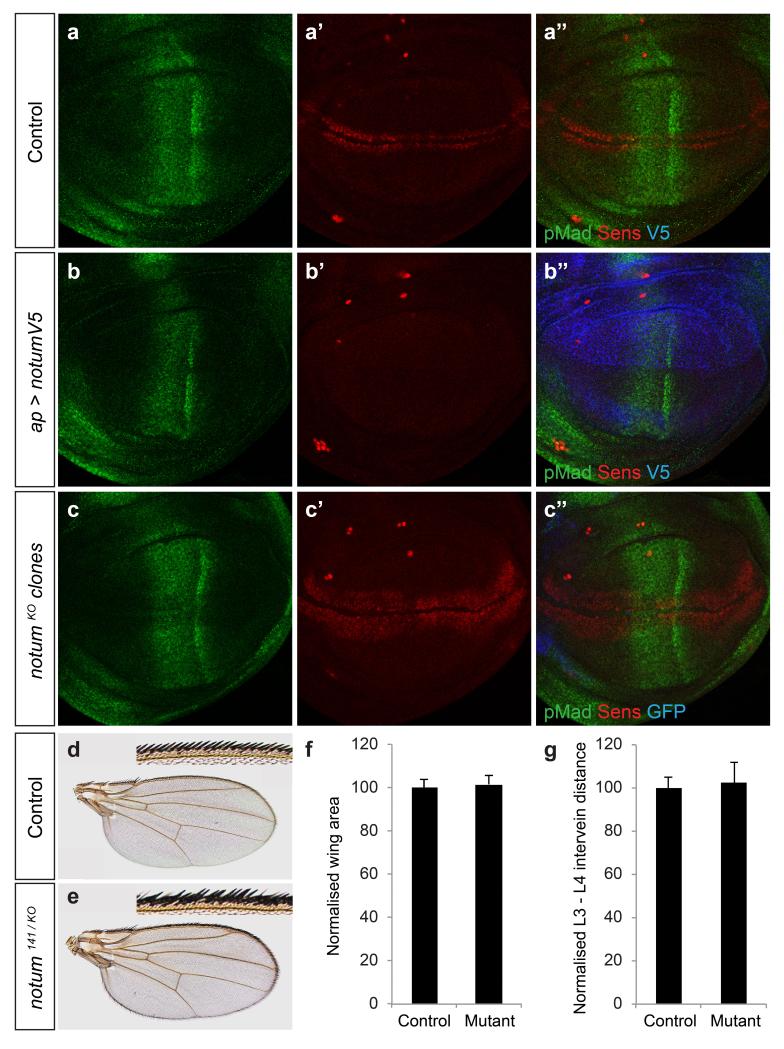
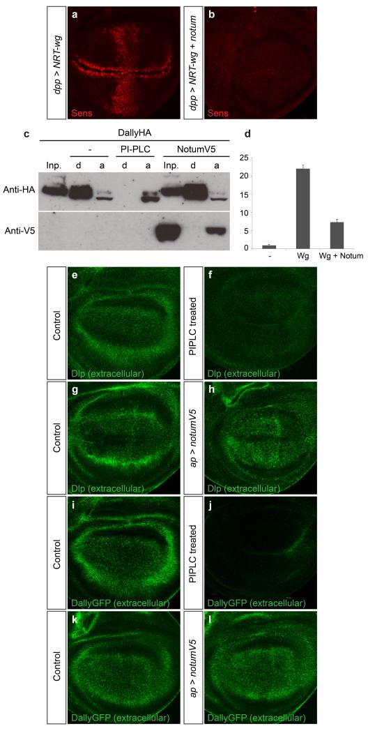
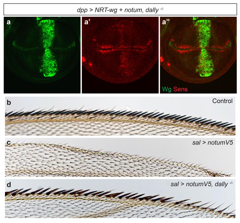
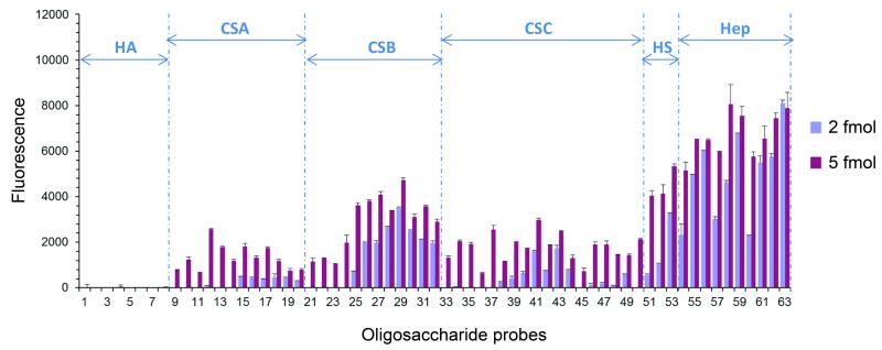
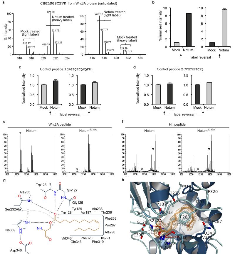
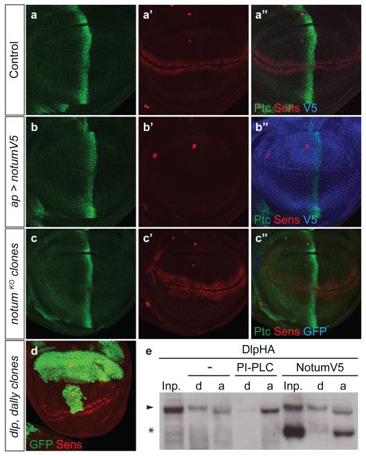
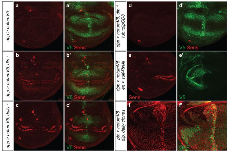
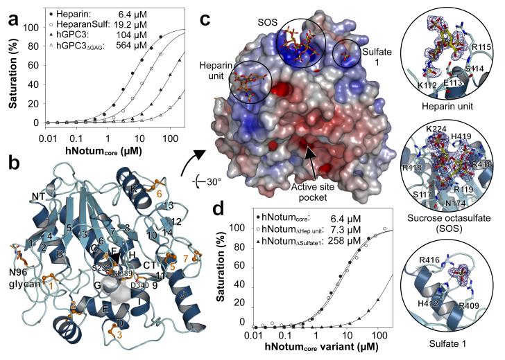
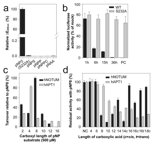
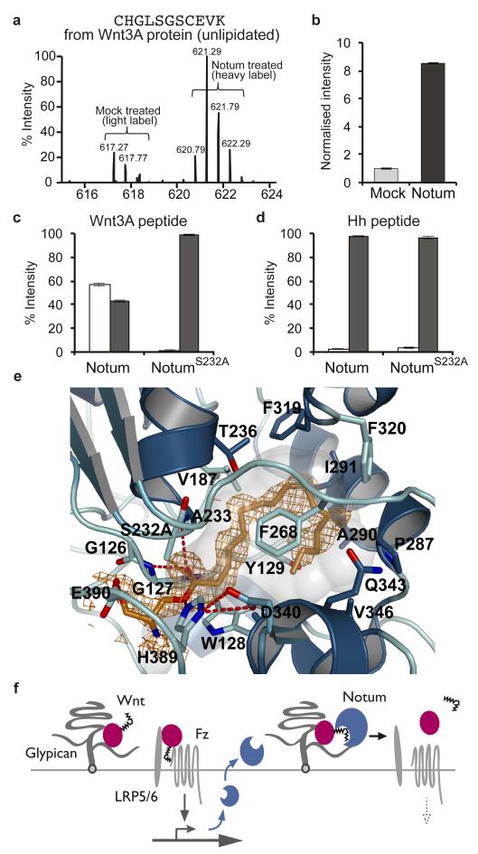
Signalling by Wnt proteins is finely balanced to ensure normal development and tissue homeostasis while avoiding diseases such as cancer. This is achieved in part by Notum, a highly conserved secreted feedback antagonist. Notum has been thought to act as a phospholipase, shedding glypicans and associated Wnt proteins from the cell surface. However, this view fails to explain specificity, as glypicans bind many extracellular ligands. Here we provide genetic evidence in Drosophila that Notum requires glypicans to suppress Wnt signalling, but does not cleave their glycophosphatidylinositol anchor. Structural analyses reveal glycosaminoglycan binding sites on Notum, which probably help Notum to co-localize with Wnt proteins. They also identify, at the active site of human and Drosophila Notum, a large hydrophobic pocket that accommodates palmitoleate. Kinetic and mass spectrometric analyses of human proteins show that Notum is a carboxylesterase that removes an essential palmitoleate moiety from Wnt proteins and thus constitutes the first known extracellular protein deacylase.


Figures

Extended data Figure 1
Extended data Figure 1. Notum modulates Wingless, but not Dpp or Hedgehog signalling
a-c, Overexpression of dNotum-V5 with the apterous-gal4 driver, which is expressed in the dorsal compartment, prevents expression of Senseless (Sens) (b’), a Wingless target gene but has no significant impact on phospho-Mad (pMad) immunoreactivity (b), an indicator of Dpp signalling. Loss of notum activity, achieved by generating large patches of notumKO tissue (See Methods), marked by the loss of GFP, leads to broadening of Senseless expression but does not affect pMad immunoreactivity. d-g, Strong, but not complete, reduction of notum activity led to ectopic wing margin bristles (compare inset in d and e) but had no significant impact on wing area, which is sensitive to Dpp signalling (f) (p = 0.26, Student’s t-Test), or on the distance between L3 and L4 veins, which is affected by changes in Hedgehog signalling (g) (p = 0.41, Student’s t-Test). 19 control (notum141 / +) and 17 mutant (notum141 / KO) wings were analysed.
Extended data Figure 2
Extended data Figure 2. dNotum does not cleave the GPI anchor of glypicans
a, b Ectopic expression of Senseless caused by NRT-Wingless, as well as endogenous Senseless, is suppressed by co-expression of dNotum. NRT-Wingless and dNotum are expressed in a vertical band under the control of dpp-gal4. c, Western blot analysis of phase-separated extracts of S2 cells transfected with a plasmid expressing HA-tagged Dally. In control extracts, Dally is found largely in the detergent (d) phase. Coexpression of dNotum-V5 from a plasmid had no impact, while treatment with PIPLC shifted all detectable Dally to the aqueous (a) phase. d, dNotum-V5 expression as in panel c was sufficient to suppress Wingless-induced TOPFlash activity. Cells were transfected with a dual luciferase TOPFlash reporter along with a mock plasmid (−), tubulin::wingless (Wg), or tubulin::wingless + actin::notum-V5 (Wg + Notum). e-h, Extracellular Dally-like protein (Dlp) in control (e, g) , PIPLC-treated (f) or apterous-Gal4 UAS-notum-V5 (h) imaginal discs. i-l, Extracellular anti-GFP staining of imaginal discs from gene trap line expressing Dally-GFP fusion protein. Discs treated with a mock solution (i) or PIPLC (j) (same discs as in e and f respectively but here showing Dally protein). In a separate experiment, dNotum was overexpressed with apterous-Gal4 in the Dally-GFP background (l). No change in the distribution of extracellular GFP could be seen compared to that in control discs (k, no apterous-Gal4)
Extended data Figure 3
Extended data Figure 3. dNotum requires Dally to inhibit Wingless signaling
a, Wingless and Senseless expression in a dally−/− wing imaginal disc expressing NRT-wingless and notum under the control of dpp-Gal4. Some senseless expression remains, indicating that, in the absence of Dally, dNotum is a poor inhibitor of NRTWingless-induced (as well as endogenous) signalling. b-d, Anterior margin of wings from control, spalt (sal)-Gal4 UAS-notum-V5, and sal-Gal4 UAS-notum-V5 dally−/− animals. Removal of dally rescues the loss of margin bristles caused by dNotum overexpression.
Extended Data Figure 4
Extended Data Figure 4. dNotum binds to sulfated glycans
Binding of dNotum-V5 to a GAG oligosaccharide array, detected by immunofluorescence. HA = Hyaluronic acid, CSA/B/C = Chondroitin Sulfate A/B/C, HS = Heparan Sulfate, Hep = Heparin. Details on the array are provided in Methods.
Extended Data Figure 5
Extended Data Figure 5. Additional structural information on Notum
a, Topology plot of hNotum. β-strands are shown as numbered triangles and α-helices as circles labelled in alphabetical order from the N to C terminus. Structural elements conserved among most α/β-hydrolases are outlined in grey. b, Comparison of the two most conformationally distinct hNotum structures (from crystal forms III and V). Crystal form III is the most structurally different. All other structures superimpose with r.m.s.d.s of <0.7Å. The circle highlights the most flexible region. c, Comparison between the structures of hNotum (Form V) and dNotum (Form I). The circle highlights the lack of a cysteine bridge in dNotum.
Extended Data Figure 6
Extended Data Figure 6. Structural and biophysical analysis of heparin binding
a, Heparin affinity chromatography of wild type hNotum and selected surface variants. b-e, Close-up views of additional sulfate binding sites on hNotum, crystal form III. Each view is accompanied with SPR heparin affinity data corresponding to each hNotum variant.
Extended Data Figure 7
Extended Data Figure 7. Relation of Notum to other Esterases of the α/β hydrolase family
a, Comparison between hNotum and human Esterase D, showing structural relatedness. hNotum is also related to APT1, a cytosolic esterase used in this study as a positive control for fatty acid esterase activity. In the views shown here, the hNotum structure has been rotated by 90° around the x-axis relative to the structure shown in Figure 3b. b, Rootless phylogenetic tree of animal Notum proteins (red) and plant pectinacetylesterases (PAE, green). Extent of sequence identity to hNotum is shown next to species name. Percentages between branches indicate sequence identity between neighbours.
Extended Data Figure 8
Extended Data Figure 8. Substrates and inhibitors of hNotum
a, Inhibition of hNotum activity on pNP-butyrate (pNP4) by PMSF (30 min preincubation with 2 mM) as well as by Triton X-100 and CHAPS (0.5%). Presence of 20mM sucrose octasulfate (SOS) and 50mg/l Heparin results in a minor increase of esterase activity. Values represent each the activity relative to the mean of four control samples lacking the additives. b, Saturable inhibition of hNotum by Triton X-100. Triton X-100 inhibits many esterases due to binding to the acyl binding pocket through its hydrophobic group. c, Lack of inhibition of Norrin-mediated β-catenin stabilization by Notum. Recombinant Norrin was pretreated with hNotumcore at a concentration sufficient to suppress Wnt3a-mediated signalling. d, e, Saturation kinetics of hNotum’s action on pNP-octanoate (pNP8, d) and pNP -butyrate (pNP4, e). The activity was normalized to the Amax calculated for hNotumcore. The activity values for the larger, full length protein were adjusted to compensate for the increased mass. Apparent Km values in (d) were corrected for the inhibition caused by Triton X-100. f, Saturation inhibition kinetics with myristoleic and palmitoleic acid. pNP8 was used at a concentration of 1mM and 250μM, respectively.
Extended Data Figure 9
Extended Data Figure 9. Additional mass spectrometric analysis of hNotum’s deacylase activity
a, Mass spectra of CHGLSGSCEVK from trypsinised Wnt3A protein mock-treated or treated with hNotumcore. Left hand graph is the same as that shown in Fig. 5a, while the right hand side shows the results of a separate experiment performed with the labels reversed. b, Triplicate LC-MS peak areas with label reversal. Irrespective of the nature of the label (grey indicates light label and black, heavy label), hNotumcore triggered an increase in peak area of the delipidated Wnt3A tryptic peptide. c, d, Two control Wnt3A cysteine-containing peptides from the same dataset were not affected by hNotumcore. e, Activity of hNotumcore and its S232A variant on a synthetic disulphide bonded Wnt3A peptide (CHGLSGSCEVK) palmitoleoylated on the first Serine. Both lipidated and unlipidated peptide could be detected by MALDI-TOF. Incubation with hNotumcore, but not its S232A variant, caused significant delipidation (peak corresponding to delipidated peptide is marked by asterisk). Quantification of duplicate such experiments is shown in Fig. 5c. f, MALDI-TOF analysis shows that neither hNotumcore nor its S232A variant delipidated a synthetic Sonic Hedgehog peptide (CGPGRGFGKRR) palmitoylated on its amino terminal Cysteine. Quantification of duplicate such experiments is shown on Fig 5d (peak corresponding to lipidated peptide is marked by black triangle). g, 2D active site schematic relating to Fig. 5e. Additional hydrogen bonds and electron pair movements thought to occur during hydrolysis by the wild type are shown in grey. h, Close-up view on the myristoleate active site complex of hNotumcore (crystal form I). The experimental omit electron density is contoured at 2σ.
Figure 1
Figure 1. Notum specifically inhibits Wnt signalling
a-c, Overexpression of V5-tagged dNotum with the apterous-gal4 driver, which is expressed in the dorsal compartment prevents expression of Senseless (Sens) but not that of Patched (Ptc) (b). Loss of notum activity, achieved by generating large patches of notumKO tissue (See Methods), marked by the loss of GFP, leads to broadening of Senseless expression but does not affect Patched expression. As in all subsequent confocal images, 3rd instar wing imaginal discs are shown with posterior to the right and dorsal up. d, Senseless is expressed seemingly normally in large patches of dlp dally mutant cells (GFP negative) e, Western blot (co-stained with anti-V5 and anti-HA) of phase separated extracts of S2 cells transfected with a plasmid expressing haemagglutinin (HA)-tagged Dlp. In control extracts, Dlp (arrowhead) is found equally in the detergent (d) and aqueous (a) phases. Coexpression of dNotum-V5 (asterisk) had no impact while treatment with PIPLC shifted Dlp to the aqueous phase.
Figure 2
Figure 2. Notum requires the GAGs of glypicans to inhibit Wingless signalling
a-c, Ectopically expressed dNotum-V5 does not suppress Senseless expression in the absence of dlp (b) or dally (c). Although dNotum is only expressed in a vertical band along the A-P boundary, it spreads along the whole A-P axis. d, Ectopic dNotum represses Senseless expression in dlp mutants that express Dlp-CD8 (tubulin promoter). e, Expression of an RNAi transgene against sulfateless in the posterior compartment prevents dNotum-V5 (expressed from dpp-lexA lex-op-notum-V5) from being retained at the cell surface and from suppressing Senseless expression. Wingless signalling is still suppressed in the anterior compartment. f, Accumulation of dNotum-V5 is reduced at the surface of dlp dally double mutant tissue (GFP-negative).
Figure 3
Figure 3. hNotum structure and GAG binding
a, Binding of hNotumcore to immobilized heparin, heparan sulfate (HeparanSulf), hGPC3 or hGPC3ΔGAG, assayed by SPR. b, Structure of hNotum. β-strands are numbered and α-helices are labelled alphabetically from N to C terminus. Disulfides are shown in orange, catalytic triad residues as sticks and the active site pocket shaded grey. N96 is glycosylated (also in dNotum). c, Heparin-mimicking ligands from three different structures are plotted onto a surface representation coloured by electrostatic potential from red (−8kbT/ec) to blue (-8kbT/ec). Close-up views of binding sites are shown on the right with experimental omit electron density contoured at 2.0σ. d, SPR assay measuring hNotumcore variant binding to immobilized heparin. Mutation of the heparin disaccharide binding site (R115S; hNotumΔHep.unit) had little effect while mutations in the sulfate binding site 1 (R409Q H412N R416Q; hNotumΔSulfate1) strongly reduced binding. For SPR (a, d), each data point is the mean result of two replicates.
Figure 4
Figure 4. Enzymatic activity of hNotum
a, Activity of hNotumcore and its S232A variant on p-nitrophenyl (pNP) acetate (pNP2) and activity of hNotumcore on other chromogenic substrates. pNPS = pNP-sulfate (sulfatase substrate); pNPP=pNP-phosphate (phosphatase substrate); pNPPC=pNP-phosphorylcholine (phospholipase C substrate); pNAA=p-Nitroacetanilide (amidase/protease substrate). b, mWnt3A inactivation by hNotum. After the indicated time, hNotumcore or its S232A variant was removed with cobalt affinity beads and residual Wnt3A activity measured with TOPFlash. PC = no hNotum removal. Results are normalised to those from identically treated mock samples. c, Activity of hNotum and hAPT1 on chromogenic p-nitrophenyl ester substrates of different lengths. d, Inhibition of hNotum by various carboxylic acids. pNP8 was used as substrate at a concentration of 1 mM as were the carboxylic acids. c, t: cis or trans C9-C10 double bond. All graphs show the mean +/− s.d. (n=4).
Figure 5
Figure 5. Wnt-deacylation by Notum
a, LC-MS analysis of mWnt3A protein treated with hNotumcore or a mock solution. By comparison to mock treatment (light label), addition of hNotum (heavy label) caused a significant increase in the signal intensity of unlipidated CHGLSGSCEVK b, LC-MS peak areas from panel a.; shown as mean +/− s.e.m. (n=3) c, d, Quantification from MALDI analysis of synthetic lipid-bearing peptides treated with hNotumcore or its S232A variant; shown as mean +/− s.e.m. (n=3). Palmitoleoylated hWnt3A peptide, but not palmitoylated hSonic Hedgehog peptide, was specifically deacylated by the wild type enzyme. e, Close-up view on the seryl-palmitoleate active site complex of hNotum. The experimental omit electron density is contoured at 2σ. f, Feedback control by Notum. Notum deacylates Wnt in a Glypican-assisted fashion.

Comment in

References

    1. Freeman M. Feedback control of intercellular signalling in development. Nature. 2000;408:313–319. - PubMed
    1. Takada R, et al. Monounsaturated fatty acid modification of Wnt protein: its role in Wnt secretion. Dev. Cell. 2006;11:791–801. - PubMed
    1. Janda CY, Waghray D, Levin AM, Thomas C, Garcia KC. Structural basis of Wnt recognition by Frizzled. Science. 2012;337:59–64. - PMC - PubMed
    1. Willert K, et al. Wnt proteins are lipid-modified and can act as stem cell growth factors. Nature. 2003;423:448–452. - PubMed
    1. Tang X, et al. Roles of N-glycosylation and lipidation in Wg secretion and signaling. Dev. Biol. 2012;364:32–41. - PMC - PubMed

Methods references

    1. Beckett K, et al. Drosophila S2 Cells Secrete Wingless on Exosome-Like Vesicles but the Wingless Gradient Forms Independently of Exosomes. Traffic. 2012;14:82–96. - PMC - PubMed
    1. Vincent J-P, Kolahgar G, Gagliardi M, Piddini E. Steep differences in wingless signaling trigger myc-independent competitive cell interactions. Dev. Cell. 2011;21:366–374. - PMC - PubMed
    1. Baena-Lopez LA, Alexandre C, Mitchell A, Pasakarnis L, Vincent JP. Accelerated homologous recombination and subsequent genome modification in Drosophila. Development. 2013;140:4818–4825. - PMC - PubMed
    1. Yagi R, Mayer F, Basler K. Refined LexA transactivators and their use in combination with the Drosophila Gal4 system. Proc Natl Acad Sci USA. 2010;107:16166–16171. - PMC - PubMed
    1. Alexandre C, Baena-Lopez A, Vincent J-P. Patterning and growth control by membrane-tethered Wingless. Nature. 2014;505:180–185. - PMC - PubMed

Extended data reference

    1. Glise B, et al. Shifted, the Drosophila ortholog of Wnt inhibitory factor-1, controls the distribution and movement of Hedgehog. Dev. Cell. 2005;8:255–266. - PubMed

Publication types

MeSH terms