Review
. 2015 Jun 1;48(8):1469-78.
doi: 10.1016/j.jbiomech.2015.02.008. Epub 2015 Feb 9.

Mechanobiology of the meniscus

Affiliations
Review

Mechanobiology of the meniscus

Amy L McNulty et al. J Biomech. .

Abstract

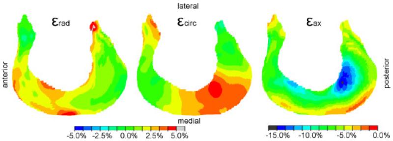
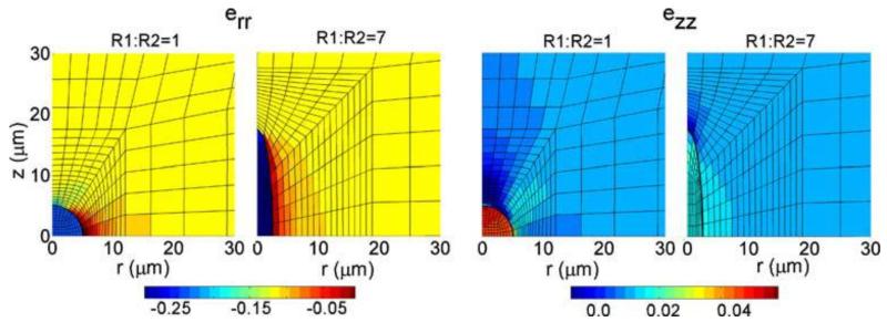
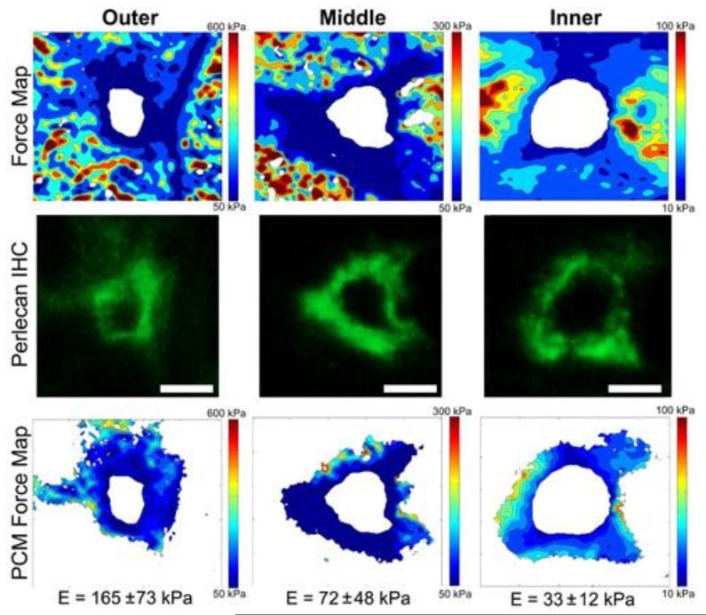
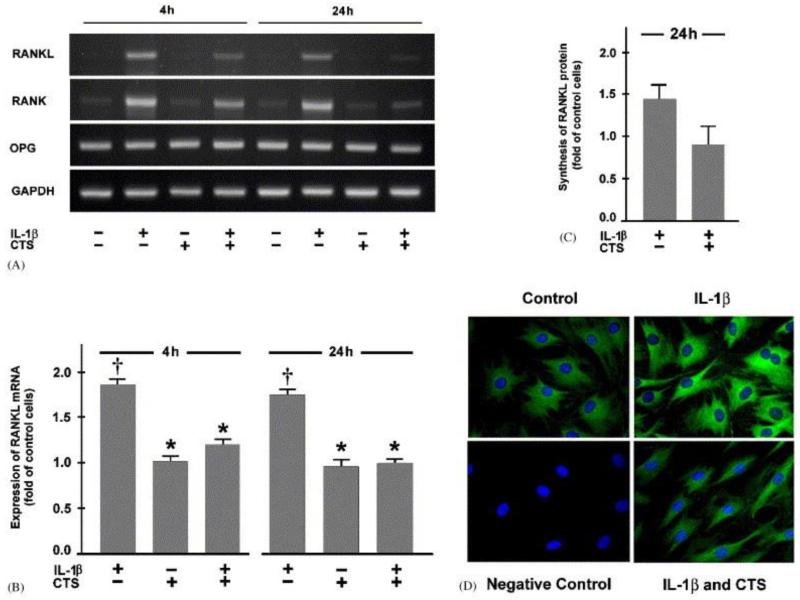
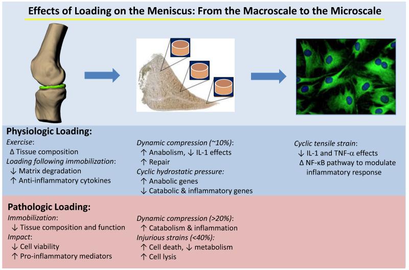
The meniscus plays a critical biomechanical role in the knee, providing load support, joint stability, and congruity. Importantly, growing evidence indicates that the mechanobiologic response of meniscal cells plays a critical role in the physiologic, pathologic, and repair responses of the meniscus. Here we review experimental and theoretical studies that have begun to directly measure the biomechanical effects of joint loading on the meniscus under physiologic and pathologic conditions, showing that the menisci are exposed to high contact stresses, resulting in a complex and nonuniform stress-strain environment within the tissue. By combining microscale measurements of the mechanical properties of meniscal cells and their pericellular and extracellular matrix regions, theoretical and experimental models indicate that the cells in the meniscus are exposed to a complex and inhomogeneous environment of stress, strain, fluid pressure, fluid flow, and a variety of physicochemical factors. Studies across a range of culture systems from isolated cells to tissues have revealed that the biological response of meniscal cells is directly influenced by physical factors, such as tension, compression, and hydrostatic pressure. In addition, these studies have provided new insights into the mechanotransduction mechanisms by which physical signals are converted into metabolic or pro/anti-inflammatory responses. Taken together, these in vivo and in vitro studies show that mechanical factors play an important role in the health, degeneration, and regeneration of the meniscus. A more thorough understanding of the mechanobiologic responses of the meniscus will hopefully lead to therapeutic approaches to prevent degeneration and enhance repair of the meniscus.

Keywords: Articular cartilage; Collagen; Fibrochondrocyte; Mechanical signal transduction; Proteoglycan.


Figures

Figure 1
Figure 1. High-resolution MRI was combined with image registration to measure the displacement and strain of the meniscus and its attachments under compression
Local strain of one representative meniscus and its attachments (view from proximal) in radial (εrad), circumferential (εcirc), and axial (εax) direction under 100% body weight (positive values indicate tension, negative values indicate compression). The meniscus and its attachments showed low average radial or circumferential stretch (less than 1%), but axial strains of nearly 12% during physiologically relevant loading of the knee. Reprinted with permission from (Freutel et al., 2014).
Figure 2
Figure 2. A finite element model of cell-matrix interactions was developed to investigate the mechanical environment of inner and outer meniscus cells with varying geometries
Pseudocolor plots depicting effects of cell geometry on cell mechanics in an isotropic extracellular matrix in response to statically applied biaxial strain (ezz = 0.01, err = −0.1). Shown are the radial (err) and axial (ezz) Eulerian finite strain components at equilibrium (t > 104 s) plotted on spatial coordinates. Compressive transverse strains in meniscus cells of both regions were approximately two to three-fold higher than those in the extracellular matrix and exhibited a modest association with cell aspect ratio. Reprinted with permission from (Upton et al., 2006).
Figure 3
Figure 3. Immunofluorescence-guided atomic force microscopy (AFM) was used to spatially map the mechanical properties of the pericellular matrix (PCM) of the meniscus
Force maps (20 × 20 μm) of pericellular sites within each meniscus region (top row) were integrated with fluorescent perlecan staining (middle row) to define PCM boundaries. The resulting PCM force maps were analyzed to yield an average elastic modulus (bottom row). The elastic modulus of the meniscus pericellular matrix was significantly higher in the outer region than the inner region, whereas extracellular matrix moduli were consistently higher than region-matched pericellular matrices in both the outer and inner regions. Adapted from (Sanchez-Adams et al., 2013) and (Wilusz et al., 2014).
Figure 4
Figure 4. Cyclic tensile strain (CTS) can exhibit an anti-inflammatory effect on meniscal cells
Fibrochondrocytes from meniscus were subjected to CTS at a magnitude of 20% and 0.05 Hz in the presence and absence of 1 ng/mL interleukin (IL)-1β. (A) mRNA expression for RANKL, RANK, and OPG at 4 and 24 h, as determined by semiquantitative RT-PCR. (B) Quantitative assessment of RANKL mRNA at 4 and 24 h, as determined by real-time PCR. Results are shown as means ± S.E.M., n=6, † significantly (p<0.05) different from control cells and stretched cells in the presence or absence of IL-1β. * significantly (p<0.05) different from unstretched IL-1β-treated cells. (C) Protein synthesis of RANKL at 24 h, as analyzed by Western blot and subsequent semiquantitative densitometry. Results are shown as means ± S.E.M., n=3. (D) Protein synthesis of RANK at 24 h, as analyzed by immunofluorescence. IL-1β treatment caused the upregulation of RANK and RANKL but did not alter OPG expression. Conversely, cyclic tensile strain suppressed the IL-1 mediated upregulation of RANK and RANKL. Reprinted with permission from (Deschner et al., 2006).
Figure 5
Figure 5. Dynamic mechanical loading enhances meniscal repair in vitro
Histological images of paraffin embedded meniscal repair model explants stained with Hematoxylin to identify the cell nuclei (black), fast green (green) to identify the collagen fibers, and Safranin O (red) to stain proteoglycans. Samples were incubated with no IL-1 or 100 pg/mL IL-1 and subjected to 0%, 1%, 10%, or 26% strain. IL-1 treatment prevented tissue repair and resulted in loss of proteoglycan staining in the meniscal extracellular matrix, whereas dynamic compression increased interfacial tissue repair in meniscal explants treated with IL-1. Scale bar is equal to 100 μm. Reprinted from (McNulty et al., 2010).
Figure 6
Figure 6. Macroscale to microscale studies have elucidated the effects of loading on the meniscus
Numerous studies have assessed the effects of physiologic and pathologic loading regimes on the meniscus at a range of different scales, including joint level studies, explant studies, and experiments using isolated meniscal cells. The meniscal cell image is reprinted with permission from (Deschner et al., 2006).

References

    1. Agarwal S, Long P, Gassner R, Piesco NP, Buckley MJ. Cyclic tensile strain suppresses catabolic effects of interleukin-1beta in fibrochondrocytes from the temporomandibular joint. Arthritis Rheum. 2001;44:608–617. - PMC - PubMed
    1. Anderson DR, Gershuni DH, Nakhostine M, Danzig LA. The effects of non-weight-bearing and limited motion on the tensile properties of the meniscus. Arthroscopy. 1993;9:440–445. - PubMed
    1. Andersson-Molina H, Karlsson H, Rockborn P. Arthroscopic partial and total meniscectomy: A long-term follow-up study with matched controls. Arthroscopy. 2002;18:183–189. - PubMed
    1. Arnoczky SP, Warren RF. The microvasculature of the meniscus and its response to injury. An experimental study in the dog. Am. J. Sports Med. 1983;11:131–141. - PubMed
    1. AufderHeide AC, Athanasiou KA. Mechanical stimulation toward tissue engineering of the knee meniscus. Ann. Biomed. Eng. 2004;32:1161–1174. - PubMed

Publication types

LinkOut - more resources