

Review
doi: 10.1101/cshperspect.a008268.
Blood and lymphatic vessel formation
Affiliations
- PMID: 25731762
- PMCID: PMC4355271
- DOI: 10.1101/cshperspect.a008268
Item in Clipboard
Review
Blood and lymphatic vessel formation
Cold Spring Harb Perspect Biol.
.
Abstract
Blood and lymphatic vessels deliver oxygen and nutrients, remove waste and CO2, and regulate interstitial pressure in tissues and organs. These vessels begin life early in embryogenesis using transcription factors and signaling pathways that regulate differentiation, morphogenesis, and proliferation. Here we describe how these vessels develop in the mouse embryo, and the signals that are important to their development.
Copyright © 2015 Cold Spring Harbor Laboratory Press; all rights reserved.
Figures
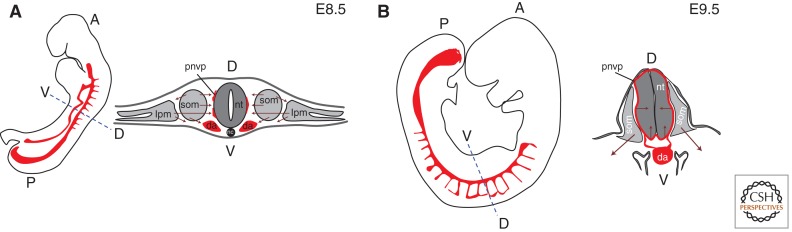
Blood vessel development in early postimplantation mouse embryos. (A) At E8.5, angioblasts and endothelial cells migrate medially and ventrally from lateral plate mesoderm and somites, and laterally from somites. (B) At E9.5, angiogenic sprouting from the perineural vascular plexus (pnvp) vascularizes the neural tube. Embryo silhouettes show pattern of trunk and intersomitic vessels in red (heart and head vessels are excluded); maroon arrows indicate migratory streams or direction of sprouting; black line denotes pnvp; da, dorsal aorta; lpm, lateral plate mesoderm; nc, notochord; nt, neural tube; som, somite; A, anterior; P, posterior; D, dorsal; V, ventral.
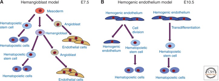
Relationships of vascular and hematopoietic lineages. (A) The hemangioblast is the hypothesized bipotential precursor of some endothelial and hematopoietic cells. (B) An alternative model has differentiated endothelial cells dividing or transdifferentiating to produce hematopoietic stem cells.
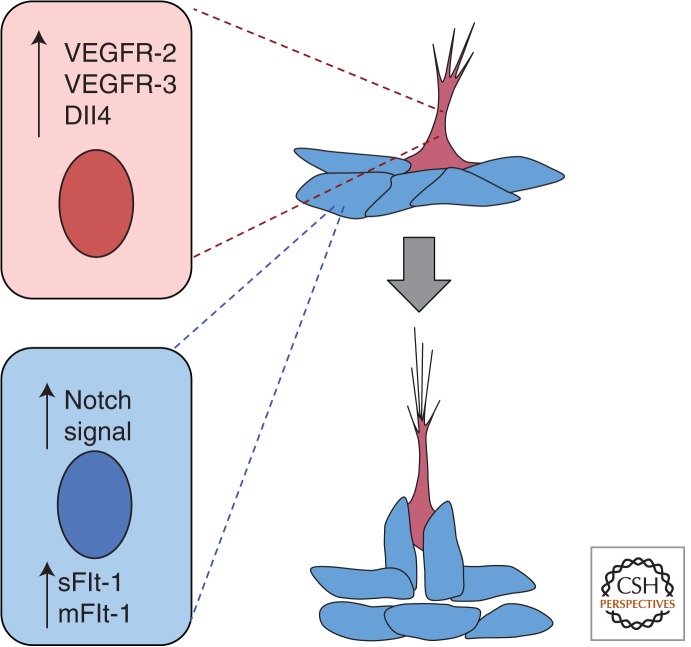
Blood vessel sprouting and lumen formation. As vessels initiate sprouting, a cell (pink) adopts a tip cell phenotype, and through signaling of the Notch ligand Dll4 to neighboring cells sets up the stalk cell phenotype (blue), which is accompanied by up-regulation of Flt-1. As the sprout extends, the lumen forms behind the tip cell. sFlt-1, soluble Flt-1; mFlt-1, membrane Flt-1.
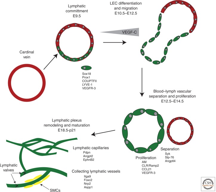
Stages of lymphangiogenesis. Lymphatic endothelial cells (LECs) are derived from venous endothelial precursors during development. The expression of lymphatic-specific receptors, like VEGFR-3, allows for LEC to migrate and separate from veins in response to chemotactic guidance cues. Following the establishment of lymphatic sacs, a cohort of growth factors drives the proliferation and remodeling of the early lymphatic plexus from mid-gestation through the early postnatal period. The maturation of large collecting lymphatic vessels, which include valves and lymphatic smooth muscle cells, also occurs during the late gestation and postnatal periods. AM, adrenomedulin; CLR/Ramp2, calcitonin-like receptor/receptor activity modifying protein-2.
References
-
- Ambler CA, Nowicki JL, Burke AC, Bautch VL 2001. Assembly of trunk and limb blood vessels involves extensive migration and vasculogenesis of somite-derived angioblasts. Dev Biol 234: 352–364. - PubMed
-
- Bahram F, Claesson-Welsh L 2010. VEGF-mediated signal transduction in lymphatic endothelial cells. Pathophysiology 17: 253–261. - PubMed
-
- Bautch VL 2011. Stem cells and the vasculature. Nat Med 17: 1437–1443. - PubMed
Publication types
MeSH terms
Grants and funding
LinkOut - more resources
Full Text Sources
Other Literature Sources