Review
. 2015 Mar 2;7(3):a019406.
doi: 10.1101/cshperspect.a019406.

Dosage compensation in mammals

Affiliations
Review

Dosage compensation in mammals

Neil Brockdorff et al. Cold Spring Harb Perspect Biol. .

Abstract

Many organisms show major chromosomal differences between sexes. In mammals, females have two copies of a large, gene-rich chromosome, the X, whereas males have one X and a small, gene-poor Y. The imbalance in expression of several hundred genes is lethal if not dealt with by dosage compensation. The male-female difference is addressed by silencing of genes on one female X early in development. However, both males and females now have only one active X chromosome. This is compensated by twofold up-regulation of genes on the active X. This complex system continues to provide important insights into mechanisms of epigenetic regulation.


Figures

Figure 1.
Figure 1.
Evolution of the Y chromosome. Early in evolution, the two sexes may have differed at only a single, autosomal locus (marked by a black box); one sex is homozygous at this locus (female) and the other sex (male) is heterozygous (designated proto-male). The “male-determining allele” is shown in yellow. If mating requires one member of each sex, then individuals homozygous for the male-determining allele cannot arise. At this early stage, physiological differences between the sexes will be subtle, comparable to those that distinguish the two mating types in yeast. To prevent the formation of intersex states, crossing-over will be suppressed within and around the male-determining locus (dark shading). Mutations, including deletions and inversions, will accumulate and cause the degenerate region in which crossing-over is suppressed to gradually expand (“Muller’s ratchet”) until the chromosome has lost most of its active, functional genes. (Mutations accumulate because suppression of crossing-over reduces the probability that they will occur in homozygous form, hence, reducing the selection pressure against them.) A small, active region must remain that is homologous to the X chromosome to allow pairing and crossing-over at meiosis (indicated by a gray ×). This is the pseudoautosomal region (PAR). The autosome, originally homologous to the future X (A in the diagram), will itself evolve sometimes through translocations from other chromosomes (shown as red shaded areas), eventually forming the distinctive X chromosome. The X, like other chromosomes, is a mosaic of DNA fragments put in place at different periods through evolution; some of these are ancient and some are relatively recent. On the human X, the more recent arrivals are enriched in genes that escape X inactivation.
Figure 2.
Figure 2.
The effects of progressive loss of X-linked alleles on a hypothetical signaling pathway. The diagram shows successive components of the pathway as colored discs, each put in place by the actions of enzymes a, b, c, and d. The size of the discs is proportional to the amount of each component (level 1). The model proposes that enzymes a, b, and c are all encoded by genes on the X chromosome, and early in evolution were encoded by genes on the two proto-X chromosomes. Alleles are lost as the proto-X progressively degenerates, eventually forming the gene-poor Y chromosome (Fig. 1). A twofold reduction in the amount of an enzyme is likely to cause a reduction in its product, although not necessarily twofold. The cell’s normal homeostatic mechanisms are likely to correct small disturbances and there is likely to be little or no effect on subsequent steps in the pathway (level 2). Even the loss of two enzymes may be corrected with no physiological effect (level 3). However, a stage will eventually be reached when the cumulative effects of enzyme (gene) depletion cause key components to decrease below a critical level and trigger an effect on phenotype (shown here on level 4). Selection pressure will be exerted to correct the phenotypic effect, most readily by up-regulating expression of one or more of the remaining, single alleles of enzymes a, b, or c.
Figure 3.
Figure 3.
Shift in median expression caused by selective up-regulation of X-linked genes. Measurement of the expression of large numbers of genes on either the X chromosome (blue) or autosomes (red) by microarrays or RNA sequencing shows that transcript levels are normally distributed with a range of expression levels spread over several orders of magnitude. If expression of genes on the single male X (or the single active X in females) is not compensated, then the X:autosome ratio of median expression levels should be 0.5, reflecting the difference in (active) copies (left panel). Alternatively, if expression of X-linked genes is up-regulated twofold to compensate for dosage differences, then the ratio of medians should be close to 1.0 (right panel).
Figure 4.
Figure 4.
The cycle of X inactivation and reactivation. The X chromosome undergoes a cycle of X inactivation and X reactivation during development. Red arrows indicate X inactivation steps and green arrows indicate X reactivation steps. Inactivation first occurs in early preimplantation embryos (imprinted X inactivation) and subsequently in cells of the epiblast at the time of gastrulation (random X inactivation). The inactive X is reactivated in ICM cells when they are first allocated at the blastocyst stage, and also in the developing germ cells.
Figure 5.
Figure 5.
Progressive chromosome-wide heterochromatinization induced by Xist RNA. (A) When the Xist gene is expressed, the RNA binds to and coats the X chromosome from which it is transcribed (green dashed line). Xist RNA is thought to trigger silencing of the chromosome by recruiting chromatin modifying activities (red and yellow circles). The initial wave of silencing, in turn, leads to recruitment of additional layers of epigenetic modification (white circles), further stabilizing the heterochromatic structure. Establishment of these different levels of epigenetic silencing is achieved in a stepwise manner through development and ontogeny. (B) Localization of Xist RNA along the X chromosomes is shown by in situ hybridization in both interphase and metaphase.
Figure 6.
Figure 6.
Xist gene regulation in early development. The figure illustrates current knowledge and models for imprinted and random Xist regulation in early XX mouse embryos. The Xm Xist allele arrives in the zygote with a repressive imprint possibly mediated through the antisense Tsix locus (black square). The Xp Xist allele is primed to be active and is expressed as soon as embryonic gene activation occurs at the two-cell stage. From the two- to four-cell stage up until morula stage, Xp Xist is expressed in all cells (expression indicated by open rectangle and arrow at 5′ end). This pattern is maintained at the early blastocyst stage and subsequently in TE and PE cells and their fully differentiated derivative (extraembryonic) tissues. In the late blastocyst, ICM Xist expression is extinguished, possibly by an ICM-specific repressor factor (blue triangle). Xist expression then commences subsequently at the time of gastrulation. Here, the blocking factor (black diamond) ensures that Xist expression cannot occur on one of the two alleles (counting).
Figure 7.
Figure 7.
Models for the regulation of random X inactivation. (A) Autosomally encoded blocking factor (yellow shapes) is produced in sufficient quantities to occupy a single Xic. Binding of blocking factor to the Xic inhibits Xist transcription, thus defining a single active X chromosome. Xist transcription occurs on any additional X chromosomes leading to X inactivation (dark green dots). Blocking factor binds to either the maternal (Xm) or paternal (Xp) X chromosome with equal probability and in a cell-autonomous manner. (B) The two-factor model invokes an X-encoded competence factor (purple triangle) and an autosomally encoded blocking factor (yellow shapes). Blocking factor titrates away competence factor (purple triangle). In cells with a single X chromosome, there is insufficient available competence factor to activate Xist, but in cells with additional X chromosomes, competence factor can activate all X chromosomes except the single X chromosome bound by blocking factor. (C) The stochastic model invokes that autosomally encoded repressors (yellow circles) and X-encoded activators (purple circles) compete with one another. All Xist alleles have an equal probability of being activated and this is increased in cells with more than one X chromosome (higher levels of Xist activators). By chance, some cells with two X chromosomes will initiate inactivation of either both or no X chromosomes. This may be dealt with by checkpoint mechanisms or cell death.
Figure 8.
Figure 8.
Genes and regulatory elements in the X inactivation-center region. The key region on the mouse X chromosome comprising known elements involved in Xist gene regulation is illustrated, showing noncoding RNA (ncRNA) genes and protein-coding genes. The Xpr region, several hundred kilobases upstream of Xist, has been implicated in trans-interaction of Xic alleles in XX cells and as such is thought to be important for initiation of X inactivation. The expanded view illustrates the intron/exon structure of the Xist and Tsix loci, including the Xite elements that function as Tsix enhancers. The network of protein factors (boxes) and ncRNAs (ovals) implicated in Xist gene regulation is shown with arrows and bars indicating repressor and activator function, respectively. Note that RNF12 mediates degradation of REX1, which functions both as a Xist repressor and a Tsix activator.
Figure 9.
Figure 9.
Factors involved in X inactivation spread. (A) The organization of LINE-1-rich (gray-shaded) and gene-rich (blue-shaded) domains is important in defining the extent of Xist RNA spreading as illustrated by the observation that large gene-rich domains attenuate Xist RNA spreading (barred arrows). YY1 is implicated in tethering Xist RNA (green wavy line) at the site of synthesis (brown circle) and interacting with Xist RNA to facilitate spreading (red circle). hnRNPU/SAFA also plays a role in localization of Xist RNA in cis, binding to Xist RNA directly. (B) Summary of characterized DNA methylation, histone modification, and histone changes at a silent gene on Xi.
Figure 10.
Figure 10.
The order of events in differentiating XX ES cells. The diagram summarizes the order in which different silencing pathways are integrated during establishment of X inactivation in differentiating XX ES cells. Early events, depletion of RNA Pol II, loss of H3K4me3/H3K9Ac, and deposition of Polycomb-associated modifications occur coincident with the onset of Xist RNA expression. H4 hypoacetylation and a transition to late replication in S-phase occur slightly later. Enrichment for macroH2A, Smchd1 Ash2l, and hnRNPU/SAF-A occur in a defined temporal window relatively late in the differentiation time course. Accumulation of DNA methylation over CpG island occurs slowly following recruitment of Smchd1, although a subset of CpG islands acquire DNA methylation more rapidly and in a Smchd1-independent manner.
Figure 11.
Figure 11.
Factors involved in Xist-mediated silencing. Depiction of changes in higher-order chromosome architecture during the establishment of X inactivation. Xist RNA initially coats repeat rich chromosomal domains; genes and other regulatory elements occupy an external position. As X inactivation proceeds, genes are internalized within the Xist territory with consequent restriction in the mobility of chromosome loops. Establishment of X inactivation is also linked to positioning of the chromosome on the nuclear and/or nucleolar periphery. Nuclear scaffold factors (SATB1) and chromosome structure factors (Smchd1) may play a role in the reorganization of chromosome architecture on Xi.
Figure 12.
Figure 12.
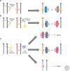
Regulation of X inactivation in cloned mouse embryos. The figure illustrates an XX donor cell with the inactive X chromosome (A) coated with Xist RNA (green line). In this model, transcription from the donor nucleus, including Xist RNA, is repressed by oocyte factors until the two-cell stage, resulting in X reactivation. Recommencement of Xist expression then occurs at the two-cell stage. Xist is then reexpressed again from the inactive X allele from the donor cell. This would be attributable to retention of a mark such as DNA methylation at the Xist promoter. This pattern is maintained in cells allocated to the TE and PE lineages, but not in pluripotent epiblast in which Xist expression is again extinguished, leading to a second reactivation event. In the ICM, erasure of the epigenetic marks governing donor Xist expression allows for subsequent random X inactivation in the embryo proper.

References

    1. Agrelo R, Souabni A, Novatchkova M, Haslinger C, Leeb M, Komnenovic V, Kishimoto H, Gresh L, Kohwi-Shigematsu T, Kenner L, et al. 2009. SATB1 defines the developmental context for gene silencing by Xist in lymphoma and embryonic cells. Dev Cell 16: 507–516. - PMC - PubMed
    1. Allis CD, Jenuwein T, Reinberg D 2014. Overview and concepts. Cold Spring Harb Perspect Biol 10.1101/cshperspect.a018739. - DOI
    1. Augui S, Filion GJ, Huart S, Nora E, Guggiari M, Maresca M, Stewart AF, Heard E 2007. Sensing X chromosome pairs before X inactivation via a novel X-pairing region of the Xic. Science 318: 1632–1636. - PubMed
    1. Bao S, Tang F, Li X, Hayashi K, Gillich A, Lao K, Surani MA 2009. Epigenetic reversion of post-implantation epiblast to pluripotent embryonic stem cells. Nature 461: 1292–1295. - PMC - PubMed
    1. Barlow DP, Bartolomei MS 2014. Genomic imprinting in mammals. Cold Spring Harb Perspect Biol 6: 018382. - PMC - PubMed

LinkOut - more resources