. 2015 Jun;138(Pt 6):1722-37.
doi: 10.1093/brain/awv024. Epub 2015 Mar 1.

Neuronal amyloid-β accumulation within cholinergic basal forebrain in ageing and Alzheimer's disease

Affiliations

Neuronal amyloid-β accumulation within cholinergic basal forebrain in ageing and Alzheimer's disease

Alaina Baker-Nigh et al. Brain. 2015 Jun.

Abstract

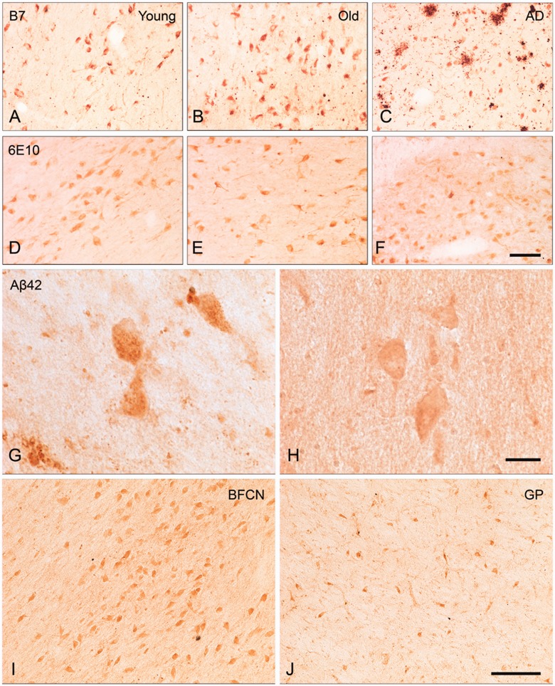
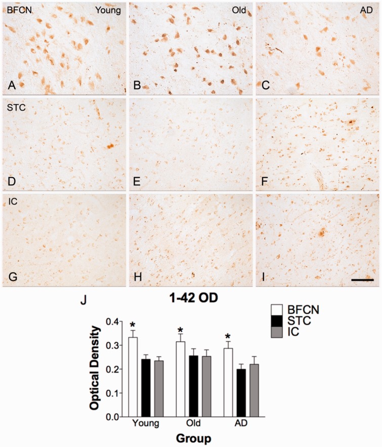
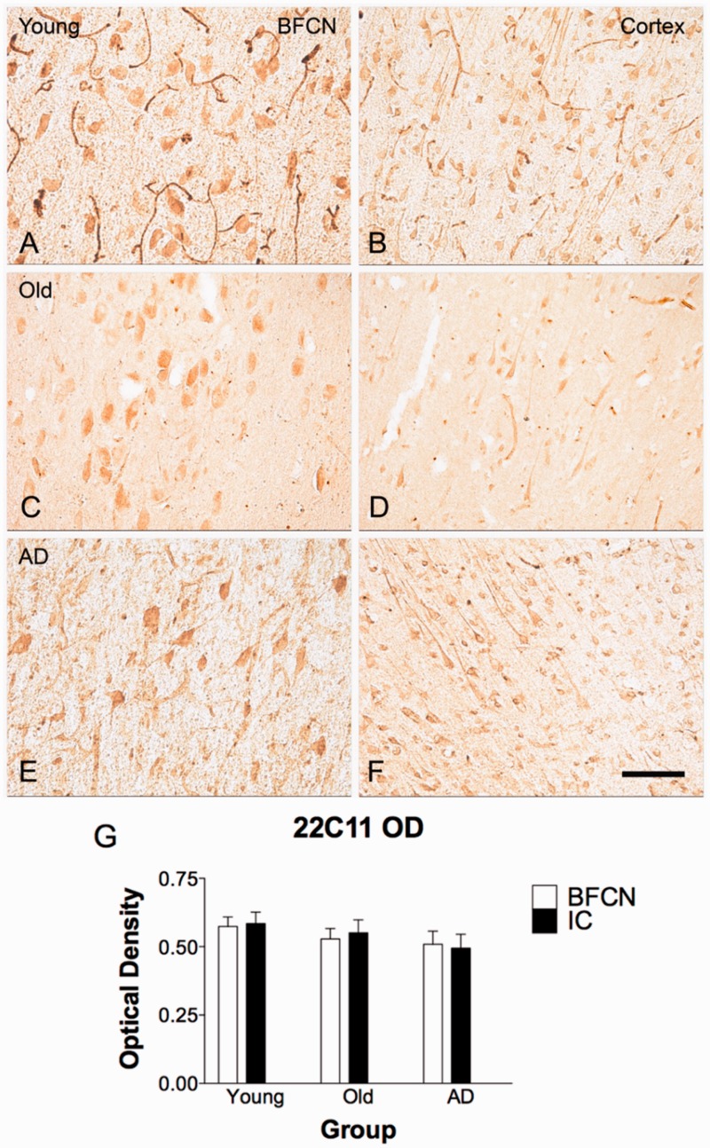
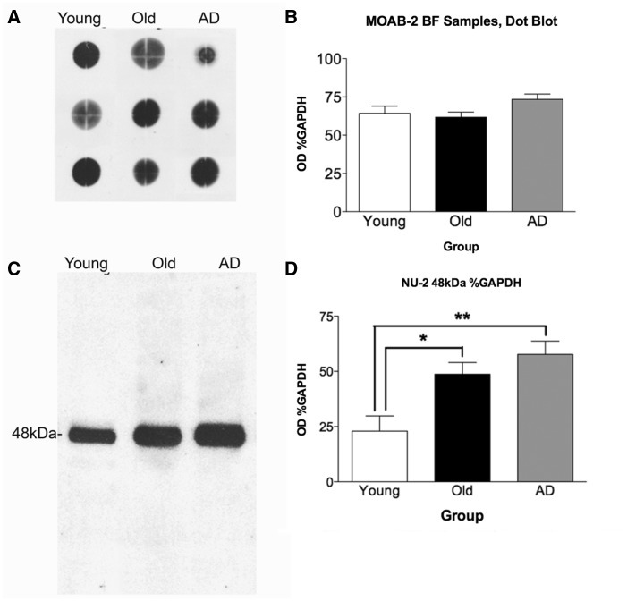
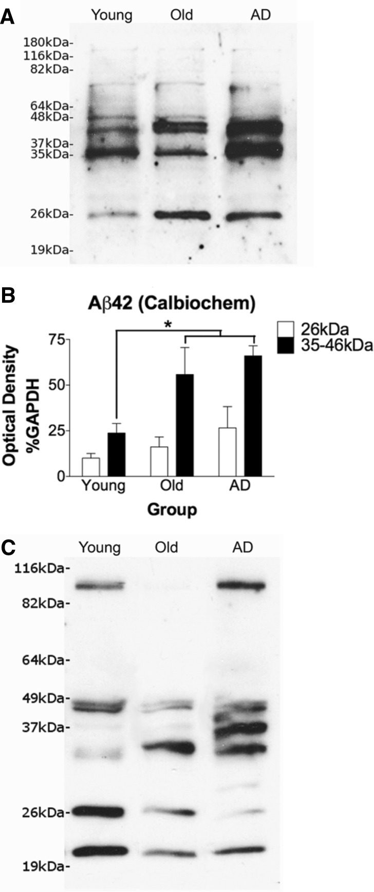
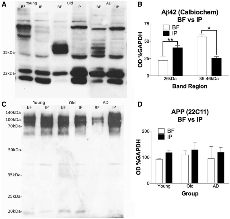
The mechanisms that contribute to selective vulnerability of the magnocellular basal forebrain cholinergic neurons in neurodegenerative diseases, such as Alzheimer's disease, are not fully understood. Because age is the primary risk factor for Alzheimer's disease, mechanisms of interest must include age-related alterations in protein expression, cell type-specific markers and pathology. The present study explored the extent and characteristics of intraneuronal amyloid-β accumulation, particularly of the fibrillogenic 42-amino acid isoform, within basal forebrain cholinergic neurons in normal young, normal aged and Alzheimer's disease brains as a potential contributor to the selective vulnerability of these neurons using immunohistochemistry and western blot analysis. Amyloid-β1-42 immunoreactivity was observed in the entire cholinergic neuronal population regardless of age or Alzheimer's disease diagnosis. The magnitude of this accumulation as revealed by optical density measures was significantly greater than that in cortical pyramidal neurons, and magnocellular neurons in the globus pallidus did not demonstrate a similar extent of amyloid immunoreactivity. Immunoblot analysis with a panel of amyloid-β antibodies confirmed accumulation of high concentration of amyloid-β in basal forebrain early in adult life. There was no age- or Alzheimer-related alteration in total amyloid-β content within this region. In contrast, an increase in the large molecular weight soluble oligomer species was observed with a highly oligomer-specific antibody in aged and Alzheimer brains when compared with the young. Similarly, intermediate molecular weight oligomeric species displayed an increase in aged and Alzheimer brains when compared with the young using two amyloid-β42 antibodies. Compared to cortical homogenates, small molecular weight oligomeric species were lower and intermediate species were enriched in basal forebrain in ageing and Alzheimer's disease. Regional and age-related differences in accumulation were not the result of alterations in expression of the amyloid precursor protein, as confirmed by both immunostaining and western blot. Our results demonstrate that intraneuronal amyloid-β accumulation is a relatively selective trait of basal forebrain cholinergic neurons early in adult life, and increases in the prevalence of intermediate and large oligomeric assembly states are associated with both ageing and Alzheimer's disease. Selective intraneuronal amyloid-β accumulation in adult life and oligomerization during the ageing process are potential contributors to the degeneration of basal forebrain cholinergic neurons in Alzheimer's disease.

Keywords: Alzheimer pathology; amyloid oligomer; amyloid-β; basal forebrain cholinergic neurons; intracellular.


Figures

None
Baker-Nigh et al. reveal accumulation of amyloid-β in basal forebrain cholinergic neurons throughout life, and the formation of large intraneuronal oligomers specifically in aged and Alzheimer brains. Accumulation and age-related aggregation of amyloid-β may contribute to the selective vulnerability of cholinergic neurons to degeneration in Alzheimer’s disease.
Figure 1
Figure 1
BFCN immunostaining with B7 and 6E10 antibodies, and amyloid-β42 staining in BFCNs compared with large neurons of globus pallidus. BFCN staining with B7 antibody was present in young (A), old (B), and Alzheimer’s disease (C) cases. No difference was observed between groups by semi-quantitative rating of staining. BFCN staining with 6E10 antibody was observed in young (D), old (E), and Alzheimer’s disease (F) cases. Quantitative analysis of optical density of staining revealed no significant differences in 6E10 immunoreactivity within the BFCNs of the three groups of subjects. Notably, while the region proximal to BFCNs in some Alzheimer’s disease brains contained a few plaques (C), other cases were completely plaque-free (F). BFCN amyloid-β42 staining with the Invitrogen antibody is presented at high magnification, demonstrating granular staining in an Alzheimer's disease case (G) and smooth staining in an old case (H). Calbiochem amyloid-β42 staining was similar between BFCNs and magnocellular globus pallidus (GP) neurons (not shown), whereas with Invitrogen amyloid-β42 staining was occasionally stronger in BFCN (I) than globus pallidus (J), as demonstrated in an old case. Scale bars: A–F and I–J = 100 µm; G and H = 25 µm.
Figure 2
Figure 2
Amyloid-β42 immunostaining in BFCN and cortical neurons. Immunoreactivity for amyloid-β42 is selective to the BFCNs. Superior temporal cortex (STC; D–F) and insular cortex (IC; G-I) from the same case and the same section as BFCNs images (A–C) exhibit minimal neuronal staining with Invitrogen amyloid-β42. (J) Quantitative determination of immunoreactivity for amyloid-β42. In a two-way mixed factorial ANOVA (SPSS), the main effect of region (BFCNs, superior temporal cortex, insular cortex) was significant; F(2,42) = 21.143, P < 0.001, with optical density measures in BFCNs significantly higher than in either cortical region (BFCNs versus insular cortex, P < 0.05, BFCNs versus superior temporal cortex, P < 0.05). Optical density of staining in cortical regions superior temporal cortex and insular cortex did not differ significantly from each other (P > 0.05). For BFCNs and superior temporal cortex, n = 7 young, 10 old, and 8 Alzheimer’s disease cases; for superior temporal cortex, n = 7 young, 10 old, and 7 Alzheimer’s disease cases. Scale bar = 100 µm.
Figure 3
Figure 3
APP immunostaining in BFCNs and cortex. APP is not differentially expressed in BFCNs compared to cortical pyramidal neurons, as indicated by immunoreactivity for 22C11. Pyramidal neurons of the insular cortex from the same section of tissue as BFCNs exhibit equivalent neuronal APP staining as in BFCNs in young (A and B), old (C and D), and Alzheimer (E and F) cases. Quantification of optical density is represented in G. One-way Kruskal-Wallis non-parametric ANOVA (InStat) revealed no differences for 22C11 optical density between groups nor by region; H(2) = 2.841, P = 0.7245, ns. n = 3 young, n = 6 old, and n = 6 Alzheimer’s disease cases. Scale bar = 100 µm. IC = insular cortex.
Figure 4
Figure 4
MOAB-2 dot blot and quantitation and NU-2 48 kDa amyloid-β oligomer western blot and quantitation of basal forebrain homogenates. (A) Representative MOAB-2 dot blot of basal forebrain (BF) samples; n = 6 young, n = 8 old, n = 7 Alzheimer’s disease cases. As observed in immunohistochemistry, there is no difference apparent between groups in total amyloid-β in basal forebrain. Quantitation of optical density of dots (B) confirmed absence of statistically significant difference between groups. The NU-2 antibody is highly specific, revealing a single band of amyloid-β oligomer at 48 kDa (C). One-way ANOVA with Student-Newman-Keuls post hoc test revealed significant difference between young cases compared to old (*P < 0.05) and Alzheimer cases [**P < 0.001; F(2,11) = 8.287, P = 0.0064]. n = 6 young, n = 8 old, and n = 7 Alzheimer’s disease cases (D).
Figure 5
Figure 5
Calbiochem and Invitrogen amyloid-β42 western blots with quantitation of the 26 kDa and 35-46 kDa banding regions. Sample blot for Calbiochem is shown in A with quantitation in B. For the 35–46 kDa band region, one-way ANOVA with Student-Newman-Keuls post hoc tests revealed a significant increase in both old cases and Alzheimer’s disease patients (AD) basal forebrain compared to young cases [P < 0.05 for each comparison; F(2,15) = 5.299, P = 0.0182]. n = 6 young, n = 6 old, and n = 6 Alzheimer cases. No significant differences were seen for the 26 kDa band. A faint band at 48 kDa was too light to assess quantitatively. All quantification was normalized to GAPDH standard. Sample blot of Invitrogen amyloid-β42 (C) shows the same pattern as the Calbiochem amyloid-β42 antibody at the intermediate molecular weight oligomer 35 kDa band region, with an apparent increase in old and Alzheimer cases when compared to the young. However, with this antibody the 26 kDa low molecular weight oligomer band trends toward a decrease.
Figure 6
Figure 6
Basal forebrain and inferoparietal cortex samples in western blot for amyloid-β42 (Calbiochem; 26 kDa and 35–46 kDa) and APP (22C11; 100 kDa). Banding regions of interest with Calbiochem amyloid-β42 were compared between basal forebrain (BF) and inferoparietal cortex (IP) samples (A). Samples across groups were pooled by region for quantitative comparison (B). T-tests revealed that the 26 kDa band was significantly lower in basal forebrain than inferoparietal cortex, t(28) = 3.480, P = 0.0017, two-tailed. The 35-46 kDa collective region was significantly higher in basal forebrain when compared with inferoparietal cortex, t(27) = 2.648, P = 0.0134, two-tailed with Welch’s correction. At 48 kDa no differences were observed (not shown). Basal forebrain samples included n = 6 young, n = 7 old, and n = 6 Alzheimer’s disease (n = 19), whereas inferoparietal cortex samples from the same cases as basal forebrain included n = 5 young, n = 3 old, and n = 3 Alzheimer cases (n = 11). APP expression in basal forebrain is not significantly different between groups or compared to cortex (C). A slight trend towards lower APP expression in basal forebrain compared to cortex is present, but likewise not statistically significant by unpaired Student’s t-test (D). These results demonstrate that APP levels are unchanged across groups in either structure, indicating that observed differences are not due to alterations in APP expression. Basal forebrain samples for n = 4 young, n = 4 old, and n = 3 Alzheimer’s disease cases were compared to inferior parietal cortex homogenate from paired cases (n = 3 young, n = 3 old, and n = 2 Alzheimer’s disease).

Comment in

References

    1. Atwood CS, Obrenovich ME, Liu T, Chan H, Perry G, Smith MA, et al. Amyloid-β: a chameleon walking in two worlds: a review of the trophic and toxic properties of amyloid-β. Brain Res Rev. 2003;43:1–16. - PubMed
    1. Bahr BA, Hoffman KB, Yang AJ, Hess US, Glabe CG, Lynch G. Amyloid β protein is internalized selectively by hippocampal field CA1 and causes neurons to accumulate amyloidogenic carboxyterminal fragments of the amyloid precursor protein. J Comp Neurol. 1998;397:139–47. - PubMed
    1. Bao F, Wicklund L, Lacor PN, Klein WL, Nordberg A, Marutle A. Different β-amyloid oligomer assemblies in alzheimer brains correlate with age of disease onset and impaired cholinergic activity. Neurobiol Aging. 2012;33:825–e1–13. - PubMed
    1. Chromy BA, Nowak RJ, Lambert MP, Viola KL, Chang L, Velasco PT, et al. Self-assembly of Abeta(1-42) into globular neurotoxins. Biochemistry. 2003;42:12749–60. - PubMed
    1. De Felice FG, Velasco PT, Lambert MP, Viola K, Fernandez SJ, Ferreira ST, et al. Abeta oligomers induce neuronal oxidative stress through an N-methyl-D-aspartate receptor-dependent mechanism that is blocked by the Alzheimer drug memantine. J Biol Chem. 2007;282:11590–601. - PubMed

Publication types

MeSH terms