Ictal SPECT in patients with rapid eye movement sleep behaviour disorder
- PMID: 25732183
- PMCID: PMC5963405
- DOI: 10.1093/brain/awv042
Ictal SPECT in patients with rapid eye movement sleep behaviour disorder
Abstract
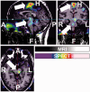
Rapid eye movement sleep behaviour disorder is a rapid eye movement parasomnia clinically characterized by acting out dreams due to disinhibition of muscle tone in rapid eye movement sleep. Up to 80-90% of the patients with rapid eye movement sleep behaviour disorder develop neurodegenerative disorders within 10-15 years after symptom onset. The disorder is reported in 45-60% of all narcoleptic patients. Whether rapid eye movement sleep behaviour disorder is also a predictor for neurodegeneration in narcolepsy is not known. Although the pathophysiology causing the disinhibition of muscle tone in rapid eye movement sleep behaviour disorder has been studied extensively in animals, little is known about the mechanisms in humans. Most of the human data are from imaging or post-mortem studies. Recent studies show altered functional connectivity between substantia nigra and striatum in patients with rapid eye movement sleep behaviour disorder. We were interested to study which regions are activated in rapid eye movement sleep behaviour disorder during actual episodes by performing ictal single photon emission tomography. We studied one patient with idiopathic rapid eye movement sleep behaviour disorder, one with Parkinson's disease and rapid eye movement sleep behaviour disorder, and two patients with narcolepsy and rapid eye movement sleep behaviour disorder. All patients underwent extended video polysomnography. The tracer was injected after at least 10 s of consecutive rapid eye movement sleep and 10 s of disinhibited muscle tone accompanied by movements registered by an experienced sleep technician. Ictal single photon emission tomography displayed the same activation in the bilateral premotor areas, the interhemispheric cleft, the periaqueductal area, the dorsal and ventral pons and the anterior lobe of the cerebellum in all patients. Our study shows that in patients with Parkinson's disease and rapid eye movement sleep behaviour disorder-in contrast to wakefulness-the neural activity generating movement during episodes of rapid eye movement sleep behaviour disorder bypasses the basal ganglia, a mechanism that is shared by patients with idiopathic rapid eye movement sleep behaviour disorder and narcolepsy patients with rapid eye movement sleep behaviour disorder.
Keywords: REM sleep behaviour disorder; basal ganglia; ictal SPECT; muscle atonia; sublaterodorsal nucleus.
© The Author (2015). Published by Oxford University Press on behalf of the Guarantors of Brain. All rights reserved. For Permissions, please email: journals.permissions@oup.com.
Figures


Comment in
-
REM sleep behaviour disorder: a window on the sleeping brain.Brain. 2015 May;138(Pt 5):1131-3. doi: 10.1093/brain/awv058. Epub 2015 Mar 19. Brain. 2015. PMID: 25792529 Free PMC article.
-
Ictal SPECT in patients with rapid eye movement sleep behaviour disorder.Brain. 2015 Nov;138(Pt 11):e390. doi: 10.1093/brain/awv146. Epub 2015 May 29. Brain. 2015. PMID: 26026164 No abstract available.
References
-
- American Academy of Sleep Medicine. The international classification of sleep disorders: diagnostic and coding manual. 2nd edn. Westchester, Illinois: American Academy of Sleep Medicine; 2005.
-
- Asenbaum S, Zeithofer J, Saletu B, Frey R, Brucke T, et al. Technetium-99m-HMPAO SPECT imaging of cerebral blood flow during REM sleep in narcoleptics. J Nucl Med. 1995;36:1150–5. - PubMed
-
- Beitz AJ. Relationship of glutamate and aspartate to the periaqueductal gray-raphe magnus projection: analysis using immunocytochemistry and microdialysis. J Histochem Cytochem. 1990;38:1755–65. - PubMed
-
- Boeve BF, Silber MH, Saper CB, Ferman TJ, Dickson DW, Parisi JE, et al. Pathophysiology of REM sleep behaviour disorder and relevance to neurodegenerative disease. Brain. 2007;130:2770–88. - PubMed
MeSH terms
LinkOut - more resources
Full Text Sources
Other Literature Sources
Medical

