. 2015 Mar 5;16(3):269-74.
doi: 10.1016/j.stem.2015.01.018. Epub 2015 Feb 26.

Successful function of autologous iPSC-derived dopamine neurons following transplantation in a non-human primate model of Parkinson's disease

Affiliations

Successful function of autologous iPSC-derived dopamine neurons following transplantation in a non-human primate model of Parkinson's disease

Penelope J Hallett et al. Cell Stem Cell. .

Abstract

Autologous transplantation of patient-specific induced pluripotent stem cell (iPSC)-derived neurons is a potential clinical approach for treatment of neurological disease. Preclinical demonstration of long-term efficacy, feasibility, and safety of iPSC-derived dopamine neurons in non-human primate models will be an important step in clinical development of cell therapy. Here, we analyzed cynomolgus monkey (CM) iPSC-derived midbrain dopamine neurons for up to 2 years following autologous transplantation in a Parkinson's disease (PD) model. In one animal, with the most successful protocol, we found that unilateral engraftment of CM-iPSCs could provide a gradual onset of functional motor improvement contralateral to the side of dopamine neuron transplantation, and increased motor activity, without a need for immunosuppression. Postmortem analyses demonstrated robust survival of midbrain-like dopaminergic neurons and extensive outgrowth into the transplanted putamen. Our proof of concept findings support further development of autologous iPSC-derived cell transplantation for treatment of PD.


Figures

Figure 1
Figure 1. Functional improvement of PD motor symptoms, increased dopamine reuptake and reinnervation of the transplanted putamen, after autologous transplantation of CM iPSC-derived dopamine neurons
Differentiated CM-iPSCs were transplanted into the right putamen of a cynomolgus monkey with stable, bilateral parkinsonism (MF25-04). A, The animal was followed for 2 years after transplantation. From 6 months post-transplantation, functional improvement was observed as determined by a sustained increase in global daytime (6AM-6PM) activity. B, Fine motor skills in MF25-04 were assessed using a computerized reaching task movement analysis panel (MAP). At 2 years post-transplantation, MAP performance in the left upper limb was significantly improved compared to pre-transplantation values (p<0.05, One-way ANOVA followed by Tukey's Multiple Comparison Test). No change in performance was observed in the right upper limb. Data shown represent averages of 5 repeated tests (baseline), 2 repeated tests (1 year post-transplantation) and 3 repeated tests (2 years post-transplantation). C, Functional analysis of dopamine reuptake was measured by PET neuroimaging for 11C-CFT, a marker of the DAT. Increased 11C-CFT binding was observed in the transplanted putamen at 2 years post-transplantation. White arrows indicate areas of hyperintense CFT PET signal. D, Low power photomicrograph of DAT immunostaining in the transplanted (right, R) and non-transplanted (left, L) putamen shows reinnervation of the transplanted side. Deposits of grafted dopamine neurons are indicated with boxes (g). Internal capsule (IC), lateral globus pallidus (LGP), lateral ventricle (LV), 3rd ventricle (3V), corpus callosum (cc) E, Grafted dopamine neurons were also labeled using tyrosine hydroxylase (TH). The boxed area is shown at higher magnification in the right panel. Robust survival of dopamine neurons with outgrowth integration into the host putamen was observed.
Figure 2
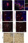
Figure 2. Phenotypes of engrafted CM iPSC-derived neurons at 2 years after autologous transplantation
A, Immunofluorescence staining confirmed that transplanted dopamine neurons were colabeled for FOXA2, TH and βIII tubulin at 2 years after transplantation. B, Labeling for dopamine transporter (DAT), TH, and TOM20 (a mitochondrial outer membrane protein) showed a punctate expression of DAT along the fibers of transplanted dopamine neurons, and a typical localization of mitochondria throughout the cell soma and neurites. C, Immunofluorescence staining for 5-HT demonstrated the presence and localization of serotonergic neurons within the graft. D, Labeling for DARPP-32, a marker of striatal GABAergic medium spiny neurons, shows robust labeling in the host putamen and occasional DARPP-32 - immunoreactive fibers at the graft-host border. E-F, Ki-67 was used to determine whether proliferating cells were present in the CM-iPSC-derived neural cell graft from MF25-04. No Ki-67-immunoreactive cells were observed in the graft (E). As a positive control, several Ki-67-immunoreactive proliferating cells were observed in the hippocampus of the same animal (F) (identified with arrows). G-H, Histological analysis of microglia using Iba1 in the host putamen (G) and graft (H), shows typical resting microglia.

Comment in

References

    1. Barker RA, Barrett J, Mason SL, Bjorklund A. Fetal dopaminergic transplantation trials and the future of neural grafting in Parkinson's disease. Lancet Neurol. 2013;12:84–91. - PubMed
    1. Brownell A-L, Jenkins BG, Elmaleh DR, Deacon TW, Spealman RD, Isacson O. Combined PET/MRS studies of the brain reveal dynamic and long-term physiological changes in a Parkinson's disease primate model. Nature Med. 1998a;4:1308–1312. - PubMed
    1. Brownell AL, Livni E, Galpern W, Isacson O. In vivo PET imaging in rat of dopamine terminals reveals functional neural transplants. Ann Neurol. 1998b;43:387–390. - PubMed
    1. Cooper O, Hargus G, Deleidi M, Blak A, Osborn T, Marlow E, Lee K, Levy A, Perez-Torres E, Yow A, et al. Differentiation of human ES and Parkinson's disease iPS cells into ventral midbrain dopaminergic neurons requires a high activity form of SHH, FGF8a and specific regionalization by retinoic acid. Mol Cell Neurosci. 2010;45:258–266. - PMC - PubMed
    1. Deleidi M, Hargus G, Hallett P, Osborn T, Isacson O. Development of histocompatible primate-induced pluripotent stem cells for neural transplantation. Stem Cells. 2011;29:1052–1063. - PMC - PubMed

Publication types