. 2015 Mar 9;212(3):333-49.
doi: 10.1084/jem.20141702. Epub 2015 Mar 2.

Targeting IL-17B-IL-17RB signaling with an anti-IL-17RB antibody blocks pancreatic cancer metastasis by silencing multiple chemokines

Affiliations

Targeting IL-17B-IL-17RB signaling with an anti-IL-17RB antibody blocks pancreatic cancer metastasis by silencing multiple chemokines

Heng-Hsiung Wu et al. J Exp Med. .

Abstract

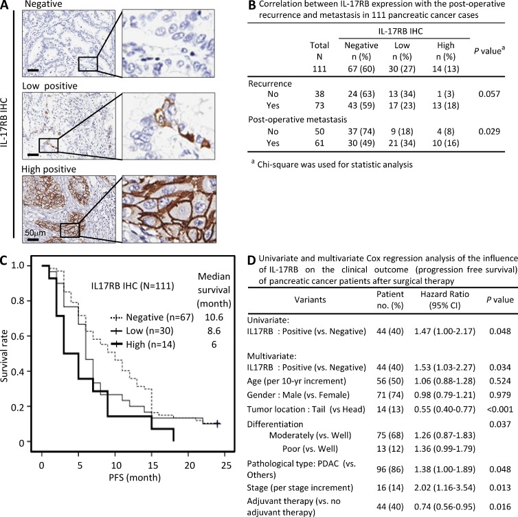
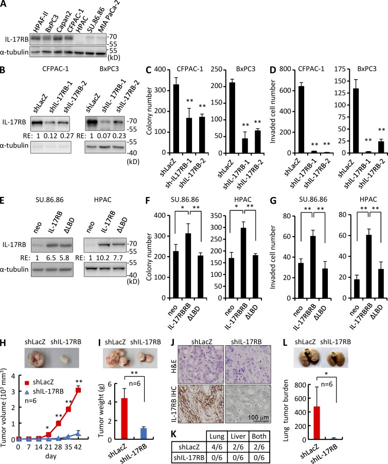
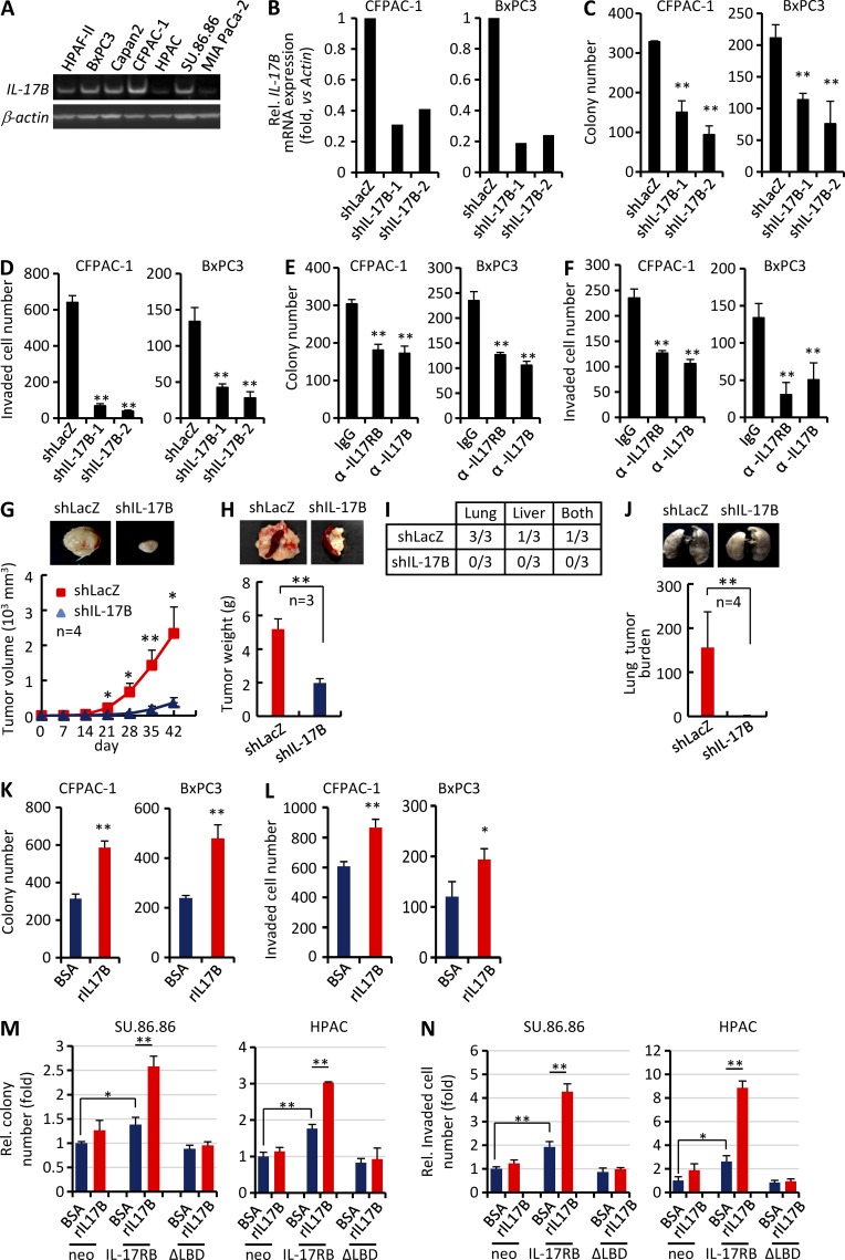
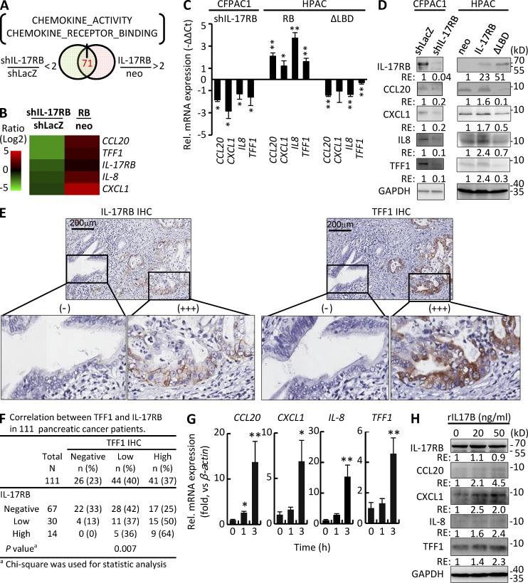
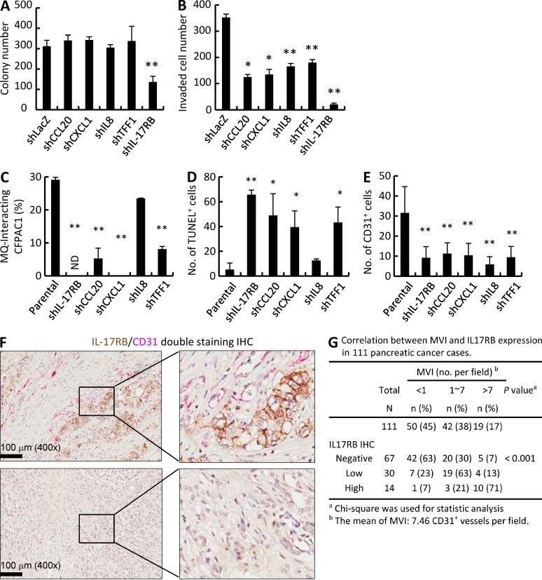
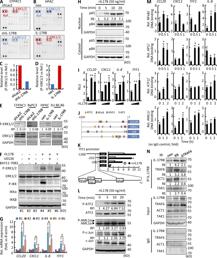
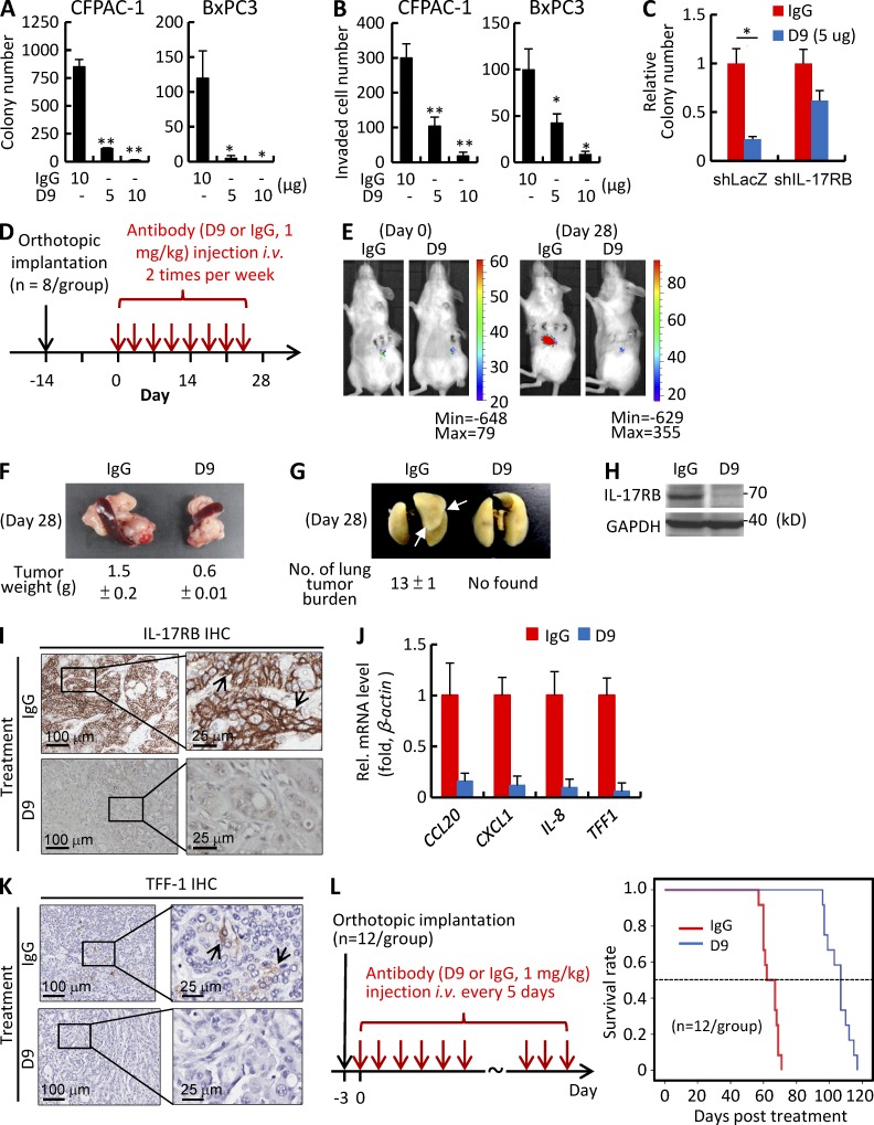
Pancreatic cancer has an extremely high mortality rate due to its aggressive metastatic nature. Resolving the underlying mechanisms will be crucial for treatment. Here, we found that overexpression of IL-17B receptor (IL-17RB) strongly correlated with postoperative metastasis and inversely correlated with progression-free survival in pancreatic cancer patients. Consistently, results from ex vivo experiments further validated that IL-17RB and its ligand, IL-17B, plays an essential role in pancreatic cancer metastasis and malignancy. Signals from IL-17B-IL-17RB activated CCL20/CXCL1/IL-8/TFF1 chemokine expressions via the ERK1/2 pathway to promote cancer cell invasion, macrophage and endothelial cell recruitment at primary sites, and cancer cell survival at distant organs. Treatment with a newly derived monoclonal antibody against IL-17RB blocked tumor metastasis and promoted survival in a mouse xenograft model. These findings not only illustrate a key mechanism underlying the highly aggressive characteristics of pancreatic cancer but also provide a practical approach to tackle this disease.


Figures

Figure 1.
Figure 1.
Overexpression of IL-17RB is associated with metastasis and poor clinical outcome in pancreatic cancer patients. (A) Representative IHC results of patients with IL-17RB–negative (0%), low positive (< 10%), and high positive (>10%) expression in cancer cells. Bar, 50 µm. Boxes show the enlarged area. (B) Correlation of IL-17RB expression in cancer cells and clinical parameters (2 yr after operation) in 111 pancreatic cancer cases. χ2 test was used. (C) Comparison of the progression free survival (PFS) of patients with different levels of IL-17RB expression using the Kaplan-Meier method. P = 0.035. (D) Univariate and multivariate Cox regression analysis of the influence of IL-17RB expression on the PFS of 111 pancreatic cancer patients after surgical therapy.
Figure 2.
Figure 2.
IL-17RB expression has an essential role in tumorigenesis and metastasis of pancreatic tumor cells. (A) Immunoblot analysis of IL-17RB in pancreatic cancer cell lines. α-Tubulin was used as a loading control. (B) IB analysis of IL-17RB in CFPAC-1 and BxPC3 cell lines transduced with lentiviral-control shRNA (shLacZ) or shIL-17RB. Tubulin was used as a loading control. RE, relative expression. (C and D) Soft agar colony formation (SACF) assay (C) and invasion assay (D) using CFPAC-1 and BxPC3 cells infected with shLacZ or shIL-17RB. (E) IB analysis of IL-17RB in control (neo), IL-17RB full-length or ΔLBD overexpressing SU86.86 and HPAC cell lines. Tubulin was used as a loading control. RE, relative expression. (F and G) Assays for soft agar colony formation (SACF; F) and invasion (G) were performed using SU.86.86 and HPAC cells overexpressing full-length or ΔLBD IL-17RB. (H) Tumorigenesis assay of NOD/SCIDγ mice subcutaneously injected with shLacZ-transduced or IL-17RB–depleted CFPAC-1 cells. Cell dose: 1 × 10⁶ cells per mouse. Six mice were used for each group. (I) Tumor weight of NOD/SCIDγ mice orthotopically implanted with shLacZ-transduced or IL-17RB–depleted CFPAC-1 cells. Cell dose: 2.5 × 10⁵ cells per mouse. Six mice were used for each group. (J) IHC of IL-17RB in tumors derived from mice orthotopically implanted with shLacZ-transduced or IL-17RB–depleted CFPAC-1 cells. (K) Summary table of lung and liver metastasis derived from orthotopic xenograft. (L) Lung metastasis of NOD/SCIDγ mice intravenously injected with shLacZ-transduced or IL-17RB–depleted CFPAC-1 cells. Cell dose: 5 × 10⁵ cells per mouse. Six mice were used for each group. Data shown are means ± SD. *, P < 0.05; **, P < 0.01 (Student’s t test). All experimental data verified in at least two independent experiments.
Figure 3.
Figure 3.
The IL-17B–IL-17RB signaling pathway is essential for tumorigenic and metastatic abilities of pancreatic cancer cell lines. (A) RT-PCR analysis of IL-17B in pancreatic cancer cell lines. β-actin was used as a loading control. (B) mRNA expression of IL-17B determined by RT-qPCR in CFPAC1 and BxPC3 cells infected with shLacZ or shIL-17RB. (C and D) SACF assay (C) and invasion assay (D) using CFPAC-1 and BxPC3 cells infected with shLacZ or shIL-17B. (E and F) SACF assay (E) and invasion assay (F) using CFPAC-1 and BxPC3 cells supplemented with IgG control, anti–IL-17RB, or anti–IL-17B (1 µg/ml). (G) Tumorigenesis assay of NOD/SCIDγ mice subcutaneously injected with shLacZ-transduced or IL-17B–depleted CFPAC-1 cells. Cell dose: 1 × 10⁶ cells per mouse. Four mice were used for each group. (H) Tumor weight of NOD/SCIDγ mice orthotopically implanted with shLacZ-transduced or IL-17B–depleted CFPAC-1 cells. Cell dose: 2.5 × 10⁵ cells per mouse. Three mice were used for each group. (I) Summary table of lung and liver metastasis derived from orthotopic xenograft. (J) Lung metastasis of NOD/SCIDγ mice intravenously injected with shLacZ-transduced or IL-17B depleted CFPAC-1 cells. Cell dose: 5 × 10⁵ cells per mouse. Four mice were used for each group. (K and L) SACF assay (K) and invasion assay (L) using CFPAC-1 and BxPC3 cells treated with BSA or rIL17B. (M and N) SACF assay (M) and invasion assay (N) using IL-17RB full-length or ΔLBD overexpressing SU.86.86 and HPAC cells treated with BSA or rIL17B. Data shown are means ± SD. *, P < 0.05; **, P < 0.01 (Student’s t test). All experimental data verified in at least two independent experiments.
Figure 4.
Figure 4.
Chemokines CCL20, CXCL1, IL-8, and TFF1 are the downstream targets of the IL-17B–IL-17RB signaling. (A) Summary of cDNA microarray analyses. Total of 71 genes expressed at least 2-fold higher in IL-17RB–overexpressing HPAC cells and 2-fold lower in IL-17RB–depleted CFPAC-1 cells compared with the proper control were identified by Affymetrix microarray analyses. (B) Four chemokines, CCL20, CXCL1, IL-8, and TFF1, were identified among the 71 genes. (C) qRT-PCR analysis to reconfirm the expression profile of the four chemokines in IL-17RB–depleted CFPAC-1, IL-17RB–, and ΔLBD-overexpressing HPAC cells. β-actin was used as an internal control. (D) Immunoblot analysis of IL-17RB and the four chemokines in shLacZ-transduced or IL-17RB–depleted CFPAC-1 cells, and lenti-neo, IL-17RB, or ΔLBD-overexpressing HPAC cells. GAPDH was used as a loading control. RE, relative expression. (E) Representative pictures of the IHC analyses of IL-17RB and TFF1 in serial sections of a PDAC case. Boxes show the enlarged area of IL-17RB high (+++) and negative (−) regions. (F) Correlation between TFF1 and IL-17RB in 111 pancreatic cancer cases from IHC assays. χ2 test was used. (G) mRNA expression of the four chemokines were measured by RT-qPCR in CFPAC-1 cells treated with 50 ng/ml rIL17B for 0, 1, and 3 h after serum-starvation. β-actin was used as internal control. Data shown are means ± SD. *, P < 0.05; **, P < 0.01 (Student’s t test). (H) Protein expression of IL-17RB and the four chemokines were measured by immunoblotting in CFPAC-1 cells treated with 0, 20, or 50 ng/ml rIL17B for 6 h after serum starvation, respectively. GAPDH were used as internal controls. RE, relative expression. All experimental data was verified in at least two independent experiments.
Figure 5.
Figure 5.
The downstream chemokines of IL-17B–IL-17RB signaling promote MQ and endothelial cell recruitment for enhancement of cancer cell survival and metastasis. (A and B) Soft agar colony formation assay (A) and invasion assay (B) using CFPAC-1 cells infected with shLacZ, shCCL20, shCXCL1, shIL8, shTFF1, and shIL-17RB. (C and D) Quantification of cancer cells, F4/80-expressing MQs (C) and TUNEL (D) staining in lung of mice 8 and 24 h, respectively, after intravenously injected with parental, or IL-17RB–depleted, GFP/LUC-tagged CFPAC-1 cells from immunofluorescence assays. (E) Quantification of CD31+ cells in tumors derived from mice 2 wk after being orthotopically injected with parental, IL-17RB, CCL20, CXCL1, IL-8, or TFF1-depleted GFP-LUC-tagged CFPAC-1 cells from IHC assays. (F) IHC of IL-17RB (brown) and CD31 (red; used as a marker for microvessel invasion [MVI]) in human pancreatic cancer. Boxes show the enlarged areas. (G) Correlation between IL-17RB expression and MVI in 111 pancreatic cancer cases. Data were derived from IHC analysis (F) and the means ± SD are shown. χ2 test was used.
Figure 6.
Figure 6.
IL-17B–IL-17RB signaling modulates CCL20, CXCL1, TFF1, and IL-8 expression through transcription factors NF-κB, ATF2, AML1, and AP1 via the TRAF6–ACT1–TAK1–ERK1/2 pathway. (A–D) Phospho-kinase array detected ERK1/2 phosphorylation in shLacZ-transduced or IL-17RB–depleted CFPAC-1 cells (A), and lenti-neo or IL-17RB overexpressing HPAC cells (B). Relative phosphorylation level of ERK1/2 protein to reference (Ref) is indicated (C and D). (E) Immunoblot analysis of ERK1/2 phosphorylation in shLacZ-transduced or IL-17RB–depleted CFPAC-1 and BxPC3 cells, and lenti-neo or IL-17RB overexpressing HPAC and SU.86.86 cells. GAPDH was used as a loading control. Relative phosphorylation (RP) level of ERK1/2 protein in IL-17RB–perturbed cells relative to control is indicated. (F) Immunoblot analysis of ERK1/2 and IKK phosphorylation in CFPAC-1 cells treated with 50 ng/ml rIL17B after pretreatment of 10 µM MEK/ERK inhibitor U0126 and/or 10 µM NF-κB inhibitor BAY117082 in a serum-free condition. GAPDH was used as a loading control. (G) mRNA expressions of CCL20, CXCL1, TFF1 and IL-8 were measured by qRT-PCR in CFPAC-1 cells treated rIL17B after pretreatment of 10 µM U0126 and/or 10 µM BAY117082 in a serum free condition. β-actin was used as an internal control. Relative expression of chemokines in rIL17B and/or kinase inhibitor treated cells relative to nontreated cells is indicated. (H) IB analysis of NF-κB subunit p65 in nuclear and cytosolic fractions of CFPAC1 cells. p84 was used as a loading control for nuclear fraction and GAPDH was used as a loading control for cytosolic fraction. (I) Reporter assay was performed using rIL17B-treated CFPAC-1 cells transfected with the promoter construct of CCL20, CXCL1, IL-8, or TFF1. Relative fold-change in luciferase activity (RLU) was shown. Data shown are means ± SD. *, P < 0.05; **, P < 0.01 (Student’s t test). (J) Diagram shows the predicted binding sites of NF-κB, ATF2, AML1 and/or AP1 on the −1-kb promoter regions of CCL20, CXCL1, TFF1, and IL-8. (K) Reporter assay using rIL17B-treated CFPAC-1 cells transfected with the segmented promoter construct of TFF1. Diagram showed the predicted binding sites of NF-κB, ATF2, and AML1 on the +1–250-bp promoter region of TFF1. (L) Time course assay using IB analysis to detect ATF2, AML1a, and c-Jun phosphorylation in CFPAC-1 cells treated with 50 ng/ml rIL17B. GAPDH was used as a loading control. Relative phosphorylation (RP) level of each protein at different time point relative to 0 time point is indicated. (M) Time-course ChIP of NF-κB, AP1, ATF2, or AML1 on the chemokine promoters in CFPAC-1 cells treated with 50 ng/ml rIL17B. Normal immunoglobulin G (IgG) was used as controls for promoter association. (N) Co-IP of IL-17RB, TRAF6, ACT1, and TAK1 in lenti-neo control, IL-17RB, or TRAF6-overexpressing HPAC cells. Normal IgG was used as a control. Relative enrichment (RE) level of IL-17RB-interacting proteins in rIL17B-treated cells at different time points relative to 0 min is indicated. Data shown are means ± SD. All experimental data were verified in at least two independent experiments.
Figure 7.
Figure 7.
Treatment with a neutralizing monoclonal antibody against IL-17RB blocks tumor growth and metastasis, and extends survivals in a mouse orthotopic xenografted model. (A and B) Soft agar colony formation (A) and invasion (B) assays using CFPAC-1 and BxPC3 cells treated with control IgG or D9 antibody. All experimental data were verified in at least two independent experiments. (C) Relative sensitivity of both control and IL-17RB–KD clones to IgG and anti–IL-17RB in colony formation activities. (D) Schematic diagram of antibody treatment in orthotopically xenografted mice. (E and F) IVIS image (E) and tumor weight (F) of antibody-treated NOD/SCIDγ mice orthotopically implanted with CFPAC-1 cells on day 28. Cell dose: 2.5 × 10⁵ cells per mouse. Eight mice were used for each group. Two mice from each group were sacrificed on day 28 for measurement of tumor weight and lung metastasis. (G) Lung metastasis of antibody-treated NOD/SCIDγ mice orthotopically injected with CFPAC-1 cells. Cell dose: 2.5 × 10⁵ cells per mouse. Six mice were used for each group. (H) Immunoblot analysis of IL-17RB using tumors derived from antibody-treated mice xenografted with CFPAC-1 cells. GAPDH was used as a loading control. All experimental data verified in at least two independent experiments. (I) Representative pictures of the IHC analyses of IL-17RB in tumors from control IgG or mAb-treated xenografted mice. Bar, 100 µm. Boxes show the enlarged area. (J) mRNA expressions of CCL20, CXCL1, IL-8, and TFF1 were measured by qRT-PCR in tumors derived from antibody-treated mice xenografted with CFPAC-1 cells. β-actin was used as an internal control. Relative expression of chemokines in D9-treated cells relative to control IgG-treated cells is shown. (K) Representative pictures of the IHC analyses of TFF-1 in tumors from control IgG or mAb-treated xenografted mice. Bar, 100 µm. Boxes show the enlarged area. All experimental data verified in at least two independent experiments. (L) Comparison of the survival periods of the antibody-treated NOD/SCIDγ mice orthotopically injected with CFPAC-1 cells using the Kaplan-Meier method. Data shown are means ± SD. P < 0.001. All xenograft experiments were performed once with CFPAC-1 cells and similar results were observed using BxPC3 cells (not depicted).

Comment in

Similar articles

Cited by

References

    1. Acharyya S., Oskarsson T., Vanharanta S., Malladi S., Kim J., Morris P.G., Manova-Todorova K., Leversha M., Hogg N., Seshan V.E., et al. . 2012. A CXCL1 paracrine network links cancer chemoresistance and metastasis. Cell. 150:165–178 10.1016/j.cell.2012.04.042 - DOI - PMC - PubMed
    1. Algül H., Treiber M., Lesina M., and Schmid R.M.. 2007. Mechanisms of disease: chronic inflammation and cancer in the pancreas—a potential role for pancreatic stellate cells? Nat. Clin. Pract. Gastroenterol. Hepatol. 4:454–462 10.1038/ncpgasthep0881 - DOI - PubMed
    1. Almoguera C., Shibata D., Forrester K., Martin J., Arnheim N., and Perucho M.. 1988. Most human carcinomas of the exocrine pancreas contain mutant c-K-ras genes. Cell. 53:549–554 10.1016/0092-8674(88)90571-5 - DOI - PubMed
    1. Arumugam T., Brandt W., Ramachandran V., Moore T.T., Wang H., May F.E., Westley B.R., Hwang R.F., and Logsdon C.D.. 2011. Trefoil factor 1 stimulates both pancreatic cancer and stellate cells and increases metastasis. Pancreas. 40:815–822 10.1097/MPA.0b013e31821f6927 - DOI - PMC - PubMed
    1. Baldwin A.S., Jr 2001. Series introduction: the transcription factor NF-kappaB and human disease. J. Clin. Invest. 107:3–6 10.1172/JCI11891 - DOI - PMC - PubMed

Publication types

MeSH terms