. 2015 Apr;25(4):412-28.
doi: 10.1038/cr.2015.29. Epub 2015 Mar 3.

Heparanase activates the syndecan-syntenin-ALIX exosome pathway

Affiliations

Heparanase activates the syndecan-syntenin-ALIX exosome pathway

Bart Roucourt et al. Cell Res. 2015 Apr.

Abstract

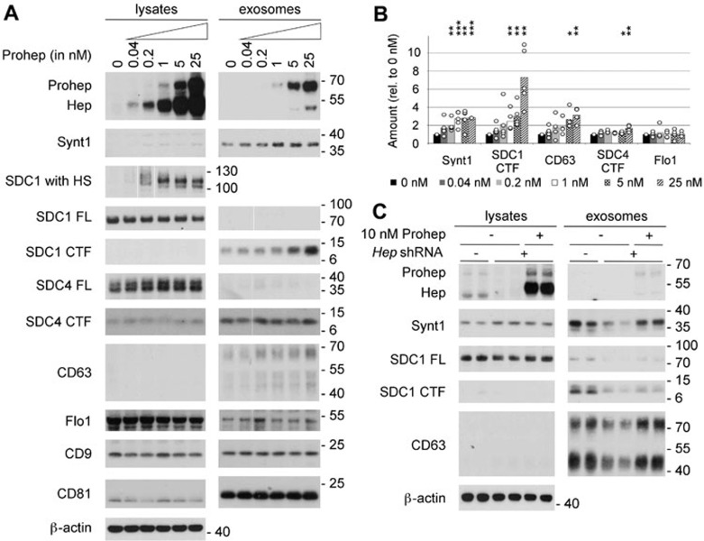
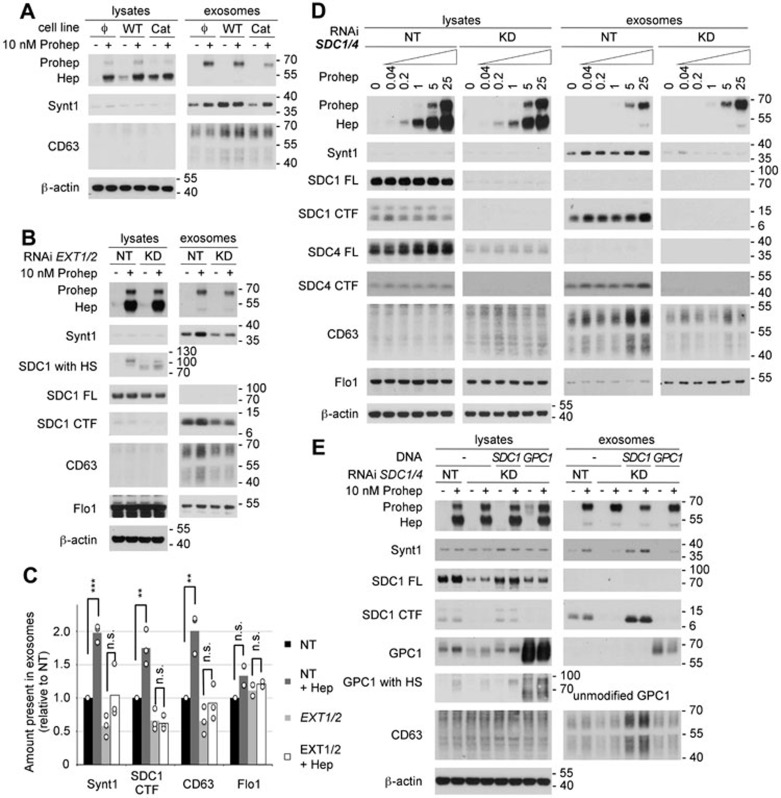
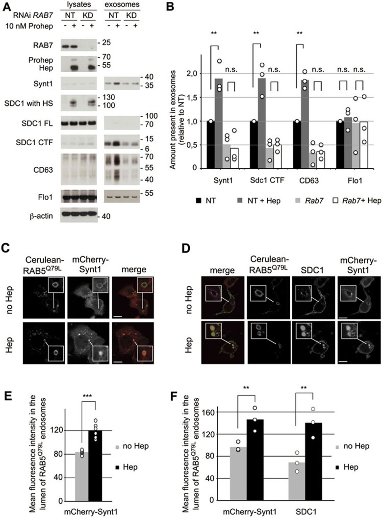
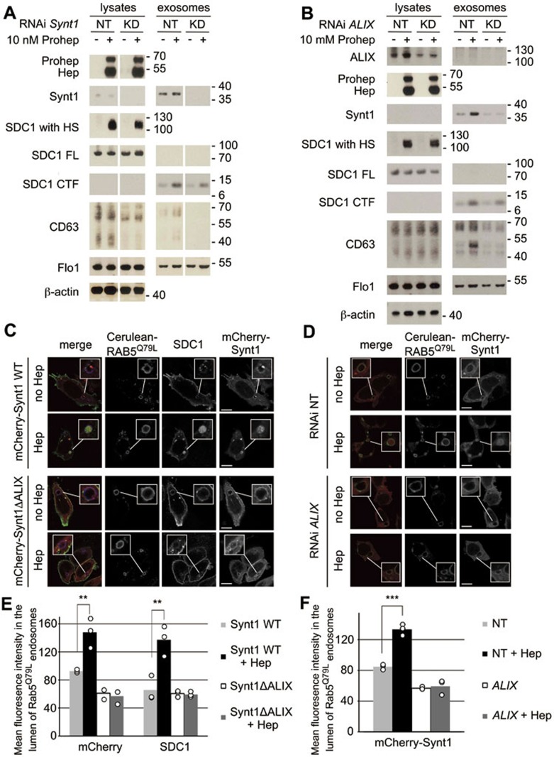
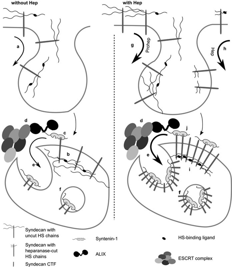
Exosomes are secreted vesicles of endosomal origin involved in signaling processes. We recently showed that the syndecan heparan sulfate proteoglycans control the biogenesis of exosomes through their interaction with syntenin-1 and the endosomal-sorting complex required for transport accessory component ALIX. Here we investigated the role of heparanase, the only mammalian enzyme able to cleave heparan sulfate internally, in the syndecan-syntenin-ALIX exosome biogenesis pathway. We show that heparanase stimulates the exosomal secretion of syntenin-1, syndecan and certain other exosomal cargo, such as CD63, in a concentration-dependent manner. In contrast, exosomal CD9, CD81 and flotillin-1 are not affected. Conversely, reduction of endogenous heparanase reduces the secretion of syntenin-1-containing exosomes. The ability of heparanase to stimulate exosome production depends on syntenin-1 and ALIX. Syndecans, but not glypicans, support exosome biogenesis in heparanase-exposed cells. Finally, heparanase stimulates intraluminal budding of syndecan and syntenin-1 in endosomes, depending on the syntenin-ALIX interaction. Taken together, our findings identify heparanase as a modulator of the syndecan-syntenin-ALIX pathway, fostering endosomal membrane budding and the biogenesis of exosomes by trimming the heparan sulfate chains on syndecans. In addition, our data suggest that this mechanism controls the selection of specific cargo to exosomes.


Figures

Figure 1
Figure 1
Heparanase stimulates the production of syntenin-1-containing exosomes. (A) Exosome production was evaluated after overnight conditioning of MCF-7 cells with increasing concentrations of proheparanase (0.04-25 nM) and compared to that of cells not receiving proheparanase (0 nM). Exosomes were collected from equivalent amounts of culture medium, conditioned by equal numbers of cells, for equal lengths of time. For each condition both the lysate and exosomal fractions were analyzed by western blot, using cognate antibodies against heparanase, monitoring the conversion of proheparanase (Prohep) into mature heparanase (Hep) and against different exosomal markers: syntenin-1 (Synt1), syndecan-1 (SDC1), syndecan-4 (SDC4), CD63, flotillin-1 (Flo1), CD9 and CD81. Syndecan-1, which is a hybrid heparan sulfate (HS)/chondroitin sulfate proteoglycan, was analyzed using two different approaches. In one approach, the samples were digested with both heparitinase and chondroitinase ABC, removing all glycosaminoglycan chains and enabling visualization of the full-length syndecan core proteins (SDC1 FL) as sharp bands. In the other approach, the samples were digested with chondroitinase ABC only, leaving the HS on the syndecans (SDC1 with HS); comparison of 'SDC1 with HS' and 'SDC1 FL' yields information on the mass of HS on syndecans. Because of the heterogeneity in HS chain length, syndecan-1 with HS is smeared over a wide mass range in the absence of heparanase activity (and is therefore hardly visible in western blot, as illustrated by lane 1 of the lysates). With increasing heparanase activity, the HS chains on syndecan-1 are trimmed to shorter chains of more or less the same length, syndecan-1 with HS migrating as one or a few bands that are readily visualized in western blot (as illustrated by lane 6 of the lysates). Note that cell lysates contain mainly full-length syndecan core proteins; the opposite is true for exosomes, where hardly any full-length syndecan is detected and C-terminal fragments (CTFs) represent the dominant form. β-actin was used as a loading control for the lysates. Western blots are representative of five independent experiments. (B) Histogram representing the quantification of the exosomal levels of syntenin-1 (Synt1), syndecan-1 CTF (SDC1 CTF), CD63, syndecan-4 CTF (SDC4 CTF) and flotillin-1 (Flo1) in response to the addition of increasing concentrations (0 nM till 25 nM) of proheparanase. Values are relative to the exosomal levels measured in absence of exogenously added proheparanase. Bar heights represent mean values, calculated from five independent experiments. Individual data points are shown as white dots on top of the corresponding bars. *P < 0.1, **P < 0.05, ***P < 0.01 (Student's t-test, assuming normal distribution of the data points). (C) Knockdown of endogenous heparanase reduces the production of syntenin-1-containing exosomes, which can be rescued by the addition of exogenous proheparanase. Duplicate lanes show the results of two independent experiments, run side by side. B16-F10 cells are sham-transfected (−) or stably transfected with a shRNA targeting murine heparanase (+). To rescue the effects of endogenous heparanase knockdown, 10 nM human proheparanase was added to the cells. Heparanase, syntenin-1, syndecan-1 full-length (SDC1 FL), syndecan 1 CTF (SDC1 CTF) and CD63 were analyzed by western blot. Positions of molecular weight markers (in kDa) are indicated on the right of each blot. Note that (because of differences in glycosylation) the Mr of human heparanase is slightly larger than that of mouse heparanase.
Figure 2
Figure 2
The effect of heparanase on exosome production depends on the modification of the heparan sulfate (HS) on syndecans. (A) The role of heparanase enzymatic activity on exosome production was investigated by comparing MCF-7 cells stably expressing wild-type heparanase (WT), catalytically dead heparanase (Cat) or empty vector (Φ) in western blot, in the absence (−) or presence (+) of 10 nM exogenously added proheparanase. (B) The importance of HS was analyzed by treating MCF-7 cells with RNAi targeting EXT1 and EXT2 (KD). Non-targeting RNAi (NT) was used as a control. Cells were challenged with 10 nM proheparanase (+) or left untreated (−). Heparanase activity, reducing the HS on syndecan, was apparent from the migration of chondroitinase ABC-treated syndecan-1 present in cell lysates (SDC1 with HS). EXT1 and EXT2 knockdown leads to the appearance of syndecan-1 that is not substituted with HS (a band running slightly > 70 kDa, after chondroitinase ABC digestion only), not detectable in cells treated with non-targeting RNAi, where all syndecan is substituted with HS (and is larger than SDC FL). A small amount of the syndecan-1 still carried HS and was affected by heparanase addition (yielding a band slightly > 100 kDa, after chondroitinase ABC digestion only), indicating an incomplete knockdown of EXT1 and EXT2. Western blots are representative of three independent experiments. (C) Quantification of the effect of EXT1 and EXT2 knockdown. Histograms representing the exosomal levels of syntenin-1, syndecan-1 CTF, CD63 and flotillin-1 in the different conditions tested (non-targeting RNAi, black bars; non-targeting RNAi and heparanase, dark gray bars; EXT1 and EXT2 RNAi, light gray bars; EXT1 and EXT2 RNAi and heparanase, white bars). Values are relative to the exosomal levels measured in cells treated with non-targeting RNAi and in the absence of exogenously added proheparanase. Bar heights represent mean values, calculated from three independent experiments. Individual data points are shown as white dots on top of the corresponding bars. **P < 0.05, ***P < 0.01, n.s., not significant (Student's t-test, assuming normal distribution of the data points). (D) Likewise, the role of syndecans was investigated by treating MCF-7 cells with RNAi targeting syndecan-1 and -4 (KD). Non-targeting RNAi (NT) was used as a control. Cells were treated with increasing concentrations of proheparanase (0 to 25 nM) and both the lysates and exosomal fractions were analyzed. Western blots are representative of three independent experiments. Note that lysate and exosome samples derived from NT-and KD-treated cells, separated by a blank space in Figure 2D, were run in the same gel, but not side by side, and that the band intensities in each row are directly comparable. (E) The ability of glypican-1 to rescue the effect of heparanase on exosome production in absence of syndecans was investigated by knocking down both syndecan-1 and -4 (KD) and overexpressing glypican-1 (GPC1) in the same cells. Glypican-1, when deglycanated by heparitinase and chondroitinase ABC treatment (GPC1) or when unmodified (because of overexpression) has a mass of ∼ 60 kDa. Treatment with chondroitinase ABC only reveals 'GPC1 with HS' of a mass of ∼ 90 kDa in heparanase-exposed cells, indicating the HS on glypican-1 is trimmed the same way as the HS on syndecans. Yet, extra glypican fails to rescue the exosomal accumulations of syntenin and CD63. Rescue by expression of mouse syndecan-1 (SDC1) was used as a positive control. Cells treated with non-targeting RNAi (NT) served as reference. Molecular weight markers (in kDa) are indicated on the right of each blot. Western blots are representative of two independent experiments. Note that MCF-7 cells use mainly Man-6-P receptors for heparanase internalization, and that neither the knock down of EXT1 and EXT2 nor the knock down of syndecans has an effect on the uptake of proheparanase and its conversion into mature active form.
Figure 3
Figure 3
Heparanase influences the biogenesis of vesicles of endosomal origin, enhancing intraluminal budding. (A) To investigate whether the extracellular vesicles affected by heparanase were of endosomal origin, RAB7 was knocked down (RAB7 RNAi) in MCF-7 cells. Non-targeting RNAi (−) served as a control. Cells were left untreated (−) or treated with proheparanase (10 nM). In both experiments, heparanase activity was evaluated using the migration pattern of chondroitinase ABC-, but not heparitinase-treated syndecan-1 present in cell lysates (SDC1 with HS). Molecular weight markers (in kDa) are indicated on the right of each blot. Western blots are representative of three independent experiments. (B) Quantification of the effect of RAB7 knockdown. Histograms representing the exosomal levels of syntenin-1, syndecan-1 CTF and CD63 in the different conditions tested (non-targeting RNAi, black bars; non-targeting RNAi with heparanase, dark gray bars; RAB7 knockdown, light gray bars; RAB7 knockdown with heparanase, white bars). Values are relative to the levels (intensities of the signals) measured in exosomes derived from cells treated with non-targeting RNAi in the absence of proheparanase. Bar heights represent mean values, calculated from three independent experiments. Individual data points are shown as white dots on top of the corresponding bars. **P < 0.05, n.s., not significant (Student's t-test, assuming normal distribution of the data points). (C) Confocal micrographs of MCF-7 cells co-transfected with mCherry-syntenin-1 (red in merge) and Cerulean-RAB5Q79L (green in merge). Note the presence of mCherry-syntenin-1, a cytosolic protein, inside the Cerulean-RAB5Q79L endosomes upon heparanase treatment (50 nM). (D) Confocal micrographs of MCF-7 cells co-transfected with mCherry-syntenin-1 (red in merge), Cerulean-RAB5Q79L (blue in merge) and syndecan-1 (green in merge), scoring the accumulations (budding) of mCherry-syntenin-1 and of syndecan-1 cytoplasmic domain inside vacuoles outlined by Cerulean-RAB5Q79L. (E) Quantification of intraluminal budding of mCherry-syntenin-1, as in Figure 3C, by measuring the fluorescence of mCherry-syntenin-1 in the lumen of RAB5Q79L-positive endosomes, corrected for the size of the RAB5Q79L-positive endosomes (mean gray value per pixel). Bar heights represent mean values calculated from six independent experiments, scoring at least 30 cells per experiment. Individual data points (mean luminal fluorescence intensity per individual experiment) are shown as white dots on top of the corresponding bars. ***P < 0.01 (Student's t-test, assuming normal distribution of the data points). (F) Quantification of intraluminal budding of mCherry-syntenin-1 and syndecan-1 cytoplasmic domain, as in Figure 3D, by measuring the fluorescence intensity of mCherry-syntenin-1 and syndecan-1 cytoplasmic domain in the lumen of RAB5Q79L-positive endosomes. Bar heights represent mean values calculated from three independent experiments, scoring at least 30 cells per experiment. Individual data points are shown as white dots on top of the corresponding bars. **P < 0.05 (Student's t-test, assuming normal distribution of the data points).
Figure 4
Figure 4
Heparanase acts through syntenin-1, ALIX and the syntenin-ALIX interaction to stimulate intraluminal budding. (A) The role of syntenin-1 and (B) the role of ALIX in the effect of heparanase on exosomes were investigated by the knockdown of syntenin-1 and ALIX (using Synt1 RNAi and ALIX RNAi, respectively) in MCF-7 cells. Non-targeting RNAi (NT) served as a control. Cells were left untreated (−) or treated with proheparanase (10 nM). In both the experiments, heparanase activity was evaluated using the migration pattern of chondroitinase ABC-, but not heparitinase-treated syndecan-1 present in cell lysates (SDC1 with HS). Molecular weight markers (in kDa) are indicated on the right of each blot. Western blots are representative of two independent experiments. Note that lysate and exosome samples derived from NT and KD cells, separated by a blank space in A, were run in the same gel, but not side by side, and that the band intensities in each row are directly comparable. (C) Confocal micrographs of MCF-7 cells co-transfected with wild-type mCherry-syntenin-1 (mCherry-Synt1 WT) or with mCherry-syntenin-1 harboring mutant LYP sequences and therefore defective in ALIX-binding (mCherry-Synt1-ΔALIX; red in merge), Cerulean-RAB5Q79L (blue in merge) and syndecan-1 (green in merge). Intraluminal budding of wild-type and mutant mCherry-syntenin-1 and of syndecan-1 cytoplasmic domain was scored in the presence (Hep) or absence (no Hep) of heparanase. (D) Confocal micrographs of MCF-7 cells co-transfected with mCherry-syntenin-1 (red in merge) and Cerulean-RAB5Q79L (blue in merge). In addition, the cells were transfected with RNAi targeting ALIX (ALIX RNAi) or with non-targeting RNAi (NT RNAi). Intraluminal budding of mCherry-syntenin-1 was evaluated in the presence (Hep) or absence (no Hep) of heparanase. (E) Quantification of intraluminal budding of mCherry-syntenin-1 and syndecan-1 cytoplasmic domain, as in C and (F) quantitation of intraluminal budding of mCherry-syntenin-1, as in D, measuring the mean fluorescence intensity of mCherry-syntenin-1 and syndecan-1 in the lumen of RAB5Q79L-positive endosomes (mean gray value per pixel). Bar heights represent mean values calculated from three independent experiments, scoring at least 30 cells per experiment. Individual data points are shown as white dots on top of the corresponding bars. **P < 0.05, ***P < 0.01 (Student's t-test, assuming normal distribution of the data points).
Figure 5
Figure 5
Schematic representation of the potential mechanism of action of heparanase in exosome biogenesis. (a) Syndecans are continuously internalized by cells. (b) Endocytosed syndecans are clustered by the ligands binding to their heparan sulfate (HS) chains, a process that might already be initiated at the cell surface. (c) These syndecan assemblies subsequently recruit syntenin, (d) ALIX and the ESCRT machinery. (e) As a result, syndecan and cargo bound to syndecan is recruited to membrane buds protruding into the lumen of MVBs. At this stage, the extracellular portion of the syndecans is cleaved by endosomal protease, generating syndecan CTFs. (f) The ESCRT machinery pinches off these buds and as such liberates intraluminal vesicles, loaded with syntenin and syndecan CTFs, into the lumen of MVBs. (g) As a ligand of HS, proheparanase might directly stimulate the clustering and subsequent endocytosis of syndecans at the cell membrane. (h) In addition, and more importantly, heparanase activity trims the HS chains on syndecan, possibly regulating the engagements and further stimulating the internalization of syndecans. Both the mechanisms lead to a higher concentration of endosomal syndecan. (i) Trimming the HS chains on syndecans might also stimulate clustering of syndecans at the limiting membrane of MVBs. Enhancement of the syndecan engagements, reduction of syndecan self-repulsion and effects on the dimensions and spacing of syndecans in syndecan-ligand complexes, by the trimming of the HS on syndecan, are several possible and non-mutually exclusive mechanisms by which heparanase may foster syndecan clustering. (j) Enhanced clustering of syndecans increases the recruitment and binding of syntenin-1 to endosomal syndecan assemblies and ultimately may cause the binding of syntenin-1 to larger associations of syndecans, increasing the syndecan-syntenin stoichiometry in syndecan-syntenin complexes and exosomes. Note that the diagram makes no statement on the accumulation of ALIX and ESCRTs in the intraluminal vesicles and exosomes, which were not investigated here.

Comment in

References

    1. Raposo G, Stoorvogel W. Extracellular vesicles: exosomes, microvesicles, and friends. J Cell Biol. 2013;200:373–383. - PMC - PubMed
    1. Valadi H, Ekstrom K, Bossios A, Sjostrand M, Lee JJ, Lotvall JO. Exosome-mediated transfer of mRNAs and microRNAs is a novel mechanism of genetic exchange between cells. Nat Cell Biol. 2007;9:654–659. - PubMed
    1. Skog J, Wurdinger T, van Rijn S, et al. Glioblastoma microvesicles transport RNA and proteins that promote tumour growth and provide diagnostic biomarkers. Nat Cell Biol. 2008;10:1470–1476. - PMC - PubMed
    1. Raposo G, Nijman HW, Stoorvogel W, et al. B lymphocytes secrete antigen-presenting vesicles. J Exp Med. 1996;183:1161–1172. - PMC - PubMed
    1. Thery C, Duban L, Segura E, Veron P, Lantz O, Amigorena S. Indirect activation of naive CD4+ T cells by dendritic cell-derived exosomes. Nat Immunol. 2002;3:1156–1162. - PubMed

Publication types

MeSH terms