. 2015 Jan-Feb;7(1):1759091415573985.
doi: 10.1177/1759091415573985.

Caspase-mediated apoptosis in the cochleae contributes to the early onset of hearing loss in A/J mice

Caspase-mediated apoptosis in the cochleae contributes to the early onset of hearing loss in A/J mice

Xu Han et al. ASN Neuro. 2015 Jan-Feb.

Abstract

A/J and C57BL/6 J (B6) mice share a mutation in Cdh23 (ahl allele) and are characterized by age-related hearing loss. However, hearing loss occurs much earlier in A/J mice at about four weeks of age. Recent study has revealed that a mutation in citrate synthase (Cs) is one of the main contributors, but the mechanism is largely unknown. In the present study, we showed that A/J mice displayed more severe degeneration of hair cells, spiral ganglion neurons, and stria vascularis in the cochleae compared with B6 mice. Moreover, messenger RNA accumulation levels of caspase-3 and caspase-9 in the inner ears of A/J mice were significantly higher than those in B6 mice at 2 and 8 weeks of age. Immunohistochemistry localized caspase-3 expression mainly to the hair cells, spiral ganglion neurons, and stria vascularis in cochleae. In vitro transfection with Cs short hairpin RNA (shRNA) alone or cotransfection with Cs shRNA and Cdh23 shRNA significantly increased the levels of caspase-3 in an inner ear cell line (HEI-OC1). Finally, a pan-caspase inhibitor Z-VAD-FMK could preserve the hearing of A/J mice by lowering about 15 decibels of the sound pressure level for the auditory-evoked brainstem response thresholds. In conclusion, our results suggest that caspase-mediated apoptosis in the cochleae, which may be related to a Cs mutation, contributes to the early onset of hearing loss in A/J mice.


Figures

Figure 1.
Figure 1.
Characterization of hearing loss in A/J mice by a time course observation. (a) 2.5% agarose gel electrophoresis of the PCR products digested with Nla III restriction endonuclease. Lane M, 50 bp DNA ladders; two lanes designated as B6: The Nla III restriction endonuclease cutting the 173 bp PCR fragment from Cs gene in B6 mice into two distinct fragments (123 bp and 50 bp); two lanes designated as A/J: Cs mutation in A/J mice alters the restriction site and leaving only one band (173 bp). (b) The mutation changes the local secondary structure of Cs from an alpha helix (h) to a random coil(c) as predicted by GOR IV methods. (c) ABR threshold detection in A/J mice at ages of 3 weeks (n = 6), 4 weeks (n = 11), 10 weeks (n = 8), 12 weeks (n = 10), 28 weeks (n = 7), and 42 weeks (n = 7) and B6 mice with the corresponding age groups (n = 6, n = 10, n = 20, n = 13, n = 6, and n = 6, respectively). Each point represents the mean ABR threshold for a group, with error bar indicating SD from the mean. The results showed that ABR thresholds were significantly higher in the A/J mice than those of the B6 mice at stimulus frequencies of 32 kHz at all points of time. **p < .01. (d) DPOAE measurement in B6 (n = 6–10) and A/J (n = 6–10) mice with age from 3 to 42 weeks at f2 frequency of 17267 kHz. DPOAE amplitudes were significantly lower in A/J mice than those of B6 mice at 3, 10, 28, and 42 weeks of age, respectively. W: weeks; *p < .05; **p < .01. PCR = polymerase chain reaction; ABR = auditory-evoked brainstem response; DPOAE = distortion product oto-acoustic emission; SPL = sound pressure level.
Figure 2.
Figure 2.
OHC loss in the cochleae of A/J mice. (a) Representative whole-mount alexa-fluor-488 phalloidin-stained preparations from basal turns of cochleae. OHC loss and abnormal morphology and arrangement of hair cells in A/J mice occur at 3 weeks of age (right), compared with the corresponding area in B6 mice (left). Both OHC and IHC loss was severe in A/J mice at age of 10 weeks or older. B6 mice also showed hair cell loss and abnormal arrangement, evidently at 28 weeks of age. Scale bars = 50 µm. (b) Progressive OHC loss in the tree turns of B6 and A/J mice at 3, 10, 16, and 28 weeks of age. OHC loss in each turn of both mouse strains increased with age. The percentages of OHC loss at 28 weeks of age were significantly more than any other time points, except for those at the apex turns in B6 mice. *p < .05; **p < .01. (c) A time course comparison of OHC loss between B6 and A/J mice. A/J mice have significantly more OHC loss beginning at 3 weeks of age at basal turns and becoming more severe at 28 weeks of age at the three turns, compared with those of B6 mice. (n = 3–5 for each group at each time point). *p < .05; **p < .01. W: weeks. OHC = outer hair cells; IHC = inner hair cell.
Figure 3.
Figure 3.
Typical pathology on HE sections from basal turns in the cochleae of A/J mice. (a) Pathological alteration in the spiral ganglion neurons (SGNs). B6 mice showed normal density of SGNs until 46 weeks of age. However, SGNs in the A/J mice were already sparse at 4 weeks of age, and loss of SGNs became severe at 46 weeks of age (indicated by circles). (b) Hair cell pathology in HE staining sections. OHC was intact before 46 weeks of age for B6 mice, whereas hair cell lesion became severe at 10 weeks of age for A/J mice (indicated by arrows). (c) Alteration of stria vascularis in A/J mice. The width of stria vascularis (indicated by arrows) of A/J mice was generally thinner than that of B6 mice at all the points of time, though there is significant difference only at 2 weeks of age (p < .05). W: weeks; scale bar = 50 µm for all panels. HE = hematoxylin and eosin; OHC = outer hair cells.
Figure 4.
Figure 4.
Relative mRNA levels of apoptosis related genes in the cochleae of A/J mice. (a) Time course mRNA accumulation level of caspase-3, caspase-9, and Aif, corrected by Gapdh, in the inner ears from A/J mice (n = 4–5 at each time point). mRNA levels of caspase-3 and caspase-9 at P1 were more than two times the levels at P56 but declined with time in this period. (b to g) Comparison of gene transcription levels between A/J mice and B6 mice. mRNA levels of caspase-3, caspase-9, and Aif in the inner ears from A/J mice (n = 4–6) were significantly higher than those from B6 mice (n = 4–6) at 2 weeks (b–d) or 8 weeks (e–g). Error bars represent the SD from the mean. W: weeks; *p < .05; **p < .01. mRNA = messenger RNA; Gapdh = glyceraldehyde 3-phosphate dehydrogenase.
Figure 5.
Figure 5.
Expression of caspase-3 in HEI-OC1 cells. (a) 2% agarose gel electrophoresis of the RT-PCR products of Gapdh, Cs, and Cdh23 in HEI-OC1 cells. Lane M, 50 bp DNA ladders; lanes 1–2, 3–4, and 5–6 represent RT-PCR products of Gapdh (100 bp), Cdh23 (228 bp), and Cs (144 bp), respectively. (b) Transcription levels of Cs and Cdh23 in HEI-OC1 cells transfected with shRNA. mRNA levels of Cs was significantly suppressed (p < .05) by Cs shRNA (Cs knockdown group) compared with that by control shRNA (control, limited to1 unit for the level of each gene). mRNA levels of Cs and Cdh23 in cotransfencted group (Cdh23 shRNA + Cs shRNA) were also lower than those of the controls. (c) Caspase-3 levels determined by Western blotting. Caspase-3 expression levels were markedly elevated in Cs knockdown group or cotransfected group compared with those of controls, whereas were not elevated in Cdh23 knockdown group. (d) The average gray intensity of caspase-3 protein detected by Western blotting was standardized to that of β-actin. Caspase-3 expression levels in Cs knockdown group or cotransfected group were significantly higher than those of the controls. Data are shown as mean ± SD of triplicate experiments. *p < .05. shRNA = short hairpin RNA; RT-PCR = reverse transcription polymerase chain reaction; mRNA = messenger RNA; Gapdh = glyceraldehyde 3-phosphate dehydrogenase.
Figure 6.
Figure 6.
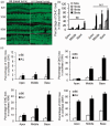
Otoprotection by pan-caspase inhibitor in A/J mice. (a) ABR thresholds in the three mouse groups measured at the stimulus frequency of 32 kHz. The number of mice tested was nine, six, and six for no treatment, DMSO and Z-VAD-FMK + DMSO groups, respectively. Each point represents the mean ABR threshold for a group, with error bar indicating SD from the mean. The results show that ABR thresholds were significantly lower in the Z-VAD-FMK + DMSO-treated mice than those of the untreated mice at the four time points (p < .01). There were significant differences for ABR thresholds between Z-VAD-FMK + DMSO-treated and DMSO-treated groups at all time points except at 4 weeks of age (p < .05); or between DMSO-treated and untreated mice at 6 weeks of age (p < .05). (b) A time course observation of DPOAE to stimulus frequency of 17672 kHz (f2) in the three mouse groups. Though gradually declined with time, the mean DPOAE amplitudes were significantly higher in Z-VAD-FMK + DMSO-treated mice than those in the untreated mice at age of 6 weeks or 8 weeks (p < .01). (c) Typical OHC distribution in the middle turns of cochleae in the three mouse groups. Loss of OHC was evident in the middle turns of cochleae of the untreated or DMSO-treated mice at 8 weeks of age, whereas only some OHC loss was seen in the corresponding areas of the Z-VAD-FMK + DMSO-treated mice. (d) Percentage of OHC loss in the three turns of cochleae in the three mouse groups at age of 8 weeks. W: weeks; Scale bar = 50 µm; *p < .05; **p < .01. ABR = auditory-evoked brainstem response; SPL = sound pressure level; DMSO = dimethyl sulfoxide; OHC = outer hair cells.

References

    1. Alam S. A., Oshima T., Suzuki M., Kawase T., Takasaka T., Ikeda K. (2001) The expression of apoptosis-related proteins in the aged cochlea of Mongolian gerbils. Laryngoscope 111: 528–534. - PubMed
    1. Angeli S., Lin X., Liu X. Z. (2012) Genetics of hearing and deafness. Anatomical Record (Hoboken) 295: 1812–1829. - PMC - PubMed
    1. Balaban R. S., Nemoto S., Finkel T. (2005) Mitochondria, oxidants, and aging. Cell 120: 483–495. - PubMed
    1. Cheng T. L., Liao C. C., Tsai W. H., Lin C. C., Yeh C. W., Teng C. F., Chang W. T. (2009) Identification and characterization of the mitochondrial targeting sequence and mechanism in human citrate synthase. Journal of Cellular Biochemistry 107: 1002–1015. - PubMed
    1. Dirks A. J., Hofer T., Marzetti E., Pahor M., Leeuwenburgh C. (2006) Mitochondrial DNA mutations, energy metabolism and apoptosis in aging muscle. Ageing Research Reviews 5: 179–195. - PubMed

Publication types

MeSH terms

LinkOut - more resources