. 2015 Mar 3;10(8):1410-21.
doi: 10.1016/j.celrep.2015.01.059. Epub 2015 Feb 26.

Plug-and-play genetic access to drosophila cell types using exchangeable exon cassettes

Affiliations

Plug-and-play genetic access to drosophila cell types using exchangeable exon cassettes

Fengqiu Diao et al. Cell Rep. .

Abstract

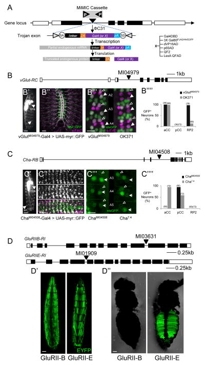
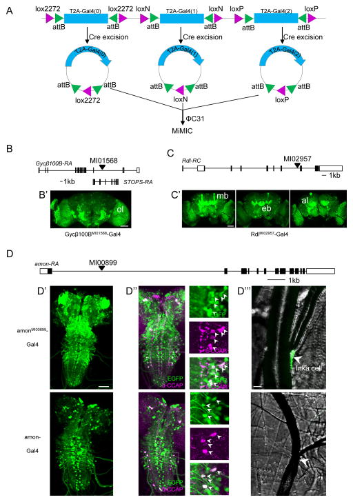
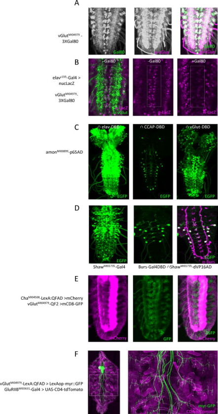
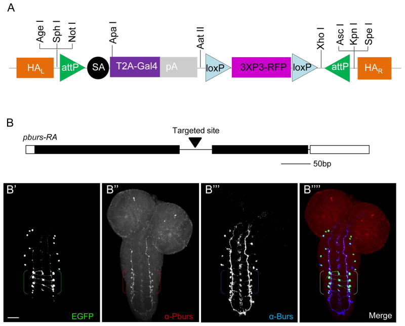
Genetically encoded effectors are important tools for probing cellular function in living animals, but improved methods for directing their expression to specific cell types are required. Here, we introduce a simple, versatile method for achieving cell-type-specific expression of transgenes that leverages the untapped potential of "coding introns" (i.e., introns between coding exons). Our method couples the expression of a transgene to that of a native gene expressed in the cells of interest using intronically inserted "plug-and-play" cassettes (called "Trojan exons") that carry a splice acceptor site followed by the coding sequences of T2A peptide and an effector transgene. We demonstrate the efficacy of this approach in Drosophila using lines containing suitable MiMIC (Minos-mediated integration cassette) transposons and a palette of Trojan exons capable of expressing a range of commonly used transcription factors. We also introduce an exchangeable, MiMIC-like Trojan exon construct that can be targeted to coding introns using the Crispr/Cas system.


Figures

Fig. 1
Fig. 1. Creating Trojan Gal4 driver lines using MiMIC insertions into coding introns
A) Trojan exon structure, integration, and expression. The flanking inverted attP sites (P) of a MiMIC cassette (gray) within a coding intron of a gene (coding exons, black) permit ΦC31 integrase-mediated exchanges for a Trojan exon cassette, via its flanking attB (B) sites. The splice acceptor site (SA) insures incorporation of the T2A-transgene sequence into the gene’s mRNA; a short linker with one of three possible lengths maintains the reading frame of the native message; the T2A sequence causes truncation of the native gene product and promotes the translation of Gal4 (or other transgene, X). pA, Hsp70 polyadenylation signal. See also Fig. S1A and S6. B) Schematic of the vGlut gene (RC isoform) and site of MiMICMI04979 insertion in a coding intron common to all splice isoforms. B) vGlutMI04979-Gal4 drives expression of a UAS- myr::GFP reporter in many neurons of the larval CNS (central nervous system, arrowhead) including motor neurons, which project axons (arrow) to bodywall muscles. Scale bar, 200 μm. (See also Fig. S2) B) Motor nerves (labeled as in B′) in an embryonic fillet showing peripheral nerves double-labeled with anti-Fas2 antibody (magenta). Scale bar: 25 μm. B′″) Comparison of the vGlutMI04979-Gal4 (left) and OK371-Gal4 (right) expression patterns (green, UAS-nlsGFP) in three hemisegments of the larval VNC, labeled with an anti-Eve antibody (magenta). Two of the three Eve-immunopositive neurons located dorsally at the ventral midline in each hemisegment, aCC (open arrowhead) and RP2 (filled arrowhead), are glutamatergic, but OK371-Gal4 consistently fails to label RP2. Scale bar, 10 μm. B″″) Bar graph summarizing the results of labeling experiments of the type shown in B′″ from vGlutMI04979-Gal4 (n=36) and OK371-Gal4 (n=72) hemisegments. C) Schematic of the Cha gene (RB isoform) and site of the MiMIC MI04508 insertion in a coding intron common to all splice isoforms. C) ChaMI04508 -Gal4 drives expression of a UAS-myr::GFP reporter in most neurons of the larval CNS (arrowhead), and sensory neurons of the peripheral nervous system (sn, arrow). Scale bar, 200 μm: C″) Co-localization of vChaT mRNA, visualized by embryonic fluorescence in situ hybridization (top) and ChaMI04508 -Gal4 driven UAS-myr::GFP (middle). Bottom, merged images: green, UAS-myr::GFP; magenta, vChaT mRNA. Scale bar: 25 μm. C′″) Comparison of the ChaMI04508 -Gal4 (left) and Cha7.4-Gal4 (right) expression patterns (green, UAS-nlsGFP) in three hemisegments of the larval VNC labeled with anti-Eve antibody. Of the three Eve+ neurons (outlined), Cha7.4-Gal4 consistently labels the glutamatergic aCC neurons (open arrowheads) but not the putative cholinergic pCC neurons. Scale bar: 10 μm. C″″) Bar graph summarizing the results of labeling experiments of the type shown in C′″ from ChaMI04508-Gal4 (n=90) and Cha7.4-Gal4 (n=72) hemisegments. D) Schematics of the GluRIIB and GluRIIE genes and the insertion sites of MiMICMI03631 and MiMICMI01909. D′) Expression of GluRIIBMI03631-Gal4 (left panel; dorsal view) and GluRIIEMI01909-Gal4 (right panel; ventral view) in larval bodywall muscles. D″) GluRIIEMI01909-Gal4 (right panel), but not GluRIIBMI03631-Gal4 (left panel) drives expression in adult ventral abdominal muscles. Scale bar in D′, D″: 150 μm. Green, UAS-EYFP.
Fig. 2
Fig. 2. Creating Gal4 drivers using the in vivo system for Trojan exon exchange
A) Schematic of the triplet donor construct consisting of three tandem Trojan Gal4 cassettes, one in each reading frame, each flanked by attB sites nested within uniquely compatible pairs of lox sites, recognized by Cre recombinase. See Figure S1B for details of genetic crosses. B) Schematic of the Gycβ100B gene locus and the site of the MiMICMI01568 insertion, which also lies within a coding intron of the STOPS gene on the opposite strand. (See also Fig. S3.) B) Expression of a UAS-EYFP reporter driven by Gycβ100BMI01568-Gal4 in the optic lobe (ol) of the adult brain. C) Schematic of the Rdl gene and the MiMICMI 02957 insertion site. C) Confocal images from an adult brain show prominent RdlMI02957-Gal4 driven expression of a UAS-EYFP reporter in the mushroom bodies (mb), ellipsoid body (eb), and antennal lobes (al). Scale bars: 50 μm. (See also Fig. S4.) D) Schematic of the amon gene and the MiMIC MI00899 insertion site. D–D″′) Comparison of larval expression patterns of amonMI00899-Gal4 (top panels) and amon-Gal4 (bottom panels). D′) Expression patterns in CNS whole mounts (green, UAS-EGFP). D″) Expression within the subset of CCAP-expressing neurons (anti-CCAP staining, magenta). Insets show higher magnification images of the boxed regions. D″′) Expression of amonMI00899-Gal4, but not amon-Gal4, in the endocrine Inka cells located at the base of the dorsal tracheal trunks (arrowheads, see also Fig. S5). Scale bars: D′, D″: 50 μm; D′″: 10 μm.
Fig. 3
Fig. 3. Combinatorial gene targeting using Gal80, Split Gal4, QF2, and LexA::QFAD Trojan exons
A–B) Suppression of Gal4 activity in glutamatergic neurons using 3XGal80FLAG/HA/EGFP. A) vGlutMI04979- 3XGal80FLAG/HA/EGFP expresses Gal80 (left; anti-GFP immunostaining) in glutamatergic motor neurons of the larval VNC labeled with anti-vGlut antibody (middle); merged images (right). Scale bar (panels A–E): 25 μm. B) elav-Gal4 drives UAS-LacZ reporter in the glutamatergic motor neurons (rectangle: anti-LacZ immunoreactivity, magenta; anti-vGlut, green), but Gal4 activity is absent when the vGlutMI04979-3XGal80 transgene is present (right). C) Identification of neuronal subsets within the amonMI00899-p65AD expression pattern. Left: The entire complement of amon-expressing neurons in the larval CNS, visualized using the pan-neuronal elav-VP16AD hemidriver and a UAS-EGFP reporter. Middle: The CCAP-expressing complement, visualized using a CCAP-Gal4DBD hemidriver. Right: The vGlut-expressing complement, visualized using a Trojan-MiMIC vGlutMI04979-Gal4DBD hemidriver. D) Split Gal4 isolation of the subset of neurons that express both the Shaw K+ channel and the hormone Bursicon. Left: neurons expressing the RA, RC, and RE splice isoforms of the Shaw gene, visualized using a Trojan-MiMICShawMI01735-Gal4 driver line and a UAS-EGFP reporter. Middle: The Shaw-expressing neurons that also express Burs, isolated using a Trojan-MiMIC ShawMI01735-dVP16AD in combination with a Burs-Gal4DBD hemidriver; green, UAS-EGFP. Right: anti-Burs immunostaining (magenta) of the section shown in middle panel (double-labeled neurons are white). E) Double-labeling of the embryonic VNC to identify subsets of cholinergic (LexAop-mCherry, magenta) and glutamatergic (QUAS-mCD8-GFP, green) neurons using orthogonal Trojan-MiMIC drivers. Left: Expression pattern of a ChaMI04508-LexA::QFAD driver in a confocal section through the ventral midline. Middle: Same section showing expression of a vGlutMI04979-QF2 driver. Right: Merged images; note the two expression patterns do not overlap. F) Double-labeling of glutamatergic neurons in the larval CNS and body wall muscles using the orthogonal Trojan-MIMC drivers vGlutMI04979-T2A-LexA::QFAD (green, LexAop2-myr::GFP) and GluRIIBMI03631-T2A-Gal4 (magenta, UAS-CD4-tdTomato). Right panel: magnified image of the boxed region at left showing neuromuscular synapses. Scale bar: 100 μm.
Fig. 4
Fig. 4. Targeting coding introns using the pT-GEM vector and the Crispr/Cas system
A) Schematic of the Trojan-Gal4 Expression Module (T-GEM), which includes a Red Fluorescent Protein (RFP) selection marker expressed in the adult eye that can be used to monitor genomic insertion. The attP sites permit cassette exchange with any other Trojan exon, and unique restriction sites at the 5′ and 3′ ends of the construct permit the insertion of suitable homologous arms (HAR and HAL) for targeting T-GEM to the desired intronic locus. B) Schematic of the pburs gene, showing the intronic site targeted by Crispr/Cas for T-GEM insertion. B-B″″) Confocal micrographs of a larval CNS whole mount triple-labeled for pbursTGEM-Gal4 (B′), Pburs (B″), and Burs, which heterodimerizes with Pburs to form the hormone Bursicon (B″′); merged image of all three labels (B″″). Brackets: Pburs-immunopositive cell bodies. Green, UAS-EGFP; red, anti-Pburs immunoreactivity; blue, anti-Burs immunoreactivity. Scale bar: 25 μm.

References

    1. Anderson MS, Halpern ME, Keshishian H. Identificiation of the Neuropeptide Transmitter Proctolin in Drosophila Larvae - Characterization of Muscle Fiber-specific Neuromuscular Endings. J Neurosci. 1988;8:242–255. - PMC - PubMed
    1. Bischof J, Maeda RK, Hediger M, Karch F, Basler K. An optimized transgenesis system for Drosophila using germ-line-specific phi C31 integrases. P Natl Acad Sci USA. 2007;104:3312–3317. - PMC - PubMed
    1. Branda CS, Dymecki SM. Talking about a revolution: The impact of site-specific recombinases on genetic analyses in mice. Dev Cell. 2004;6:7–28. - PubMed
    1. Buckingham SD, Biggin PC, Sattelle BM, Brown LA, Sattelle DB. Insect GABA receptors: Splicing, editing, and targeting by antiparasitics and insecticides. Mol Pharmacol. 2005;68:942–951. - PubMed
    1. Daniels RW, Gelfand MV, Collins CA, Diantonio A. Visualizing glutamatergic cell bodies and synapses in Drosophila larval and adult CNS. J Comp Neurol. 2008;508:131–152. - PubMed

Publication types