Review
. 2015 Apr;22(2):229-45.
doi: 10.1007/s12350-015-0096-5. Epub 2015 Mar 3.

Myocardial viability as integral part of the diagnostic and therapeutic approach to ischemic heart failure

Affiliations
Review

Myocardial viability as integral part of the diagnostic and therapeutic approach to ischemic heart failure

Jeroen J Bax et al. J Nucl Cardiol. 2015 Apr.

Abstract

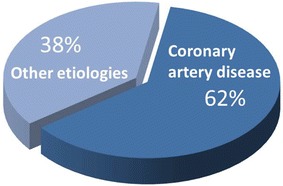
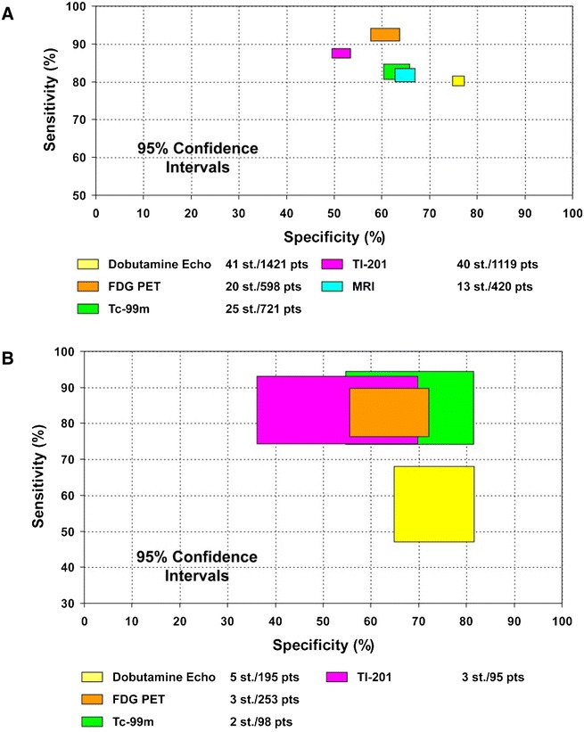
Chronic heart failure is a major public-health problem with a high prevalence, complex treatment, and high mortality. A careful and comprehensive analysis is needed to provide optimal (and personalized) therapy to heart failure patients. The main 4 non-invasive imaging techniques (echocardiography, magnetic resonance imaging, multi-detector-computed tomography, and nuclear imaging) provide information on cardiovascular anatomy and function, which form the basis of the assessment of the pathophysiology underlying heart failure. The selection of imaging modalities depends on the information that is needed for the clinical management of the patients: (1) underlying etiology (ischemic vs non-ischemic); (2) in ischemic patients, need for revascularization should be evaluated (myocardial ischemia/viability?); (3) left ventricular function and shape assessment; (4) presence of significant secondary mitral regurgitation; (5) device therapy with cardiac resynchronization therapy and/or implantable cardiac defibrillator (risk of sudden cardiac death). This review is dedicated to assessment of myocardial viability, however "isolated assessment of myocardial viability" may be clinically not meaningful and should be considered among all those different variables. This complete information will enable personalized treatment of the patient with ischemic heart failure.


Figures

Figure 1
Figure 1
Heart failure etiology. From 24 multicenter heart failure trials, including 43,568 heart failure patients, 62% of patients had an ischemic etiology
Figure 2
Figure 2
Prediction of functional recovery post-revascularization in dysfunctional segments with subendocardial scar is difficult. When the epicardial (non-infarcted) region is normal, no recovery will occur (left panel). However when the epicardial region contains jeopardized (viable) myocardium, the likelihood of recovery is high (right panel). Reproduced with permission from Kaandorp et al
Figure 3
Figure 3
Example of a 76-year-old patient with previous anterior myocardial infarction and 2-vessel coronary artery disease on invasive coronary angiography (significant long lesion on the proximal left anterior descending coronary artery and dominant circumflex coronary artery with a significant lesion proximal). Left ventricular ejection fraction was 27%. Selected short-axis views of myocardial perfusion 99technetium tetrofosmin SPECT images show a perfusion defect in the anteroseptal wall. On fluorine18-deoxyglucose (FDG) SPECT images segments, uptake of radiopharmaceutical in the anteroseptal wall is visualized indicating perfusion-metabolic mismatch, pattern of myocardial viability
Figure 4
Figure 4
Contrast-enhanced magnetic resonance imaging for characterization of myocardial scar. Selected 4-chamber views on contrast-enhanced MRI of two patients with ischemic cardiomyopathy show non-transmural subendocardial scar (A arrow) and transmural scar (B arrow)
Figure 5
Figure 5
Comparison of sensitivities and specificities with 95% confidence intervals of the various techniques for the prediction of recovery of regional (A) and global (B) left ventricular function following coronary revascularization. Data based on Schinkel et al. Echo, echocardiography; FDG, fluorine18-deoxyglucose; MRI, magnetic resonance imaging; PET, positron emission tomography; Tc-99m, 99technetium; Tl-201, 201thallium
Figure 6
Figure 6
Annualized mortality rate of patients with and without significant viable myocardium according to treatment strategy. Results of a pooled analysis from 28 prognostic studies using different imaging techniques. Patients with viable myocardium who underwent coronary revascularization had the lowest mortality rate. Data based on Schinkel et al
Figure 7
Figure 7
Assessment of left ventricular systolic function and geometry with current imaging modalities. A 2-dimensional transthoracic echocardiography; B 3-dimensional transthoracic echocardiography; C magnetic resonance imaging; D multi-detector row computed tomography; E ECG-gated single-photon emission-computed tomography
Figure 8
Figure 8
Example of a patient with large anterior myocardial infarction and subsequent formation of a large apical aneurysm. On transthoracic 2D echocardiography (A), the arrow points to the large apical aneurysm with thrombus formation. Magnetic resonance imaging shows a thin-walled apical aneurysm (arrow) extending from the mid septum to the apical lateral wall (B). On contrast-enhanced MRI (C), the short-axis view of the aneurysm shows hyperenhanced transmural scar (white) with a large apical thrombus (black, arrow)
Figure 9
Figure 9
Assessment of secondary mitral regurgitation. Example of a 56-year-old patient with previous inferior myocardial infarction and chronically occluded right coronary artery who presented with dyspnea on exertion. On transthoracic color Doppler echocardiography, severe mitral regurgitation with an eccentric jet (green) adhering to the lateral left atrial wall and reaching the pulmonary veins (due to restriction of the posterior mitral leaflet) is shown (A left). Continuous wave Doppler shows a holosystolic dense signal of the regurgitant jet (A right). On transesophageal echocardiography, the lack of coaptation between the leaflets can be appreciated leading to large regurgitant jet on color Doppler view (B). On 3-dimensional transesophageal echocardiography, the en-face view of the mitral valve shows lack of coaptation at all the segments of the mitral leaflets (left) and the multiplanar reconstructions of the 3-dimensional color Doppler data show a large elongated effective regurgitant orifice (C right)
Figure 10
Figure 10
Myocardial scar and viability for assessment of risk of arrhythmic death. Example of a 57-year-old patient with anterior myocardial infarction and a left ventricular ejection fraction of 17%. Contrast-enhanced magnetic resonance images show large transmural scar in the anterior and septal walls and subendocardial (50% of the wall) in the inferoseptal wall. Selected vertical and horizontal long-axis and short-axis views of the left ventricle on 99mTechnetium tetrofosmin SPECT show a perfusion defect of the septal, anterior and apical segments that match the defects on fluorine18-deoxyglucose SPECT. The patient received an implantable cardiac defibrillator (ICD) for primary prevention of sudden cardiac death. Two months later, the patient was admitted with an appropriate ICD shock
Figure 11
Figure 11
Cardiac innervation imaging with 123-iodine (123I)-metaiodobenzylguanidine (MIBG). The planar images show the global myocardial uptake (red circles) of 123I-MIBG of two patients with ischemic heart failure: patient A had a heart-to-mediastinum (H/M) ratio of 1.54, indicating low cardiac MIBG uptake, while patient B had an H/M ratio of 1.64, indicating more preserved MIBG uptake. Both patients received an implantable cardiac defibrillator (ICD). At follow-up, only patient A had appropriate ICD shocks while patient B remained free of ventricular arrhythmias
Figure 12
Figure 12
Phase analysis of ECG-gated SPECT myocardial perfusion imaging to assess LV dyssynchrony. From the reconstructed and reoriented ECG-gated SPECT myocardial perfusion imaging data, a gated short-axis image is obtained. On each temporal frame of the gated short-axis image, 3-dimensional sampling is performed to detect the regional maximum counts which represent regional wall thickening data. The first harmonic Fourier function is used to approximate the regional wall thickening data to calculate the phase angle for each region. From the regional phase angles, the phase distribution is derived and presented in a polar map or a histogram. Reproduced with permission from Chen et al
Figure 13
Figure 13
Fusion imaging of cardiac venous anatomy and left ventricular site of latest activation to guide left ventricular lead position. The SPECT-vein navigation tool kit permits fusion imaging of fluoroscopic venograms into a 3-dimensional LV epicardial surface extracted from ECG-gated SPECT myocardial perfusion imaging (A). The mid part of the anterior vein (AV, blue line) was aligned with the optimal site (white segment). B shows the final position of the LV lead on the left anterior oblique (LAO) and right anterior oblique (RAO) projections. C shows the post-implantation ECG with significant reduction of the QRS duration when CRT was activated (from 168 to 140 ms). Reproduced with permission from Zhou et al

Similar articles

Cited by

References

    1. Go AS, Mozaffarian D, Roger VL, Benjamin EJ, Berry JD, Borden WB, et al. Heart disease and stroke statistics–2013 update: A report from the American Heart Association. Circulation. 2013;127:e6–e245. doi: 10.1161/CIR.0b013e31828124ad. - DOI - PMC - PubMed
    1. Go AS, Mozaffarian D, Roger VL, Benjamin EJ, Berry JD, Blaha MJ, et al. Heart disease and stroke statistics–2014 update: A report from the American Heart Association. Circulation. 2014;129:e28–e292. doi: 10.1161/01.cir.0000441139.02102.80. - DOI - PMC - PubMed
    1. Mozaffarian D, Benjamin EJ, Go AS, Arnett DK, Blaha MJ, Cushman M, et al. Heart disease and stroke statistics-2015 update: A report from the american heart association. Circulation. 2015;131:e29–e322. doi: 10.1161/CIR.0000000000000152. - DOI - PubMed
    1. Yancy CW, Jessup M, Bozkurt B, Butler J, Casey DE, Jr, Drazner MH, et al. 2013 ACCF/AHA guideline for the management of heart failure: A report of the American College of Cardiology Foundation/American Heart Association Task Force on Practice Guidelines. J Am Coll Cardiol. 2013;62:e147–e239. doi: 10.1016/j.jacc.2013.05.019. - DOI - PubMed
    1. Djousse L, Driver JA, Gaziano JM. Relation between modifiable lifestyle factors and lifetime risk of heart failure. JAMA. 2009;302:394–400. doi: 10.1001/jama.2009.1062. - DOI - PMC - PubMed

Publication types