Review
. 2015:112:129-96.
doi: 10.1016/bs.ctdb.2014.11.018. Epub 2015 Feb 12.

Nuclear signaling from cadherin adhesion complexes

Affiliations
Review

Nuclear signaling from cadherin adhesion complexes

Pierre D McCrea et al. Curr Top Dev Biol. 2015.

Abstract

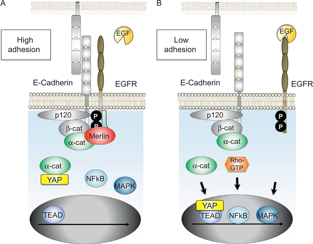
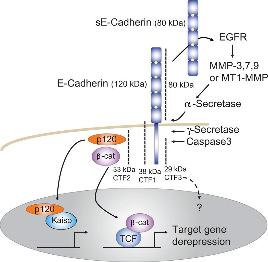
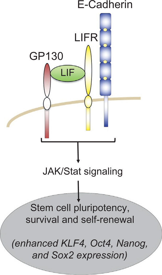
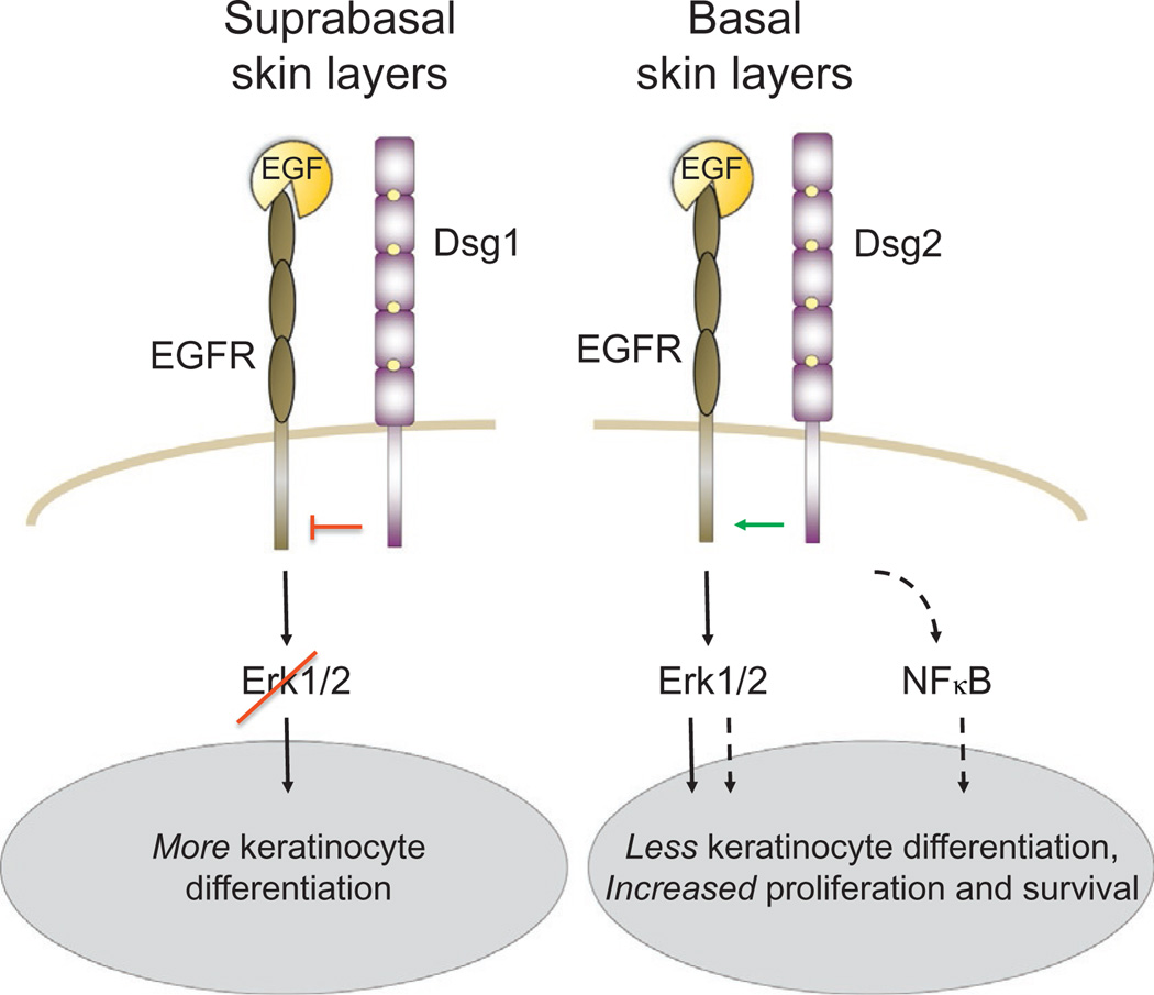
The arrival of multicellularity in evolution facilitated cell-cell signaling in conjunction with adhesion. As the ectodomains of cadherins interact with each other directly in trans (as well as in cis), spanning the plasma membrane and associating with multiple other entities, cadherins enable the transduction of "outside-in" or "inside-out" signals. We focus this review on signals that originate from the larger family of cadherins that are inwardly directed to the nucleus, and thus have roles in gene control or nuclear structure-function. The nature of cadherin complexes varies considerably depending on the type of cadherin and its context, and we will address some of these variables for classical cadherins versus other family members. Substantial but still fragmentary progress has been made in understanding the signaling mediators used by varied cadherin complexes to coordinate the state of cell-cell adhesion with gene expression. Evidence that cadherin intracellular binding partners also localize to the nucleus is a major point of interest. In some models, catenins show reduced binding to cadherin cytoplasmic tails favoring their engagement in gene control. When bound, cadherins may serve as stoichiometric competitors of nuclear signals. Cadherins also directly or indirectly affect numerous signaling pathways (e.g., Wnt, receptor tyrosine kinase, Hippo, NFκB, and JAK/STAT), enabling cell-cell contacts to touch upon multiple biological outcomes in embryonic development and tissue homeostasis.

Keywords: Atypical cadherins; Desmosomal cadherins; Hippo signaling; Outside-in signaling; Protocadherins; RTK signaling; Wnt signaling.


Figures

Figure 1
Figure 1
Cadherins as competitive stoichiometric inhibitors of p120ctn and β-catenin signaling. The model presented in (A) and (B) reflects evidence that cells with greater cadherin abundance (A=high levels of cadherin expression; B=lower cadherin expression) can sequester and thereby inhibit the ability of p120ctn and β-catenin to derepress activities of their respective DNA-binding factors, Rest/CoRest and TCF/LEF.
Figure 2
Figure 2
Cadherin-based adhesion as an enhancer of β-catenin destruction. In densely confluent cells with mature junctions, cadherins promote a faster turnover of β-catenin than in less adhesive (subconfluent) cells with immature junctions (A). This may explain why cells migrating adjacent to a wound appear sensitized to Wnt signals (Howard, Deroo, Fujita, & Itasaki, 2011; Maher et al., 2009). Mechanistically, the latter study suggests that enhanced β-catenin signaling is due to an activation step that depends on cadherins and a signaling endosome (B), while the former suggests that these cells show reduced capacity to degrade β-catenin (A).
Figure 3
Figure 3
Cadherins as facilitators of Wnt/β-catenin signaling. A number of studies show that E- and N-cadherins are required for efficient Wnt/β-catenin signaling, working at the level of the Wnt receptor complex. Work from the Dunach group shows that p120ctn facilitates Lrp5/6 phosphorylation and signaling by functioning as a proximal scaffold for CK1ε. p120ctn also contributes to the inhibition of GSK3 by multivesicular bodies leading to enhanced signaling from an endosome.
Figure 4
Figure 4
Nuclear roles for catenins. The armadillo family of catenin proteins (β-catenin, p120ctn, δ-catenin, ARVCF, and plakophilins (PKP) 2 and 3 (shades of purple; light gray in the print version)) modulate gene expression via association with cognate DNA-binding factors (shades of blue; gray in the print version). In most instances characterized to date, derepression occurs to activate the target genes. However, Pkp3 appears to further activate gene targets already positively modulated by ETV1, and δ-catenin's action upon ZIFCAT is not yet known. Lacking an armadillo domain and homology to the other catenins, α-catenin (green; gray in the print version) is structurally related to vinculin and may impact transcription through its actin-binding function, inhibiting RNA polymerase II (RNA Pol II). PKP2 can interact with a component of the RNA Pol III complex RPC155.
Figure 5
Figure 5
Cadherin signaling via RTKs, NFκB, and Hippo pathways. E-Cadherin in densely packed epithelial monolayers can inhibit access of EGF to the EGFR as well as down-stream signaling from the EGFR via Merlin (A), as contrasted with less mature contacts characterized by lowered extents of trans-E-cadherin interactions, where the indicated nuclear signaling trajectories are enhanced (B). Cadherin engagement can also limit the nuclear accumulation of YAP and NFκB through α-catenin and Rho-dependent mechanisms, respectively.
Figure 6
Figure 6
Cadherin signaling via proteolysis. Cadherins are cleaved at specific sites by proteases to generate fragments that are capable of transducing signals, either to the extracellular or to the intracellular space. The soluble extracellular E-cadherin fragment (sE-cadherin) can associate with intact E-cadherin present on other cells to alter cadherin-dependent cell properties inclusive of intracellular signaling (not shown). It also interacts with EGFR family members to activate MAPK signaling or metalloproteases, with the latter directly or indirectly resulting in the production of defined E-cadherin fragments. The intracellular C-terminal fragment number 2 (CTF2) is not membrane associated, so has been proposed to assist in the protection or translocation of p120ctn or β-catenin to the nucleus. Here, p120ctn displaces and thereby derepresses Kaiso-mediated repression of its gene targets, whereas β-catenin derepresses TCF/LEF-mediated gene repression via the recruitment of transcriptional coactivators. In the context of such selective proteolysis of E-cadherin, each catenin is thereby assisted in activating its respective gene targets.
Figure 7
Figure 7
Cadherin signaling promotes JAK/STAT signaling for stem cell maintenance. Mouse and human ESCs require E-cadherin for pluripotency, self-renewal, and long-term survival. The transmembrane and ectodomain of E-cadherin interact with the LIF receptor (LIFR) and GP130 coreceptor. This association stabilizes the complex and thereby promotes JAK/Stat signaling and consequent stemness properties to the stem cells.
Figure 8
Figure 8
Desmosomal cadherin signaling in skin. In the suprabasal skin layers, the cytoplasmic domain of Desmoglein1 inhibits EGFR signaling via the Erk1/2 signaling trajectory (Getsios et al., 2009). This might occur via a direct interaction of the Desmoglein1 cytodomain with EGFR. Ectopic Desmoglein2 expression (involucrin promoter) in the suprabasal keratinocyte layer increases proliferation and cell survival, apparently via effects upon EGFR and NFκB signaling (Brennan et al., 2007). Misexpression of desmocollin3a and 3b in subrabasal keratinocytes leads to proliferation and differentiation defects, with increased β-catenin signaling normally seen only in basal keratinocytes (Hardman et al., 2005) (not shown).

References

    1. Abe K, Takeichi M. NMDA-receptor activation induces calpain-mediated beta-catenin cleavages for triggering gene expression. Neuron. 2007;53:387–397. - PubMed
    1. Abreu-Blanco MT, Verboon JM, Liu R, Watts JJ, Parkhurst SM. Drosophila embryos close epithelial wounds using a combination of cellular protrusions and an actomyosin purse string. Journal of Cell Science. 2012;125:5984–5997. - PMC - PubMed
    1. Aktary Z, Kulak S, Mackey J, Jahroudi N, Pasdar M. Plakoglobin interacts with the transcription factor p53 and regulates the expression of 14-3-3 sigma. Journal of Cell Science. 2013;126:3031–3042. - PubMed
    1. Aktary Z, Pasdar M. Plakoglobin: Role in tumorigenesis and metastasis. International Journal of Cell Biology. 2012;2012:189–521. - PMC - PubMed
    1. Aktary Z, Pasdar M. Plakoglobin represses SATB1 expression and decreases in vitro proliferation, migration and invasion. PLoS One. 2013;8:e78388. - PMC - PubMed

Publication types

LinkOut - more resources