. 2015 May;197(9):1632-9.
doi: 10.1128/JB.00009-15. Epub 2015 Mar 2.

Identification and characterization of mutations conferring resistance to D-amino acids in Bacillus subtilis

Affiliations

Identification and characterization of mutations conferring resistance to D-amino acids in Bacillus subtilis

Sara A Leiman et al. J Bacteriol. 2015 May.

Abstract

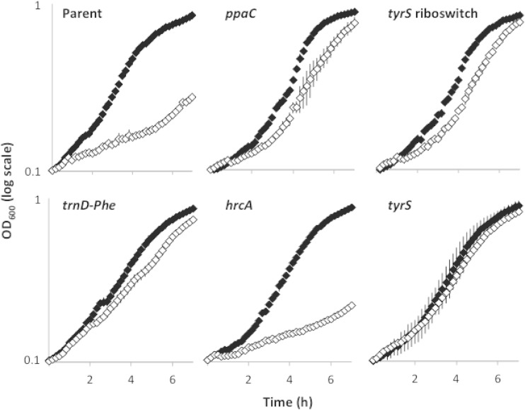
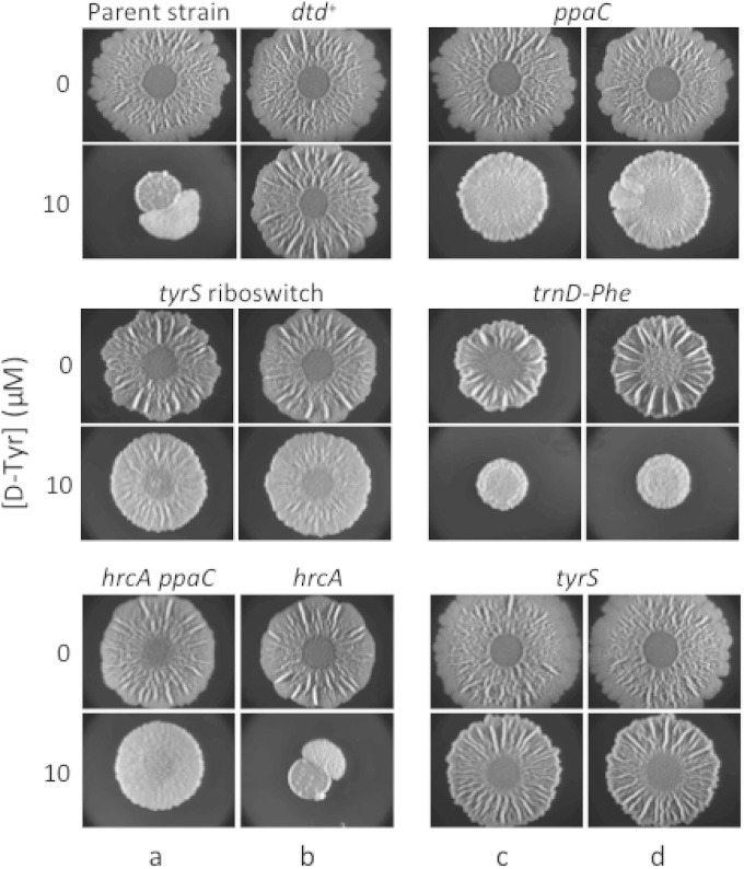
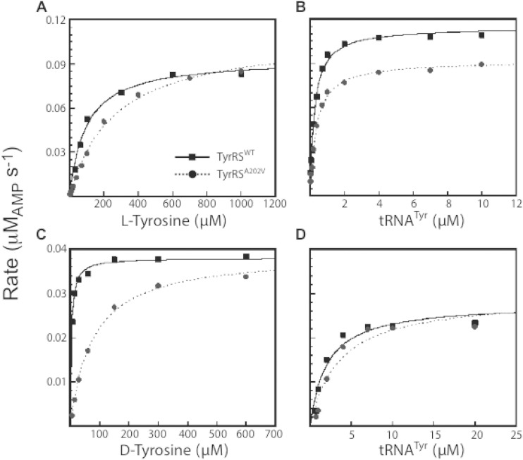
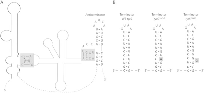
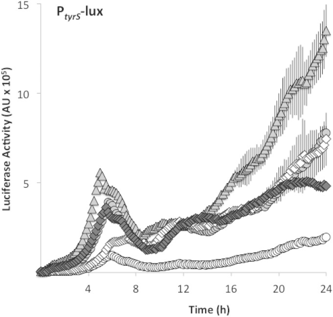
Bacteria produce d-amino acids for incorporation into the peptidoglycan and certain nonribosomally produced peptides. However, D-amino acids are toxic if mischarged on tRNAs or misincorporated into protein. Common strains of the Gram-positive bacterium Bacillus subtilis are particularly sensitive to the growth-inhibitory effects of D-tyrosine due to the absence of D-aminoacyl-tRNA deacylase, an enzyme that prevents misincorporation of D-tyrosine and other D-amino acids into nascent proteins. We isolated spontaneous mutants of B. subtilis that survive in the presence of a mixture of D-leucine, D-methionine, D-tryptophan, and D-tyrosine. Whole-genome sequencing revealed that these strains harbored mutations affecting tRNA(Tyr) charging. Three of the most potent mutations enhanced the expression of the gene (tyrS) for tyrosyl-tRNA synthetase. In particular, resistance was conferred by mutations that destabilized the terminator hairpin of the tyrS riboswitch, as well as by a mutation that transformed a tRNA(Phe) into a tyrS riboswitch ligand. The most potent mutation, a substitution near the tyrosine recognition site of tyrosyl-tRNA synthetase, improved enzyme stereoselectivity. We conclude that these mutations promote the proper charging of tRNA(Tyr), thus facilitating the exclusion of D-tyrosine from protein biosynthesis in cells that lack D-aminoacyl-tRNA deacylase.

Importance: Proteins are composed of L-amino acids. Mischarging of tRNAs with D-amino acids or the misincorporation of D-amino acids into proteins causes toxicity. This work reports on mutations that confer resistance to D-amino acids and their mechanisms of action.


Figures

FIG 1
FIG 1
Mutations from spontaneously arising mutants confer resistance to growth inhibition by d-amino acids when moved to the parental strain. Optical density of cells grown in shaking MSgg medium was measured in the presence (white diamonds) or absence (black diamonds) of 10 μM d-Tyr. The ppaCA145V, tyrS riboswitch (tyrS−38C>T), tyrSA202V, trnD-Phe35A>T, and hrcA−8A>G mutants were constructed by reconstituting select spontaneously arising mutations (Table 1) in the parent strain 3610. Results are shown as a semilog plot and indicate the averages from four replicates. Error bars show the standard deviations.
FIG 2
FIG 2
Congenic mutants exhibiting resistance to biofilm inhibition by d-amino acids. Spontaneous mutants (columns a and c) and their reconstituted counterparts (columns b and d) were spotted on solid MSgg medium alone or solid MSgg medium containing 10 μM d-Tyr. The reconstituted spontaneous mutants are as follows: ppaCA145V, tyrS riboswitch (tyrS−38C>T), tyrSA202V, trnD-Phe35A>T, and hrcA−8A>G. The parental strain of the spontaneous mutants (3610, which is mutant for dtd) and a dtd+ strain (SLH31) are included as a negative control and positive control, respectively. Images were taken after 72 h of incubation at 30°C.
FIG 3
FIG 3
Representative steady-state kinetics of B. subtilis TyrRS. Representative data from eight independent trials are shown for the measurement of binding affinity (Km) and turnover rate (kcat) of wild-type TyrRS (TyrRSWT) and mutant TyrRS (TyrRSA202V) for l-Tyr, d-Tyr, and tRNATyr. The experimental conditions were as follows: (A) 5 nM TyrRS and 2.5 μM tRNATyr, (B) 5 nM TyrRS and 1 mM l-Tyr, (C) 125 nM TyrRS and 10 μM tRNATyr, and (D) 125 nM TyrRS and 800 μM d-Tyr. All experiments included Mg-ATP at 10 mM. The results are summarized in Table 2.
FIG 4
FIG 4
Schematic of the B. subtilis tyrS riboswitch. (A) The interaction sites of a tRNATyr (gray) with the tyrS riboswitch (black) are highlighted with gray boxes. The gray box on the left shows binding between the tRNATyr anticodon (AUG) and the riboswitch specifier sequence (UAC). The gray box on the right shows binding between the 3′-end sequence of the tRNATyr (ACCA) and the antiterminator. The mutant tRNAPhe resulting from trnD-Phe35A>T harbors the AUG anticodon as well as the ACCA sequence at its 3′ end and may therefore bind the tyrS riboswitch at the same locations as does tRNATyr. Dashed lines represent riboswitch sequence that is not significant for binding tRNATyr. (B) Comparison of the wild-type (WT) tyrS terminator hairpin with the proposed structures of mutant terminators. The relevant mutations are in bold and highlighted with gray boxes. In panels A and B, bases shown in italics are shared between the antiterminator and terminator hairpins. The secondary structures in panel A and the WT tyrS structure in panel B were originally depicted by Grundy et al. (28) and Gerdeman et al. (29).
FIG 5
FIG 5
Mutations in the tyrS riboswitch and trnD-Phe cause readthrough. Expression of a luciferase transcriptional fusion to the tyrS riboswitch was measured in shaking MSgg medium. Riboswitch readthrough is indicated by white circles for the parental strain (SLH57) and by diamonds for the trnD-Phe35A>T mutant (SLH58). Luciferase fusions were also constructed with the tyrS riboswitch harboring either a C-to-T point mutation or an insertion mutation in the terminator hairpin. Readthrough of the tyrS riboswitch point mutant (SLH61) is shown as triangles, whereas readthrough of the insertion mutant (SLH59) is shown as gray circles. Readthrough of a tyrS riboswitch lacking the terminator hairpin sequence (SLH75) is indicated by white diamonds. Luciferase activity was determined by normalizing luminescence to optical density. Results shown are the averages from three replicates, and error bars show the standard deviations.
FIG 6
FIG 6
tapA mutations do not confer resistance to d-Tyr. A B. subtilis strain lacking the tapA operon (SLH63) was compared with a tapA+ strain (3610) as well as with strains complemented at amyE for the tapA operon containing wild-type (SLH64) tapA or mutant tapA genes harboring the previously described frameshift mutation tapA2 (SLH65) or tapA6 (SLH66). All strains were spotted on solid MSgg medium lacking l-FTW and containing the indicated concentration of d-Tyr. Images were taken after 72 h of incubation at 30°C.

References

    1. Lam H, Oh DC, Cava F, Takacs CN, Clardy J, de Pedro MA, Waldor MK. 2009. d-Amino acids govern stationary phase cell wall remodeling in bacteria. Science 325:1552–1555. doi:10.1126/science.1178123. - DOI - PMC - PubMed
    1. Vollmer W, Blanot D, de Pedro MA. 2008. Peptidoglycan structure and architecture. FEMS Microbiol Rev 32:149–167. doi:10.1111/j.1574-6976.2007.00094.x. - DOI - PubMed
    1. Lupoli TJ, Tsukamoto H, Doud EH, Wang TS, Walker S, Kahne D. 2011. Transpeptidase-mediated incorporation of d-amino acids into bacterial peptidoglycan. J Am Chem Soc 133:10748–10751. doi:10.1021/ja2040656. - DOI - PMC - PubMed
    1. Ongena M, Jacques P. 2008. Bacillus lipopeptides: versatile weapons for plant disease biocontrol. Trends Microbiol 16:115–125. doi:10.1016/j.tim.2007.12.009. - DOI - PubMed
    1. Perego M, Glaser P, Minutello A, Strauch MA, Leopold K, Fischer W. 1995. Incorporation of d-alanine into lipoteichoic acid and wall teichoic acid in Bacillus subtilis. Identification of genes and regulation. J Biol Chem 270:15598–15606. - PubMed

Publication types

MeSH terms

LinkOut - more resources