. 2015 May;35(10):1660-72.
doi: 10.1128/MCB.01478-14. Epub 2015 Mar 2.

Promyelocytic Leukemia Protein Isoform II Promotes Transcription Factor Recruitment To Activate Interferon Beta and Interferon-Responsive Gene Expression

Affiliations

Promyelocytic Leukemia Protein Isoform II Promotes Transcription Factor Recruitment To Activate Interferon Beta and Interferon-Responsive Gene Expression

Yixiang Chen et al. Mol Cell Biol. 2015 May.

Abstract

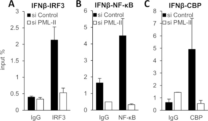
To trigger type I interferon (IFN) responses, pattern recognition receptors activate signaling cascades that lead to transcription of IFN and IFN-stimulated genes (ISGs). The promyelocytic leukemia (PML) protein has been implicated in these responses, although its role has not been defined. Here, we show that PML isoform II (PML-II) is specifically required for efficient induction of IFN-β transcription and of numerous ISGs, acting at the point of transcriptional complex assembly on target gene promoters. PML-II associated with specific transcription factors NF-κB and STAT1, as well as the coactivator CREB-binding protein (CBP), to facilitate transcriptional complex formation. The absence of PML-II substantially reduced binding of these factors and IFN regulatory factor 3 (IRF3) to IFN-β or ISGs promoters and sharply reduced gene activation. The unique C-terminal domain of PML-II was essential for its activity, while the N-terminal RBCC motif common to all PML isoforms was dispensable. We propose a model in which PML-II contributes to the transcription of multiple genes via the association of its C-terminal domain with relevant transcription complexes, which promotes the stable assembly of these complexes at promoters/enhancers of target genes, and that in this way PML-II plays a significant role in the development of type I IFN responses.


Figures

FIG 1
FIG 1
Depletion of PML-II reduces IFN-β expression. (A) HEK293 cells were transfected with 125 pmol/ml siRNA as indicated and, after 48 h, transfected with 1 μg/ml poly(I·C) for 16 h. PML-II mRNA levels in total RNA were determined by RT-qPCR and normalized to β-actin mRNA levels and are shown here relative to values obtained from control siRNA-treated cells. (B) HEK293 cells grown on coverslips were treated with siRNA and poly(I·C) as described for panel A and then fixed and stained with PML-II antibody. Images shown are overlays of DAPI (4′,6-diamidino-2-phenylindole) (blue) and PML-II staining (green) and are representative of multiple fields that were randomly selected based on DAPI fluorescence. Scale bar, 20 μm. (C) HEK293 cells were cotransfected with 450 ng pCI-neo and either 50 ng Flag-PML-II (upper panels) or Flag-PML-V (lower panels), alongside 75 pmol/ml or 125 pmol/ml of the siRNAs indicated. Control siRNA cells were treated with 125 pmol/ml siRNA. Forty-eight hours later, cells were harvested and analyzed by SDS-PAGE and Western blotting with anti-FLAG and antiactin antibodies. Protein sizes are indicated in kDa. Empty vector (EV) lane, background detection by the anti-FLAG antibody. (D) HEK293 cells were treated with the siRNAs indicated for 24 h, cotransfected with IFN-β-Luc and CMV-βgal plasmids for a further 24 h, and then transfected with 1 μg/ml poly(I·C) for 16 h before being harvested for reporter assays. Error bars indicate the standard deviations (SD) from the means for at least three biological replicates within an experiment. (E) HEK293 cells were treated with the siRNAs indicated for 48 h and then stimulated with poly(I·C) as described for panel A. IFN-β mRNA levels in total RNA were determined by RT-qPCR and normalized to β-actin mRNA levels and are shown here relative to values obtained from control siRNA-treated, poly(I·C)-induced cells. In this experiment, poly(I·C) treatment achieved an 8,000-fold stimulation of IFN-β mRNA levels. Data are the means ± SD of three replicate values from one representative of three experiments. (F) HeLa cells were treated and assayed for IFN-β mRNA levels, and data were analyzed and presented as described for panel E. Poly(I·C) treatment achieved a 1,000-fold stimulation of IFN-β mRNA levels. (G) MRC5 cells were treated with 50 pmol/ml siRNA for 72 h prior to transfection with 1 μg/ml poly(I·C). Twenty-four hours later, total RNA was analyzed for IFN-β expression by RT-qPCR and data were analyzed as described for panel E. Data are the means ± SD from five replicates in two independent experiments. A fold stimulation value for the effect of poly(I·C) could not be obtained, as basal IFN-β expression was undetectable.
FIG 2
FIG 2
Depletion of PML-II impairs activities of IRF3 and NF-κB. (A, E) HEK293 cells were treated with siRNA as indicated for 24 h, cotransfected with CMV-βgal and PRDIII/I-Luc (A) or PRDII-Luc (E) for 24 h, stimulated with 1 μg/ml poly(I·C) or control treated for 16 h, and then assayed for reporter activity. Error bars indicate the standard deviations from the means for at least three replicates. (B to D, F to H) HEK293 cells were treated with siRNA for 48 h and stimulated with poly(I·C) as described for panel A. Total RNA was analyzed for specific mRNA levels by RT-qPCR. mRNA levels are displayed relative to those observed in control siRNA-treated cells without stimulation. Error bars indicate the standard deviations of the means from at least three replicates within an experiment. (I) As for panels B to D and F to H, except values were normalized in each case to the level observed in control siRNA-, poly(I·C)-treated cells; there was no significant difference in expression levels with or without stimulation.
FIG 3
FIG 3
PML-II depletion reduces expression of ISGs. (A, B) HEK293 cells were transfected with siRNA as indicated for 24 h and then cotransfected with ISRE-Luc and CMV-βgal plasmids for 24 h. Following either poly(I·C) transfection for 16 h or IFN-α treatment for 8 h, cell lysates were assayed for reporter activity. Data shown are the means ± SD from at least three replicates. (C to J) HEK293 cells were treated with siRNA for 48 h and stimulated with either poly(I·C) for 16 h (C to F) or 1,000 units/ml IFN-α for 8 h (G to J). Total RNA was analyzed for specific mRNA levels by RT-qPCR. mRNA levels are displayed relative to those observed in control siRNA-treated cells without stimulation. Quantification was performed as described for Fig. 1E. Data shown are the means ± SD from at least three replicates within an experiment.
FIG 4
FIG 4
PML-II has limited effects on cytoplasmic events in IFN-β induction. (A) HEK293 cells were transfected with 125 pmol/ml PML-II or control siRNA for 48 h and then stimulated by transfection with 1 μg/ml poly(I·C) (t = 0) before collection at the indicated time points. Samples were separated by SDS-PAGE, blotted, and probed with antibody to phosphorylated IRF3, total IRF3, or β-actin. Band intensities in digitized images were quantified using QuantityOne software (Bio-Rad); the amounts of pIRF3 detected are shown relative to total IRF3. (B) HEK293 cells grown on coverslips were treated with siRNA as described for panel A and then transfected with 1 μg/ml poly(I·C) or mock transfected for a further 16 h before fixation and detection of IRF3 by immunofluorescence. Arrows indicate examples of nuclei that showed positive staining for IRF3 following poly(I·C) stimulation. (C) As for panel B but detecting NF-κB. (D) Quantification of IRF3 and NF-κB nuclear staining. All nuclei in a field were detected by DAPI staining, and the proportion showing positive staining for IRF3 or NF-κB was determined manually over 2 or 3 fields of view.
FIG 5
FIG 5
PML-II interacts with transcriptional complexes. (A, B) HEK293 cells were mock stimulated or transfected with 1 μg/ml poly(I·C) for 16 h and then lysed with 0.5% NP-40 lysis buffer. A portion of each cell lysate was retained for direct analysis by Western blotting, while equal parts of the remainder were incubated with IRF3, NF-κB (p65), or CBP antibodies or with control IgG and precipitated with protein A-Sepharose beads; proteins in total or immunoprecipitated samples were separated by SDS-PAGE, and IRF3, NF-κB, or CBP was detected by Western blotting. (C) HEK293 cells were transfected with 250 ng/ml Flag-PML-II plasmid or pCI-neo empty vector for 24 h and then stimulated with poly(I·C); lysates were prepared and immunoprecipitated with anti-Flag beads, and precipitates and total lysates were analyzed for Flag-PML, CBP and NF-κB, and STAT1 as described for panel A. (D, E) HEK293 cells were treated with siRNA and then stimulated with poly(I·C) as described for Fig. 1A. Lysates were prepared and immunoprecipitated with IRF3 or CBP antibodies, and precipitates and total lysates were analyzed for IRF3, STAT1, and CBP as described for panel A.
FIG 6
FIG 6
Knockdown of PML-II impairs IRF3, NF-κB, and CBP recruitment to the IFN-β promoter. HEK293 cells were transfected with PML-II or control siRNA for 48 h and stimulated with poly(I·C) for 16 h for IRF3, 10 h for NF-κB, or 10 h for CBP. Cells were then fixed and subjected to ChIP with IRF3 (A), NF-κB (B), or CBP (C) antibodies, in each case in parallel with a normal IgG control precipitation on an equal volume of extract. Precipitated DNA was assayed for IFN-β promoter sequences by SYBR green qPCR. Precipitated DNA is expressed as the mean percentage of the amount of that DNA present in the extract volume subjected to precipitation ± SD from one representative of at least two experiments.
FIG 7
FIG 7
Removal of PML-II affects transcription factor binding at the ISRE elements of ISGs. (A) HEK293 cells were transfected with PML-II or control siRNA for 48 h and then transfected with poly(I·C) for 16 h. Cells were fixed and subjected to ChIP with STAT1 antibodies in parallel with a normal IgG control precipitation on an equal volume of extract. Precipitated DNA was assayed for ISG15, ISG56, and PKR promoter sequences by SYBR green qPCR, and data were processed as for Fig. 6. (B) HEK293 cells treated with siRNA as described for panel A were fixed and subjected to ChIP analysis using p53 antibodies and qPCR primers specific for the p21 promoter.
FIG 8
FIG 8
PML-II functions in gene expression via its unique C-terminal domain. (A) Representation of the C-terminal region of PML-II showing the positions of deletion mutations (47). (B) HEK293 cells were transfected with 250 ng/ml Flag-PML-II wild-type or deletion mutant plasmid or pCI-neo empty vector as indicated for 48 h and then stimulated with poly(I·C), and lysates were prepared and immunoprecipitated with anti-Flag beads. Precipitates and total lysates were analyzed for Flag-PML, NF-κB (p65), and STAT1 by Western blotting. (C) As described for panel B but using ΔRBCC variants of each PML-II plasmid. (D, E) HEK293 cells were transfected with plasmids as described for panels B and C. Following 1 μg/ml poly(I·C) (B) or 1,000 units/ml IFN-α stimulation (C) for 16 h, total RNA was analyzed for specific mRNA levels by RT-qPCR. mRNA levels are displayed relative to those observed in control siRNA-treated cells without stimulation. Quantification was performed as described for Fig. 1. Data shown are the means ± SD for at least three replicates within an experiment.
FIG 9
FIG 9
PML-II associates with target promoters. HEK293 cells were transfected with 250 ng/ml Flag-PML-II-ΔRBCC, Flag-PML-V-ΔRBCC, or pCI-neo plasmids, and 48 h later, cells were transfected with poly(I·C) for 16 h. Cell lysates were subjected to ChIP with normal IgG or anti-Flag antibody. Precipitated DNA was assayed by SYBR green qPCR for ISG15 and ISG56 promoter or nonpromoter sequences. Results are presented as means ± SD from triplicate determinations within an experiment.

Similar articles

Cited by

References

    1. Randall RE, Goodbourn S. 2008. Interferons and viruses: an interplay between induction, signalling, antiviral responses and virus countermeasures. J Gen Virol 89:1–47. doi:10.1099/vir.0.83391-0. - DOI - PubMed
    1. Mogensen TH. 2009. Pathogen recognition and inflammatory signaling in innate immune defenses. Clin Microbiol Rev 22:240–273. doi:10.1128/CMR.00046-08. - DOI - PMC - PubMed
    1. Jacobs BL, Langland JO. 1996. When two strands are better than one: the mediators and modulators of the cellular responses to double-stranded RNA. Virology 219:339–349. doi:10.1006/viro.1996.0259. - DOI - PubMed
    1. Yoneyama M, Fujita T. 2009. RNA recognition and signal transduction by RIG-I-like receptors. Immunol Rev 227:54–65. doi:10.1111/j.1600-065X.2008.00727.x. - DOI - PubMed
    1. Wathelet MG, Lin CH, Parekh BS, Ronco LV, Howley PM, Maniatis T. 1998. Virus infection induces the assembly of coordinately activated transcription factors on the IFN-beta enhancer in vivo. Mol Cell 1:507–518. doi:10.1016/S1097-2765(00)80051-9. - DOI - PubMed

Publication types

MeSH terms