. 2015 May;14(5):1323-33.
doi: 10.1074/mcp.M114.047837. Epub 2015 Mar 2.

Glycan motif profiling reveals plasma sialyl-lewis x elevations in pancreatic cancers that are negative for sialyl-lewis A

Affiliations

Glycan motif profiling reveals plasma sialyl-lewis x elevations in pancreatic cancers that are negative for sialyl-lewis A

Huiyuan Tang et al. Mol Cell Proteomics. 2015 May.

Abstract

The sialyl-Lewis A (sLeA) glycan forms the basis of the CA19-9 assay and is the current best biomarker for pancreatic cancer, but because it is not elevated in ∼25% of pancreatic cancers, it is not useful for early diagnosis. We hypothesized that sLeA-low tumors secrete glycans that are related to sLeA but not detectable by CA19-9 antibodies. We used a method called motif profiling to predict that a structural isomer of sLeA called sialyl-Lewis X (sLeX) is elevated in the plasma of some sLeA-low cancers. We corroborated this prediction in a set of 48 plasma samples and in a blinded set of 200 samples. An antibody sandwich assay formed by the capture and detection of sLeX was elevated in 13 of 69 cancers that were not elevated in sLeA, and a novel hybrid assay of sLeA capture and sLeX detected 24 of 69 sLeA-low cancers. A two-marker panel based on combined sLeA and sLeX detection differentiated 109 pancreatic cancers from 91 benign pancreatic diseases with 79% accuracy (74% sensitivity and 78% specificity), significantly better than sLeA alone, which yielded 68% accuracy (65% sensitivity and 71% specificity). Furthermore, sLeX staining was evident in tumors that do not elevate plasma sLeA, including those with poorly differentiated ductal adenocarcinoma. Thus, glycan-based biomarkers could characterize distinct subgroups of patients. In addition, the combined use of sLeA and sLeX, or related glycans, could lead to a biomarker panel that is useful in the clinical diagnosis of pancreatic cancer. Précis: This paper shows that a structural isomer of the current best biomarker for pancreatic cancer, CA19-9, is elevated in the plasma of patients who are low in CA19-9, potentially enabling more comprehensive detection and classification of pancreatic cancers.


Conflict of interest statement

Conflicts of Interest: None declared.

Figures
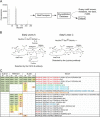
Fig. 1.
Fig. 1.
Identifying binders to sialyl-Lewis A. (A) We used a database of glycan array data to search for binders to sialyl-Lewis A. To develop the database, we processed all the available datasets from the Consortium for Functional Glycomics using the motif segregation algorithm, and stored the metadata, raw data, analyzed data, and motifs. (B) Two motifs up-regulated in pancreatic cancer are sialyl-Lewis A and sialyl-Lewis C, each detectable by a monoclonal antibody. (C) We selected anti-sLeA clones SLE121, M081221, and 9L426 for use in profiling experiments based on their diversity in specificity. The numbers are derived from glycan array experiments and represent binding to the indicated glycan in fluorescence unit. The number above each column is the concentration of the antibody incubated on the array, in μg/ml. Green cells indicate high binding, yellow is medium binding, and orange is low binding.
Fig. 2.
Fig. 2.
Capture:detection combinations potentially associated with sLeA-low cancers. (A) We used antibody arrays to examine multiple capture:detection combinations. A sample is incubated on an antibody array to allow glycan capture by the immobilized antibodies, and then a single detection antibody is incubated to probe its target at each capture antibody. This process is repeated for multiple detection antibodies. The notation sLeA(clone ID):sLeA(clone ID) indicates capture by the first sLeA antibody and detection by the second sLeA antibody. (B) The patient samples are grouped according to diagnosis and CA19–9 level. The graphs depict results from the capture:detection combinations that showed significantly different levels in the CA19–9-low cancers relative to the control samples. Each column is the average of two replicates. In the sample labels, “Mix” refers to a mixture of two different samples of the same classification, which we did because of low volume available from the individual samples.
Fig. 3.
Fig. 3.
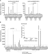
Testing predictions with sialyl-Lewis X antibodies. (A) The graphs depict results from assays that use the anti-sLeX antibody (clone CSLEX1) as the capture. Detection with two of the sLeA antibodies showed elevations in 3 of the 6 sLeA-low cancers, relative to the controls and other cancers. (B) The measurements were obtained using anti-sLeX as both the capture and detection antibody. The values represent the average of three independent incubations, with the standard deviations indicated by the error bars. The dashed line indicates a threshold chosen to maximize detection in the low-sLeA group while minimizing false-positive detections, and the asterisks indicate the samples that are elevated. The sLeX assay showed elevations in seven of the 15 sLeA-low cancer group and only two of the 26 controls. The inset compares sLeA to sLeX levels and shows distinct groups with elevations in either marker.
Fig. 4.
Fig. 4.
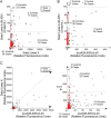
sLeX and dual expression of sLeA and sLeX are elevated in some sLeA-low cancers. (A) The graph shows the correlation between sLeA (using anti-sLeA clone 9L426 as capture and detection, y axis) and sLeX (using anti-sLeX clone CSLEX1 as capture and detection, x axis). Each point represents a patient sample, and each value is the average of three replicates. The dashed lines represent thresholds defining elevations for each assay. (B) The y axis indicates the sLeA measurements, and the y axis indicates measurements of dual expression of sLeA and sLeX, using anti-sLeA capture and anti-sLeX detection. At the thresholds defined by the dashed lines, many cancers are elevated only in the sLeA sandwich assay or the sLeA:sLeX sandwich assay. (C) The graph depicts measurements from the capture:detection combinations indicated on the axis labels, and the right graph is a zoomed portion of the left. At the thresholds defined by the dashed lines, many cancers are elevated only in the sLeX sandwich assay or the sLeA:sLeX sandwich assay.
Fig. 5.
Fig. 5.
Relationships between plasma levels and tissue staining. (A) The sLeA and sLeX plasma levels are depicted for 14 pancreatic cancer patients who had pancreatic resections and for 3 healthy control subjects. Each point is the average of three replicate measurements, and a patient ID is shown for selected samples. The dashed lines indicate thresholds defining elevation. (B–G) Each panel shows adjacent, or near adjacent, sections stained with anti-sLeA or anti-sLeX. The inset numbers correspond to the IDs in panel A. Note the well-defined ducts in cases high in plasma sLeA (B and C) and the various other histomorphologies in cases with low plasma sLeA, including adenosquamous formations (D); clusters of invasive cells without mucin-producing ducts (E); and intestinal differentiation arising in the ampulla (F). Neuroendocrine tumors (G) showed low sLeA and high sLeX staining in the cancer cells, with corresponding plasma levels.

References

    1. Abraham S. C., Wilentz R. E., Yeo C. J., Sohn T. A., Cameron J. L., Boitnott J. K., Hruban R. H. (2003) Pancreaticoduodenectomy (whipple resections) in patients without malignancy: Are they all “chronic pancreatitis”? Am. J. Surg. Pathol. 27, 110–120 - PubMed
    1. Adsay N. V., Bandyopadhyay S., Basturk O., Othman M., Cheng J. D., Klöppel G., Klimstra D. S. (2004) Chronic pancreatitis or pancreatic ductal adenocarcinoma? Semin. Diagn. Pathol .21, 268–276 - PubMed
    1. Kennedy T., Preczewski L., Stocker S. J., Rao S. M., Parsons W. G., Wayne J. D., Bell R. H., Talamonti M. S. (2006) Incidence of benign inflammatory disease in patients undergoing whipple procedure for clinically suspected carcinoma: A single-institution experience. Am. J. Surg. 191, 437–441 - PubMed
    1. Klöppel G., Adsay N. V. (2009) Chronic pancreatitis and the differential diagnosis versus pancreatic cancer. Arch. Pathol. Lab. Med. 133, 382–387 - PubMed
    1. Pitman M. B., Centeno B. A., Ali S. Z., Genevay M., Stelow E., Mino-Kenudson M., Castillo C. F., Schmidt C. M., Brugge W. R., Layfield L. J. (2014) Standardized terminology and nomenclature for pancreatobiliary cytology: The papanicolaou society of cytopathology guidelines. Cytojournal 11, 3. - PMC - PubMed

Publication types

MeSH terms