. 2015 Mar 10;112(10):3092-7.
doi: 10.1073/pnas.1419388112. Epub 2015 Mar 2.

MinD-like ATPase FlhG effects location and number of bacterial flagella during C-ring assembly

Affiliations

MinD-like ATPase FlhG effects location and number of bacterial flagella during C-ring assembly

Jan S Schuhmacher et al. Proc Natl Acad Sci U S A. .

Abstract

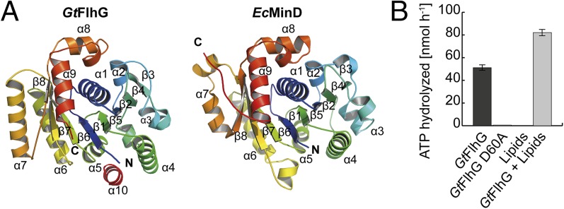
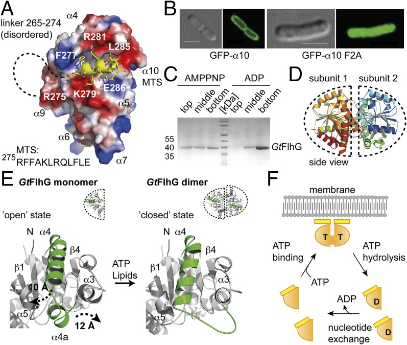
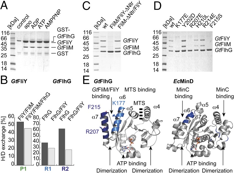
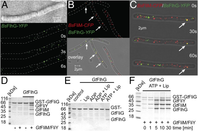
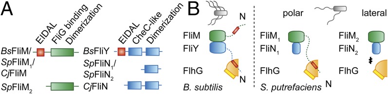
The number and location of flagella, bacterial organelles of locomotion, are species specific and appear in regular patterns that represent one of the earliest taxonomic criteria in microbiology. However, the mechanisms that reproducibly establish these patterns during each round of cell division are poorly understood. FlhG (previously YlxH) is a major determinant for a variety of flagellation patterns. Here, we show that FlhG is a structural homolog of the ATPase MinD, which serves in cell-division site determination. Like MinD, FlhG forms homodimers that are dependent on ATP and lipids. It interacts with a complex of the flagellar C-ring proteins FliM and FliY (also FliN) in the Gram-positive, peritrichous-flagellated Bacillus subtilis and the Gram-negative, polar-flagellated Shewanella putrefaciens. FlhG interacts with FliM/FliY in a nucleotide-independent manner and activates FliM/FliY to assemble with the C-ring protein FliG in vitro. FlhG-driven assembly of the FliM/FliY/FliG complex is strongly enhanced by ATP and lipids. The protein shows a highly dynamic subcellular distribution between cytoplasm and flagellar basal bodies, suggesting that FlhG effects flagellar location and number during assembly of the C-ring. We describe the molecular evolution of a MinD-like ATPase into a flagellation pattern effector and suggest that the underappreciated structural diversity of the C-ring proteins might contribute to the formation of different flagellation patterns.

Keywords: Bacillus; C-ring; FlhG; Shewanella; flagellum.


Conflict of interest statement

The authors declare no conflict of interest.

Figures

Fig. 1.
Fig. 1.
Crystal structure of the MinD ATPase FlhG. (A) Cartoon representation of the crystal structures of GtFlhG (this study, Left) and EcMinD (PDB ID: 3Q9L, Right). Both structures are rainbow-colored from the N to the C terminus as indicated by “N” and “C,” respectively. (B) ATPase activity of GtFlhG and the GtFlhG D60A variant (in nanomoles per hour) in the absence or presence of lipids. GtFlhG (20 µM) was incubated with 2 mM ATP at 37 °C for 1 h.
Fig. 2.
Fig. 2.
Lipid- and ATP-dependent homodimerization of FlhG. (A) Electrostatic surface view of GtFlhG with the MTS shown in yellow. Dashed lines indicate the disordered linker (residues 265–274) connecting ATPase and the MTS. (B) In vivo fluorescence micrographs of GFP-α10 and GFP-α10-F2A show that α10 of GtFlhG is a functional MTS. (Scale bars, 2 µm.) (C) Coomassie-stained SDS/PAGE of the flotation assay of GtFlhG with LUVs in the presence of ADP and AMPPNP (from top to bottom fractions). Note: GtFlhG interacting with LUVs is found in the top fraction. (D) Cartoon representation of the GtFlhG homodimer. Dashed lines indicate each monomer. Note: Although ATP was added before crystallization, the crystal structure of the GtFlhG homodimer has only ADP bound in its active sites, likely because of residual ATPase activity during crystal growth (3–4 wk). (E) Structural differences between the monomeric and dimeric states of GtFlhG. Major conformational changes are shown in green. (F) Model of the FlhG ATPase mechanism (orange) showing the ATP (T)-dependent homodimerization and expulsion of the MTS (yellow), membrane interaction of the homodimer through the MTS, and ATP hydrolysis-dependent dissociation of the homodimer.
Fig. 3.
Fig. 3.
FlhG interacts with the flagellar C-ring proteins FliM/FliY. (A) Coomassie-stained SDS/PAGE of an in vitro pulldown assay of GST-GtFlhG and GtFliM/FliY in the absence and presence of ADP, ATP, or AMPPNP. (B) Deuterium incorporation of depicted peptides of free protein and the dimeric GtFlhG/FliY complex are given in percent H/D exchange. Decreased deuterium content upon complex formation indicates potential interfaces (peptides GtFliY R1: DALLRGMDDSDHVPALH; GtFlhG P1: TDAYAMMKYMHAAGSEAPFSV and P2: VFERLKHVTGRFLNKD). (C) Coomassie-stained SDS/PAGE of an in vitro pulldown assay using (His)6-tagged GtFlhG, GtFliM/FliY, and GtFliM/FliY variants lacking the FliY-Ntr and FliM-Ntr. (D) Coomassie-stained SDS/PAGE of an in vitro pulldown assay using different (His)6-tagged GtFlhG variants and the GtFliM/GtFliY complex. (E) Major differences that allow FlhG (Left) and MinD (Right) to bind FliM/FliY and MinC, respectively, are shown in blue.
Fig. 4.
Fig. 4.
Physiological role of FlhG. (A) Localization of a BsFlhG-YFP fusion protein in B. subtilis displays distinct foci at the membrane. (B and C) Localization of FlhG and FliM was detected in B. subtilis carrying BsFlhG-YFP (green) and BsFliM-CFP (red). (D) Coomassie-stained SDS/PAGE of an in vitro pulldown assay of GST-GtFliG with GtFliM/FliY in the absence and presence of GtFlhG. (E) Coomassie-stained SDS/PAGE investigating the ability of GtFlhG to bind to GST-GtFliG and its dependence on lipids, ADP, ADP+lipids, ATP, and ATP+lipids. (F) Coomassie-stained SDS/PAGE of a time-resolved pulldown assay (0, 1, 5, 10, and 30 min) investigating the binding of GtFliM/FliY and GtFlhG to GST-GtFliG in presence of ATP and lipids.
Fig. 5.
Fig. 5.
Role of FlhG in the polar- and lateral-flagellated S. putrefaciens. (A) Electron micrographs of S. putrefaciens (Upper) and its ΔflhG mutant (Lower). (B) SpFlhG ATPase activity (in nanomoles per hour). FlhG (100 µM) was incubated with 1 mM ATP at 37 °C for 30 min. (C) Coomassie-stained SDS/PAGE of an in vitro pulldown assay shows that (His)6-tagged SpFlhG interacts with SpFliM1/FliN1 but not with SpFliM2/FliN2. (D) Coomassie-stained SDS/PAGE of an in vitro pulldown assay shows that (His)6-tagged SpFlhG does not interact with an SpFliM1/FliN1 variant lacking the N-terminal 27 amino acids of FliM1 (FliM1-ΔNtr). (E, Left) FliM1-mCherry localizes in distinct foci at the cell pole of S. putrefaciens in 41% of the cells (n = 576). (Right) FliM1-ΔNtr-mCherry displays decreased polar localization (22% of cells; n = 456).
Fig. 6.
Fig. 6.
FlhG and the diversity of flagellar C-ring proteins. (A) Diversity in the architecture of the C-ring proteins FliM (Left, green) and FliY/FliN (Right, blue) from B. subtilis, S. putrefaciens, and C. jejuni. The conserved EIDAL motif is shown in red. (B) Interaction of FlhG with flagellar C-ring complexes in B. subtilis (Left) and S. putrefaciens (Right). The EIDAL motifs are shown in red. “N” indicates N termini.

References

    1. Chevance FF, Hughes KT. Coordinating assembly of a bacterial macromolecular machine. Nat Rev Microbiol. 2008;6(6):455–465. - PMC - PubMed
    1. Altegoer F, Schuhmacher J, Pausch P, Bange G. From molecular evolution to biobricks and synthetic modules: A lesson by the bacterial flagellum. Biotechnol Genet Eng Rev. 2014;30:49–64. - PubMed
    1. Kusumoto A, et al. Regulation of polar flagellar number by the flhF and flhG genes in Vibrio alginolyticus. J Biochem. 2006;139(1):113–121. - PubMed
    1. Campos-García J, Nájera R, Camarena L, Soberón-Chávez G. The pseudomonas aeruginosa motR gene involved in regulation of bacterial motility. FEMS Microbiol Lett. 2000;184(1):57–62. - PubMed
    1. Dasgupta N, Arora SK, Ramphal R. fleN, a gene that regulates flagellar number in Pseudomonas aeruginosa. J Bacteriol. 2000;182(2):357–364. - PMC - PubMed

Publication types

Substances

LinkOut - more resources