. 2015 Mar 17;112(11):3380-5.
doi: 10.1073/pnas.1502214112. Epub 2015 Mar 2.

Nanomechanics and intermolecular forces of amyloid revealed by four-dimensional electron microscopy

Affiliations

Nanomechanics and intermolecular forces of amyloid revealed by four-dimensional electron microscopy

Anthony W P Fitzpatrick et al. Proc Natl Acad Sci U S A. .

Abstract

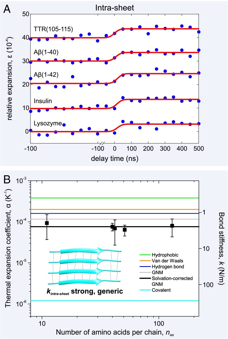
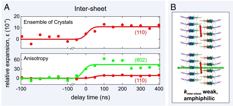
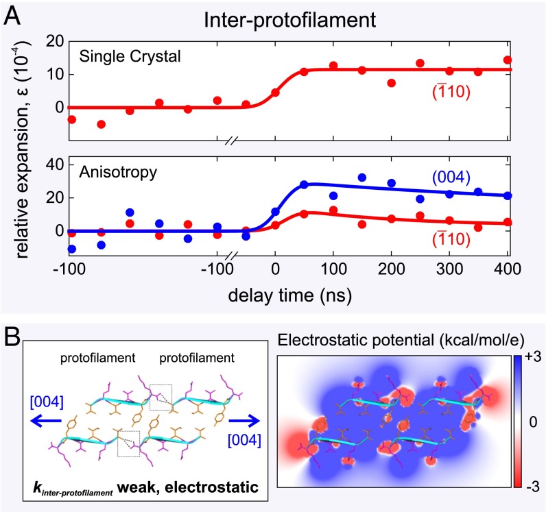
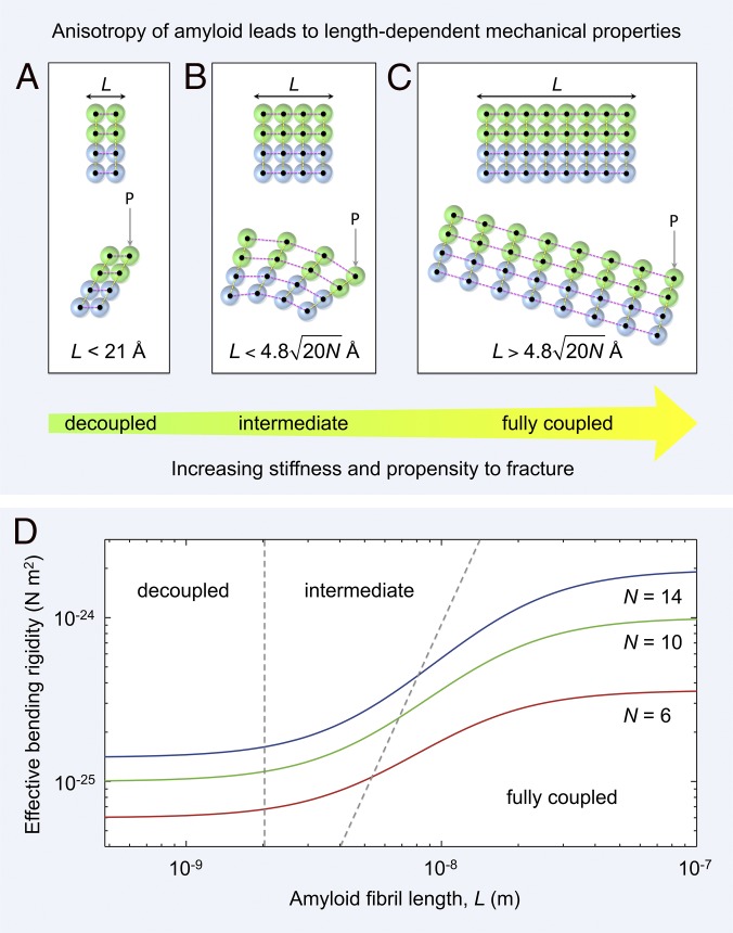
The amyloid state of polypeptides is a stable, highly organized structural form consisting of laterally associated β-sheet protofilaments that may be adopted as an alternative to the functional, native state. Identifying the balance of forces stabilizing amyloid is fundamental to understanding the wide accessibility of this state to peptides and proteins with unrelated primary sequences, various chain lengths, and widely differing native structures. Here, we use four-dimensional electron microscopy to demonstrate that the forces acting to stabilize amyloid at the atomic level are highly anisotropic, that an optimized interbackbone hydrogen-bonding network within β-sheets confers 20 times more rigidity on the structure than sequence-specific sidechain interactions between sheets, and that electrostatic attraction of protofilaments is only slightly stronger than these weak amphiphilic interactions. The potential biological relevance of the deposition of such a highly anisotropic biomaterial in vivo is discussed.

Keywords: 4D electron diffraction; nanomechanics; proteins; structural dynamics.


Conflict of interest statement

The authors declare no conflict of interest.

Figures

Fig. 1.
Fig. 1.
Images, selected area diffraction patterns, and simulated diffraction patterns of (A) a network of amyloid fibrils, (B) an ensemble of amyloid-like microcrystals, and (C) a single microcrystal taken using our 4D electron microscope. Protein density is shown in white on (A and B) lacey carbon substrate and (C) holey silicon nitride substrate, respectively. (A) Image and diffraction patterns of a network of amyloid fibrils. Note the strong 4.8-Å reflection––a hallmark of amyloid structure corresponding to the interstrand separation within β-sheets. (B) Image and diffraction patterns of an ensemble of amyloid-like microcrystals. The 4.8-Å (1 1 0) Bragg peaks give rise to the most intense Debye–Scherrer ring. Other reflections can be used to gain a more complete picture of lattice dynamics. (C) Image and diffraction patterns of a single amyloid microcrystal. Note the intense, paired (1¯ 1 0) and (1 1¯ 0) spots at 4.8 Å, and the (0 0 4) and (0 0 4¯) spots at 5.4 Å. The scale bar in the real-space images corresponds to a distance of 1 μm and in the diffraction images it is equal to 1 nm−1.
Fig. 2.
Fig. 2.
Atomic expansion dynamics of amyloid fibrils as a function of amino acid sequence and chain length. (A) Plots of the relative expansion of the amyloid fibrils formed by five peptides and proteins as a function of time (curves have been shifted for clarity). Upon initiation of the T-jump, there is a rapid (Fig. S2) expansion of between 3.2–4.2 × 10⁻⁴ by all of the amyloid fibril networks, irrespective of sequence or chain length, naa. (B) By determining the T-jump for each of the fibril networks (Fig. S1), the thermal expansion coefficients, α, can be plotted, along with experimental error bars. This physical quantity is inversely proportional to the square of the bond stiffness, k (right axis), and a simple GNM, together with values of α from the literature (–21), can be used to explain the experimental results (see main text). A schematic of the fibril’s constituent β-sheets is shown (Inset) with individual β-strands, connected by interbackbone hydrogen bonds (black dashed lines), shown as cyan ribbons. The representative β-sheet image was created using Protein Data Bank (PDB) ID code 2M5N.
Fig. 3.
Fig. 3.
Atomic expansion dynamics of an ensemble of amyloid-like microcrystals measured using 4D electron microscopy. (A) Plots of the relative expansion of the amyloid-like microcrystals as a function of time. (B) Schematics of the expansion of the amyloid microcrystals' paired β-sheet structure. Individual β-strands, connected by interbackbone hydrogen bonds (black dashed lines), are shown as cyan ribbons. The interstrand separation is 4.8 Å, whereas the intersheet separation is 10.4 Å. The ensemble of 3D microcrystals displays an expansion of the β-sheets (along the [1 1 0] direction) of 9.2 ± 0.8×10⁴ (red arrow), whereas the dynamics along the [8 0 2] direction shows that there is a much larger expansion of 46 ± 11×104 (green arrow) in the sheet–sheet direction.
Fig. 4.
Fig. 4.
Atomic expansion dynamics of a single amyloid-like microcrystal measured using 4D electron microscopy. (A) The single 3D microcrystal displays an expansion of the β-sheets of 11 ± 4.0×10⁴, whereas the dynamics along the [0 0 4] direction shows that there is a much larger expansion of 28 ± 8.0×10⁴ in the interprotofilament direction. (B, Left) Schematic of the expansion of the protofilament–protofilament interface with the stabilizing bifurcated hydrogen bonds (dashed black lines) highlighted by two gray boxes. Individual β-strands are shown as cyan ribbons. (B, Right) A cross-section of the electrostatic potential surface of the protofilament–protofilament complex is shown ranging from +3 kcal⋅mol−1 per electron (blue) to −3 kcal⋅mol−1 per electron (red), with white representing uncharged regions of the constituent peptides. For clarity, an overlaid ribbon and stick representation makes individual sidechains more identifiable. The cross-β structure was created using PDB ID code 3OVL.
Fig. 5.
Fig. 5.
Mechanical anisotropy of amyloid leads to length-dependent material properties. (AC) An amyloid fibril is a network of rigid β-strands (colored spheres) interconnected via elastic (strong) longitudinal (magenta dashed lines) and (weak) lateral bonds (yellow dashed lines). Amyloid fibrils of different lengths, L, along the hydrogen-bonding axis are shown schematically as two laterally connected protofilaments (green and blue spheres represent the first and second protofilament, respectively). (A) A short fibril (Upper) bends under a load P through shearing of lateral intersheet and interprotofilament bonds [Lower, decoupled regime (36)]. (B) Fibrils of intermediate length (Upper) bend through a combination of extension or compression of longitudinal bonds and shearing of lateral intersheet and interprotofilament bonds [Lower, intermediate regime (36)]. (C) For long fibrils (Upper), longitudinal bonds stretch or compress during bending, with shear contributions becoming negligible [Lower, fully coupled regime (36)]. (D) The predicted shear-weakening effect (36) on the effective bending rigidity of fibrils is plotted as a function of fibril length (SI Methods). Data are plotted for doublet (red line), triplet (green line), and quadruplet (blue line) fibril polymorphs formed by TTR(105-115) (4). The boundaries between decoupled, intermediate, and fully coupled bending are shown as gray dashed lines.

References

    1. Knowles TP, Buehler MJ. Nanomechanics of functional and pathological amyloid materials. Nat Nanotechnol. 2011;6(8):469–479. - PubMed
    1. Sunde M, et al. Common core structure of amyloid fibrils by synchrotron X-ray diffraction. J Mol Biol. 1997;273(3):729–739. - PubMed
    1. Chiti F, Dobson CM. Protein misfolding, functional amyloid, and human disease. Annu Rev Biochem. 2006;75:333–366. - PubMed
    1. Fitzpatrick AW, et al. Atomic structure and hierarchical assembly of a cross-β amyloid fibril. Proc Natl Acad Sci USA. 2013;110(14):5468–5473. - PMC - PubMed
    1. Gras SL, et al. Functionalised amyloid fibrils for roles in cell adhesion. Biomaterials. 2008;29(11):1553–1562. - PubMed

Publication types

LinkOut - more resources