

Ionizing Radiation Stimulates Expression of Pro-Osteoclastogenic Genes in Marrow and Skeletal Tissue
- PMID: 25734366
- PMCID: PMC4490751
- DOI: 10.1089/jir.2014.0152
Ionizing Radiation Stimulates Expression of Pro-Osteoclastogenic Genes in Marrow and Skeletal Tissue
Abstract
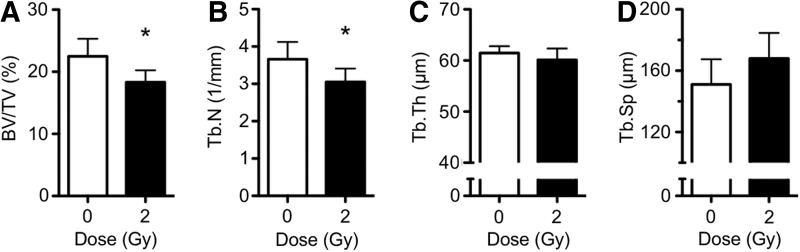
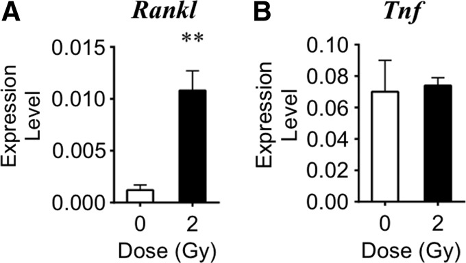
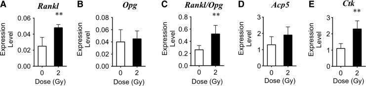
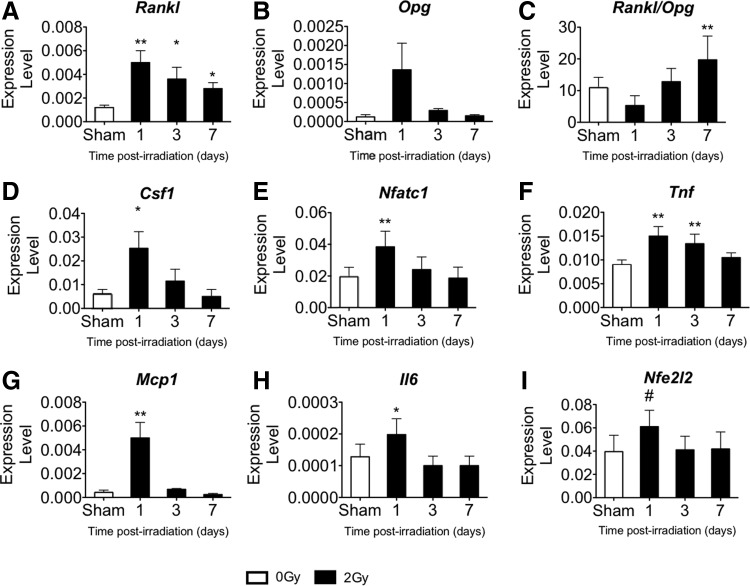
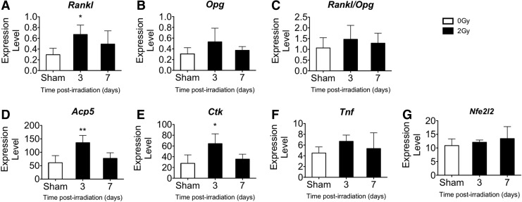
Exposure to ionizing radiation can cause rapid mineral loss and increase bone-resorbing osteoclasts within metabolically active, cancellous bone tissue leading to structural deficits. To better understand mechanisms involved in rapid, radiation-induced bone loss, we determined the influence of total body irradiation on expression of select cytokines known both to stimulate osteoclastogenesis and contribute to inflammatory bone disease. Adult (16 week), male C57BL/6J mice were exposed to either 2 Gy gamma rays ((137)Cs, 0.8 Gy/min) or heavy ions ((56)Fe, 600MeV, 0.50-1.1 Gy/min); this dose corresponds to either a single fraction of radiotherapy (typical total dose is ≥10 Gy) or accumulates over long-duration interplanetary missions. Serum, marrow, and mineralized tissue were harvested 4 h-7 days later. Gamma irradiation caused a prompt (2.6-fold within 4 h) and persistent (peaking at 4.1-fold within 1 day) rise in the expression of the obligate osteoclastogenic cytokine, receptor activator of nuclear factor kappa-B ligand (Rankl), within marrow cells over controls. Similarly, Rankl expression peaked in marrow cells within 3 days of iron exposure (9.2-fold). Changes in Rankl expression induced by gamma irradiation preceded and overlapped with a rise in expression of other pro-osteoclastic cytokines in marrow (eg, monocyte chemotactic protein-1 increased by 11.9-fold, and tumor necrosis factor-alpha increased by 1.7-fold over controls). The ratio, Rankl/Opg, in marrow increased by 1.8-fold, a net pro-resorption balance. In the marrow, expression of the antioxidant transcription factor, Nfe2l2, strongly correlated with expression levels of Nfatc1, Csf1, Tnf, and Rankl. Radiation exposure increased a serum marker of bone resorption (tartrate-resistant acid phosphatase) and led to cancellous bone loss (16% decrement after 1 week). We conclude that total body irradiation (gamma or heavy-ion) caused temporal elevations in the concentrations of specific genes expressed within marrow and mineralized tissue related to bone resorption, including select cytokines that lead to osteoclastogenesis and elevated resorption; this is likely to account for rapid and progressive deterioration of cancellous microarchitecture following exposure to ionizing radiation.
Figures
References
-
- Alwood JS, Yumoto K, Mojarrab R, Limoli CL, Almeida EA, Searby ND, Globus RK. 2010. Heavy ion irradiation and unloading effects on mouse lumbar vertebral microarchitecture, mechanical properties and tissue stresses. Bone 47:248–255 - PubMed
-
- Baxter NN, Habermann EB, Tepper JE, Durham SB, Virnig BA. 2005. Risk of pelvic fractures in older women following pelvic irradiation. JAMA 294(20):2587–2593 - PubMed
-
- Bouxsein ML, Boyd SK, Christiansen BA, Guldberg RE, Jepsen KJ, Muller R. 2010. Guidelines for assessment of bone microstructure in rodents using micro-computed tomography. J Bone Miner Res 25(7):1468–1486 - PubMed
Publication types
MeSH terms
Substances
LinkOut - more resources
Full Text Sources
Other Literature Sources
Research Materials
Miscellaneous