

Positive effects of bFGF modified rat amniotic epithelial cells transplantation on transected rat optic nerve
- PMID: 25734497
- PMCID: PMC4347977
- DOI: 10.1371/journal.pone.0119119
Positive effects of bFGF modified rat amniotic epithelial cells transplantation on transected rat optic nerve
Erratum in
-
Correction: positive effects of bFGF modified rat amniotic epithelial cells transplantation on transected rat optic nerve.PLoS One. 2015 Apr 7;10(4):e0124713. doi: 10.1371/journal.pone.0124713. eCollection 2015. PLoS One. 2015. PMID: 25850015 Free PMC article. No abstract available.
Abstract
Purpose: Effective therapy for visual loss caused by optic nerve injury or diseases has not been achieved even though the optic nerve has the regeneration potential after injury. This study was designed to modify amniotic epithelial cells (AECs) with basic fibroblast growth factor (bFGF) gene, preliminarily investigating its effect on transected optic nerve.
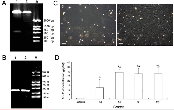
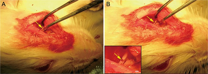
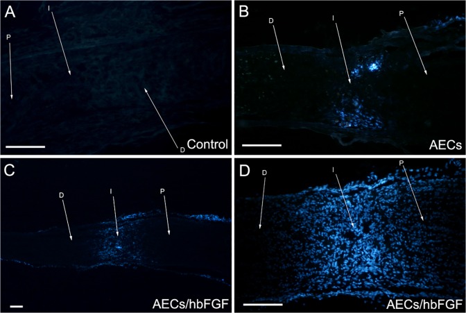
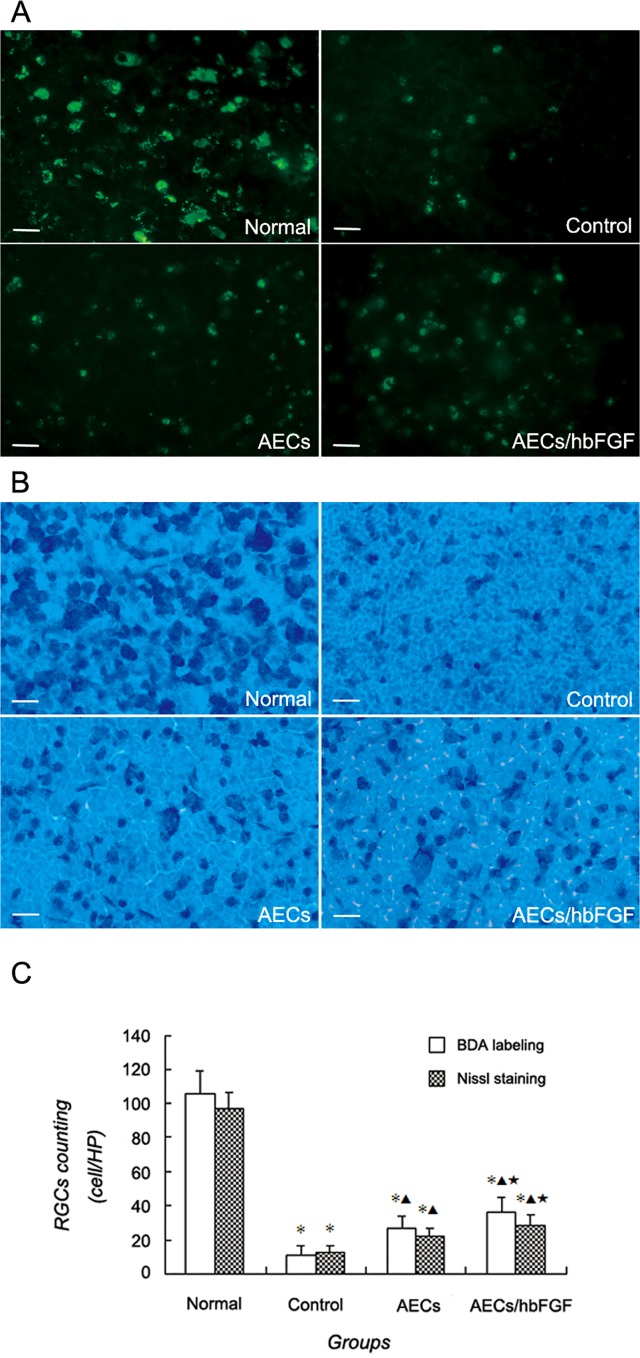
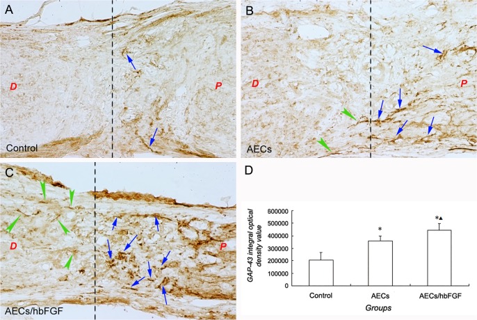
Methods: A human bFGF gene segment was delivered into rat AECs (AECs/hbFGF) by lentiviral vector, and the gene expression was examined by RT-PCR and ELISA. The AECs/hbFGF and untransfected rat AECs were transplanted into the transected site of the rat optic nerve. At 28 days post transplantation, the survival and migration of the transplanted cells was observed by tracking labeled cells; meanwhile retinal ganglion cells (RGCs) were observed and counted by employing biotin dextran amine (BDA) and Nissl staining. Furthermore, the expression of growth associated protein 43 (GAP-43) within the injury site was examined with immunohistochemical staining.
Results: The AECs/hbFGF was proven to express bFGF gene and secrete bFGF peptide. Both AECs/hbFGF and AECs could survive and migrate after transplantation. RGCs counting implicated that RGCs numbers of the cell transplantation groups were significantly higher than that of the control group, and the AECs/hbFGF group was significantly higher than that of the AECs group. Moreover GAP-43 integral optical density value in the control group was significantly lower than that of the cell transplantation groups, and the value in the AECs/hbFGF group was significantly higher than that of the AECs group.
Conclusions: AECs modified with bFGF could reduce RGCs loss and promote expression of GAP-43 in the rat optic nerve transected model, facilitating the process of neural restoration following injury.
Conflict of interest statement
Figures
References
-
- So KF, Aguayo AJ. Lengthy regrowth of cut axons from ganglion cells after peripheral nerve transplantation into the retina of adult rats. Brain Res. 1985;328: 349–354. - PubMed
-
- Berry M, Ahmed Z, Lorber B, Douglas M, Logan A. Regeneration of axons in the visual system. Restor Neurol Neurosci. 2008;26: 147–174. - PubMed
-
- Hicks D. Putative functions of fibroblast growth factors in retinal development, maturation and survival. Semin Cell Dev Biol. 1998;9: 263–269. - PubMed
-
- Blanco RE, López-Roca A, Soto J, Blagburn JM. Basic fibroblast growth factor applied to the optic nerve after injury increases long-term cell survival in the frog retina. J Comp Neurol. 2000;423: 646–658. - PubMed
MeSH terms
Substances
LinkOut - more resources
Full Text Sources
Other Literature Sources
Medical
Miscellaneous
